a smaller, second peak between the ages of 40 and 70, “late- onset IBD.”
1-4IBD flares do not exhibit any monthly or sea- sonal associations.
5Approximately 10% of patients at the first flare of IBD are 60 years or older, with similar distributions between UC and CD; 50% of them are diagnosed between 60 and 70 years of age.
6,7As the number of elderly people in the total population continues to increase, the proportion of late-onset IBD also appears to be increasing globally.
7-9Previous studies have suggested that late-onset IBD differs from early-onset IBD. Several studies have identified lower requirements for steroids, immunomodulators, and surgery, and fewer admissions for flares in late-onset IBD, suggesting a milder disease course.
10,11Several other studies have em-
Old Age at Diagnosis Is Associated With Favorable
Outcomes in Korean Patients With Inflammatory Bowel Disease
Jae Hyuk Choi 1 , Eun Soo Kim 1 , Kwang Bum Cho 1 , Kyung Sik Park 1 , Yoo Jin Lee 1 , Sang Min Lee 1 , Yu Jin Kang 1 , Byung Ik Jang 2 , Kyeong Ok Kim 2 ; Daegu-Gyeongbuk Gastrointestinal Study Group (DGSG)
Division of Gastroenterology and Hepatology, Department of Internal Medicine, Keimyung University School of Medicine
1, Division of Gastroenterology and Hepatology, Department of Internal Medicine, Yeungnam University College of Medicine
2, Daegu, Korea
Received March 31, 2014. Revised September 4, 2014.
Accepted September 5, 2014.
Correspondence to Eun Soo Kim, Division of Gastroenterology and Hepatology, Department of Internal Medicine, Keimyung University School of Medicine, 56 Dalseong-ro, Jung-gu, Daegu 700-712, Korea. Tel: +82-53- 250-8096, Fax: +82-53-250-7088, E-mail: [email protected] Financial support: None. Conflict of interest: None.
INTRODUCTION
Patients are most often diagnosed with IBD in the second or third decades of life; however, epidemiological studies of IBD have revealed a bimodal distribution of disease onset, with an initial peak in the third decade, “early-onset IBD,” and
Background/Aims: Despite the rising incidence and prevalence of inflammatory bowel disease (IBD) in Asian populations, data regarding clinical characteristics of patients in Asia based on age at diagnosis are relatively sparse. The aim of this study was to compare clinical characteristics based on the age at diagnosis according to the Montreal Classification in Korean IBD patients. Methods: We recruited consecutive patients with IBD at two tertiary hospitals and retrospectively reviewed their medical information. Patients were divided into three groups according to their age at diagnosis: youth (<17 years), young adult (17−40 years), and middle-old (>40 years). The main clinical characteristics for comparison were the achievement of a remis- sion state at the last follow-up visit, cumulative rate of surgery, and cumulative use of immunomodulators and tumor necrosis factor-α (TNFα) blockers during the follow-up period. Results: In total, 346 IBD patients were included (Crohn’s disease [CD]
146 and ulcerative colitis 200; 36 youth, 202 young adult, and 113 middle-old). The middle-old group with CD was character- ized by a predominance of uncomplicated behavior ( P=0.013) and a lower frequency of perianal disease (P=0.009). The mid- dle-old group was associated more with a less aggressive disease course than the younger group, as shown by more frequent remission (P=0.004), being less likely to undergo surgery (P<0.001), and lower cumulative use of immunomodulators and TNFα blockers ( P<0.001). Conclusions: Age at diagnosis according to the Montreal Classification is an important prognostic factor for Korean IBD patients. (Intest Res 2015;13:60-67)
Key Words: Inflammatory bowel diseases; Age of onset; Prognosis; Crohn disease; Colitis, ulcerative ISSN 1598-9100(Print) • ISSN 2288-1956(Online)
http://dx.doi.org/10.5217/ir.2015.13.1.60
Intest Res 2015;13(1):60-67
phasized that middle-old age IBD patients have an increased rate of postoperative complications, along with an increased length of admission and increased operation durations.
1,12It is important to understand the differences between early and late-onset IBD as a first step in improving the quality of care delivered to late-onset IBD patients.
To address aspects of clinical definitions and classifica- tion within IBDs and the current status of genetic and se- rological studies, the World Congress of Gastroenterology introduced the Montreal Classification system. Using the Montreal Classification system, IBD is classified based on age at diagnosis, disease extent, and severity.
13Previous stud- ies that compared clinical characteristics between early and late-onset IBD yielded conflicting results.
9,11,14-16One of the reasons for these mixed results may be that the prior studies did not consistently classify IBD according to a unified clas- sification system.
10-12Thus, in our study, the Montreal Clas- sification system was used to classify the clinical spectrum of IBD. There is little information available on the clinical characteristics of IBD based on the age at diagnosis in Asian populations according to the Montreal Classification system.
Therefore, the aim of this study was to compare the clinical characteristics of IBD based on age at diagnosis in Korean patients according to the Montreal Classification system.
METHODS
1. Subjects
To assess whether the age at which patients had been diagnosed was associated with the clinical characteristics of IBD, the age at diagnosis was divided using the Montreal Classification system into youth (<17 years), young adult (17−40 years), and middle-old (>40 years) groups.
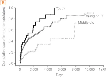
13The main clinical characteristics compared were the achieve- ment of a remission state at the last follow-up visit, cumula- tive rate of surgery, and cumulative use of immunomodula- tors and tumor necrosis factor-α (TNFα) blockers during the follow-up period.
2. Design and Setting
This was a retrospective study conducted at Keimyung University Dongsan Hospital and Yeungnam University Hospital in Daegu Gyeongbuk province, in South-East Ko- rea. The diagnosis of IBD was confirmed by standard clini- cal, radiological, endoscopic, and pathological features.
13,17,18Complete medical records for at least one year after diagno-
sis were required for inclusion in this study. All records were reviewed for accuracy and completeness, and validated by the responsible investigator (ESK). Patients with indetermi- nate colitis, segmental colitis associated with diverticulosis, ischemic colitis, and primary neoplasms were excluded. The study protocol was approved by the Institutional Review Boards of both hospitals.
3. Variable Definitions
According to the Montreal Classification system, the maxi- mum extent and severity of clinical characteristics of IBD at any time point from the time of diagnosis to the most recent clinical evaluation was used for the clinical characteristics of IBD.
13CD location was classified as L1 (ileal), L2 (colonic), L3 (ileocolonic), or L4 (isolated upper disease). Behavior was classified as B1 (non-stricturing, non-penetrating), B2 (stric- turing), B3 (penetrating), or perianal disease. B2 (strictur- ing) and B3 (penetrating) were combined and defined as
“complicated behavior.” Perianal disease included abscesses and/or fistulas. Extent of UC was classified as E1 (ulcerative proctitis), E2 (left-sided UC), or E3 (extensive UC).
Clinical remission was defined as an absence of corticoste- roid treatment and complete relief of symptoms for one year, based on the physician’s global assessment and patient report.
Regarding surgery, our study included procedures involving incision, excision, and anastomosis in small bowel surgery, other large bowel surgery, rectal and perirectal surgery, and other abdominal surgery.
19Use of immunomodulators (aza- thioprine) or TNFα blockers (infliximab) was included if prescribed at least once during follow-up. TNFα blockers were supplied via 5 mg/kg infusions at weeks 0, two, and six, followed by every eight weeks. Follow-up duration was mea- sured in months from the time of diagnosis to the time of the last patient visit.
4. Data Analysis
Statistical analyses were performed using SPSS (version
17.0; IBM Inc., Chicago, IL, USA). We compared the unad-
justed association between the categorical outcome vari-
ables using the chi-squared test and Fisher’s exact test. Dif-
ferences between the mean scores on continuous variables
were assessed using one-way ANOVA. Surgery and cumu-
lative use of immunomodulators or TNFα blockers during
follow-up, based on the age at diagnosis, were estimated by
Kaplan-Meier survival analysis. The log-rank test was used
to measure significant differences according to the Montreal Classification system. P-values <0.05 were considered to be statistically significant.
RESULTS
1. Patient Demographics
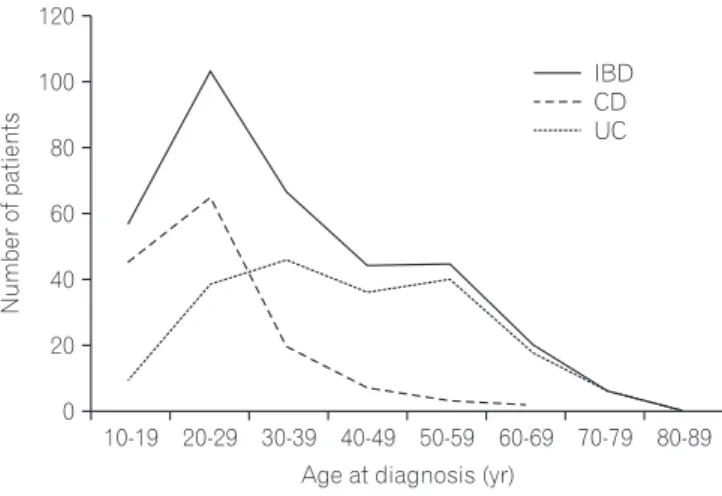
In total, 146 patients with CD and 200 patients with UC were enrolled. The median age at diagnosis was 32 years (range, 12−81 years). The age distribution of UC at diagnosis showed a bimodal distribution with two peaks correspond- ing to the characterizations of early and late-onset UC. The two peak ages were in the third and sixth decades of life.
In the subgroup analysis of CD, however, we did not find a second peak. The distribution of age at diagnosis is shown in Fig. 1.
The youth, young adult, and middle-old groups contained 31 (9.0%), 202 (58.4%), and 113 (32.7%) patients, respec-
tively. The median follow-up duration in patients with IBD was 52 months (range, 27−86 months). There was no sig- nificant difference in the follow-up duration in the youth, young adult, and middle-old groups (median, 64, 50, and 51 months; P=0.604).
The most common manifestations at diagnosis in patients with IBD were hematochezia (39.9%), followed by abdomi- nal pain (32.7%), and diarrhea (23.4%). The younger group had more abdominal pain as a presenting manifestation compared to the other groups (youth, 41.9%; young adult, 33.2%; middle-old, 29.2%), while the older group had more hematochezia (youth, 29.0%; young adult, 33.7%; middle- old, 54.0%; P<0.001; Table 1). However, no such difference was observed when subjects were divided into CD and UC groups (data not shown).
2. Locations and Behaviors of CD According to Age at Diagnosis
The median age of the patients with CD at diagnosis was 23 years (range, 12−66 years). The young adult group (72.6%) had the largest proportion of CD patients, followed by the youth group (18.5%) and the middle-old group (8.9%).
Overall, CD patients were predominantly male (68.5%).
When compared by age at diagnosis, the younger groups (youth and young adult) had more male patients compared with the middle-old group (70.4, 72.6, and 30.8%, respective- ly; P=0.013). In all groups, L1 (ileal) was the most frequent location of CD, and there was no significant difference in locations of CD among the age groups (P=0.933).
Behaviors of CD were as follows: 72 (49.3%) in B1 (non- stricturing, non-penetrating), 33 (22.6%) in B2 (stricturing), and 41 (28.1%) in B3 (penetrating). When compared based on the age of diagnosis, there was a significant difference (P=0.030). B3 (penetrating) was the most common type in the youth group (51.9%), while B1 (non-stricturing, non- penetrating) was the most common type in the young adult Fig. 1. The age distribution of IBD at diagnosis. Showing a bimodal dis-
tribution with the two peaks corresponding to the characterizations of early-onset and late-onset UC. The two peak ages were in the third and sixth decade of life. In CD, however, we did not find a second peak.
10-19 20-29 30-39 40-49 50-59 60-69 70-79 80-89 120
100
80
60
40
20
Number of patients
Age at diagnosis (yr) 0
IBD CD UC
Table 1. The Most Common Manifestations at Diagnosis in Patients With IBD
Variables Youth group (n=31) Young adult group (n=202) Middle-old group (n=113) P-value*
Hematochezia 9 (29.0) 68 (33.7) 61 (54.0) <0.001
Abdominal pain 13 (41.9) 67 (33.2) 33 (29.2)
Diarrhea 6 (19.4) 58 (28.7) 17 (15.0)
Anal pain 3 (9.7) 9 (4.5) 0
Tenesmus 0 0 2 (1.8)
Values are presented as n (%).
*Chi-squared, Fisher’s exact test.
(51.9%) and middle-old groups (46.2%).
Additionally, perianal disease, which is known to be related to a poor prognosis in CD, was more frequently ob- served in the youth group than the young adult and middle- old groups (59.3, 44.3, and 7.7%; P=0.009). The phenotypic presentation of CD is shown in Table 2.
3. Extent of UC According to Age at Diagnosis
The median age of the patients with UC at diagnosis was 41 years (range, 15−81 years). The middle-old group (50.0%) had the highest proportion, followed by the young adult group (48.0%), and the youth group (2.0%). Males were predominant (59.0%) in UC patients and there were no
significant differences among the groups in terms of gender (P=0.804), unlike CD.
The extents of UC were as follows: 87 (43.5%) in E1 (ul- cerative proctitis), 63 (31.5%) in E2 (left sided UC), and 50 (25.0%) in E3 (extensive UC). There were no significant differences in extent of UC according to age at diagnosis (P=0.349). The phenotypic presentation of UC is shown in Table 3.
4. Main Clinical Outcomes According to Age at Diagno- sis
We compared disease courses of IBD, including achieving a remission state at last follow-up, cumulative rate of surgery, Table 2. Clinical Characteristics According to Age of Diagnosis in Patients With CD
Variables Youth group (n=27) Young adult group (n=106) Middle-old group (n=13) P-value*
Male gender 19 (70.4) 77 (72.6) 4 (30.8) 0.013
Location NS
L1 14 (51.9) 53 (50.0) 5 (38.5)
L2 7 (25.9) 26 (24.5) 4 (30.8)
L3 6 (22.2) 27 (25.5) 4 (30.8)
Behavior 0.030
B1 11 (40.7) 55 (51.9) 6 (46.2)
B2 2 (7.4) 27 (25.5) 4 (30.8)
B3 14 (51.9) 24 (22.6) 3 (23.1)
Perianal disease 16 (59.3) 47 (44.3) 1 (7.7) 0.009
Follow-up duration, month (median) 55 (18−76) 54 (33−87) 47 (35−79) NS
†Values are presented as n (%).
Location and behavior in CD based on the Montreal classification.
L1, ileal; L2, colonic; L3, ileocolonic; B1, non-stricturing, non-penetrating; B2, stricturing; B3, penetrating.
*Chi-squared, Fisher’s exact test.
†
ANOVA test.
Table 3. Clinical Characteristics According to Age of Diagnosis in Patients With UC
Variables Youth group (n=4) Young adult group (n=96) Middle-old group (n=100) P-value*
Male/Female 3/1 55/41 60/40 NS
Extent, n (%) NS
E1 1 (25) 42 (43.8) 44 (44.0)
E2 0 31 (32.3) 32 (32.0)
E3 3 (75.0) 23 (24.0) 24 (24.0)
Follow-up duration, mo (median) 84 (79−163) 50 (26−93) 51 (21−79) NS
†Location in UC based on the Montreal classification.
E1, ulcerative proctitis; E2, left sided UC; E3, extensive UC.
*Chi-squared, Fisher’s exact test.
†