• 검색 결과가 없습니다.

Analysis of MAPK Signaling Pathway Genes in the Intestinal Mucosal Layer of Necrotic Eenteritis-Afflicted Two Inbred Chicken Lines

N/A
N/A
Protected

Academic year: 2021

Share "Analysis of MAPK Signaling Pathway Genes in the Intestinal Mucosal Layer of Necrotic Eenteritis-Afflicted Two Inbred Chicken Lines"

Copied!
11
0
0

로드 중.... (전체 텍스트 보기)

전체 글

(1)

Analysis of MAPK Signaling Pathway Genes in the Intestinal Mucosal Layer of Necrotic Eenteritis-Afflicted Two Inbred Chicken Lines

Anh Duc Truong

1,3

, Yeojin Hong

1

, Janggeun Lee

1

, Kyungbaek Lee

1

, Hyun S. Lillehoj

2

and Yeong Ho Hong

1

1

Department of Animal Science and Technology, Chung-Ang University, Anseong 17546, Republic of Korea,

2

Animal Biosciences and Biotechnology Laboratory, Agricultural Research Services, United States Department of Agriculture, MD 20705, USA

3

National Institute of Veterinary Research, 86 Truong Chinh, Dong Da, Hanoi, Vietnam.

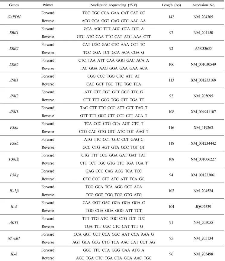
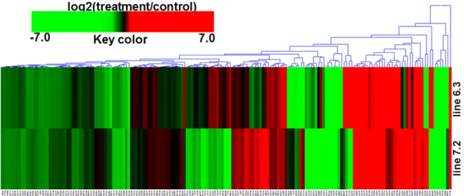
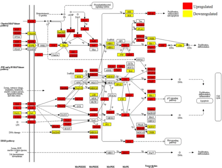
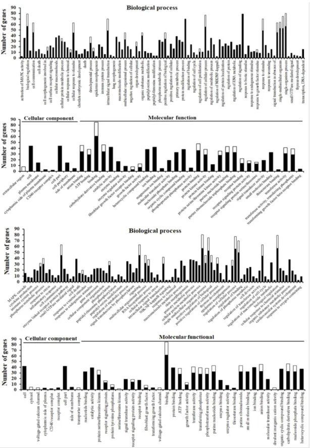
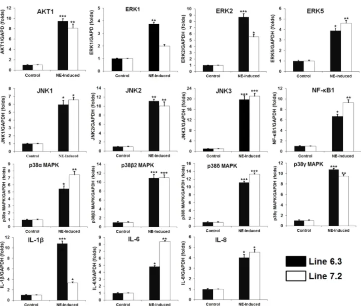
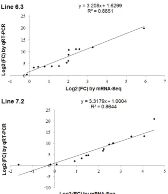
ABSTRACT Mitogen-activated protein kinase (MAPK) signaling pathways play a key role in innate immunity, inflammation, cell proliferation, cell differentiation, and cell death. The main objective of this study was to investigate the expression level of candidate MAPK pathway genes in the intestinal mucosal layer of two genetically disparate chicken lines (Marek’s disease- resistant line 6.3 and Marek’s disease-susceptible line 7.2) induced with necrotic enteritis (NE). Using high-throughput RNA sequencing, we investigated 178 MAPK signaling pathway related genes that were significantly and differentially expressed between the intestinal mucosal layers of the NE-afflicted and control chickens. In total, 15 MAPK pathway genes were further measured by quantitative real-time PCR(qRT-PCR) and the results were consistent with the RNA-sequencing data. All 178 identified genes were annotated through Gene Ontology and mapped onto the KEGG chicken MAPK signaling pathway. Several key genes of the MAPK pathway, ERK1/2, JNK1-3, p38 MAPK, MAP2K1-4, NF-κB1/2, c-Fos, AP-1, Jun-D, and Jun, were differentially expressed in the two chicken lines. Therefore, we believe that RNA sequencing and qRT-PCR analysis provide resourceful information for future studies on MAPK signaling of genetically disparate chicken lines in response to pathogens.

(Key words: chicken, DEG, MAPK pathway, RNA-seq, qRT-PCR)

To whom correspondence should be addressed : [email protected]

INTRODUCTION

Mitogen-activated protein kinase (MAPK) signaling pathway, a family of serine/threonine protein kinases widely conserved in eukaryotes, plays a key role in innate immunity and is in- volved in many cellular functions such as inflammation, cell proliferation, cell differentiation, and cell death (Krishna and Narang, 2008; Yang et al., 2003). To date, several key genes of MAPK pathway have been well-characterized in mammals, such as extracellular regulated kinases (ERK1-5, ERK7-8), c-Jun NH2 terminal kinases (JNK1-3), p38 MAPK (p38α/β/γ/

δ), and Nemo-like kinase (NLK) (Cargnello and Roux, 2011;

Krens et al., 2006; Schaeffer and Weber, 1999). Among them, the most extensively studied MAPK pathway genes are ERK1/2, JNKs and p38MAPK (Garrington and Johnson, 1999; Krishna and Narang, 2008; Yang et al., 2003). The role of MAPK sig- naling pathway in mammals, which has already been studied and published by researchers, is gaining more attention in recent years due to its complex roles in different cancers,

including breast, lung, colon, and skin cancer, as well as can- cers of hematopoietic origin (Chen et al., 2014; Dhillon et al., 2007; Hirakawa et al., 2014; Neuzillet et al., 2014) and rheu- matoid arthritis (Gonzalo-Gil and Galindo-Izquierdo, 2014).

Moreover, the function of the MAPK pathway depends on its

constitutive and extensive communication with the TGF-β

signaling pathway, the phosphatidylinositol-3 kinase/Akt path-

way, the Wnt pathway, the Hedgehog pathway, the Notch

pathway, and the interleukin/interferon-gamma/tumor necrosis

factor-alpha cytokines interaction (Elumalai et al., 2014; Guo

and Wang, 2009; Huang et al., 2014; Roskoski, 2012). Previous

studies have shown that influenza virus, herpes simplex virus

type 2, and human immunodeficiency virus type 1 (HIV-1) can

activate MAPK pathway genes including ERK1/2 (p42/p44

MAPK), JNK, or p38 MAPK, which further increase the ex-

pressions of cytokines or affect the growth of viruses (Andrus

et al., 2011; Horton et al., 2006; Wang et al., 2012). In addition,

regulation of many genes by MAPK pathway are dependent

on NF-κB, c-Fos, and c-Jun transcription (Das et al., 2013;

(2)

Huang et al., 2014). However, the expression of MAPK signa- ling pathway-related genes in chicken co-infected by protozoan parasite and bacteria is not yet studied.

Therefore, the aim of this study is to analyze the MAPK signaling pathway cascade that mediates immune-related gene responses in the intestinal mucosal layer of two genetically disparate chicken lines (namely, Marek’s disease (MD)-resistant line 6.3 and MD-susceptible line 7.2) afflicted with necrotic enteritis (NE), the result of co-infecting Eimeria maxima and Clostridium perfringens type A.

MATERIALS AND METHODS

1. Experimental Birds and Care

The two White Leghorn chicken lines, the MD-resistant line 6.3 and MD-susceptible line 7.2, were obtained from the Avian Disease and Oncology Laboratory (East Lansing, MI, USA) of the Agricultural Research Service of the United Sta- tes Department of Agriculture (USDA). Care and experimental use of the birds were approved by the Beltsville Area Insti- tutional Animal Care and Use Committee of the USDA-ARS.

All the experiments including bird management, experimental induction of NE, and sample collection were performed at USDA-ARS, Beltsville, USA.

2. Experimental Induction of NE and Sample Collection for RNA Sequencing

Twenty chickens per group were randomly selected and in- fected with E. maxima strain 41A (1.0 × 10

4

oocysts/chicken) by oral gavage on day 14 post-hatch, followed by oral gavage with C. perfringens strain Del-1 (1.0 × 10

9

CFU/chicken) on day 18 post-hatch, as previously reported (Jang et al., 2012).

For the development of NE, the birds were fed a non-medi- cated commercial basal ration of 17% crude protein from 1 to 18 days of age, and the feed was replaced with a commercial non-medicated feed containing 24% crude protein at 18∼20 days of age. Chickens were raised in Petersime starter brooder units, provided ad libitum access to water, and maintained under uniform standard management conditions. The experi- mental and control groups were kept in separate rooms to prevent cross-contamination throughout the course of the ex- periment. The intestinal mucosal layers were collected from

NE-afflicted chickens of both lines and from their respective controls (five each) on day 20 post-hatch. Total RNA was extracted from all samples and used for RNA-Seq and data analyses as previously described (Truong et al., 2015).

3. MAPK Signaling Pathway Enrichment Analysis The previous study reported that the expression levels of 7,841 and 6,043 genes were significantly upregulated in line 6.3 and line 7.2, respectively, and 2,919 and 2,764 genes were downregulated in line 6.3 and line 7.2 when afflicted with NE compared with their uninfected controls, respectively (Truong et al., 2015). All transcript coding genes were alig- ned with the Kyoto Encyclopedia of Genes and Genomes (KEGG: http://www.kegg.jp/) pathway database using DAVID Bioinformatics Resources version 6.7 (National Institute of Allergy and Infectious Diseases/National Institutes of Health, USA; http://david.abcc.ncifcrf.gov/tools.jsp) to find the genes involved with the MAPK signaling pathway of chicken.

4. Hierarchical Clustering and Gene Ontology (GO) Analysis

The genes identified in MAPK pathway were analyzed and subjected to hierarchical clustering using Cluster (MeV v4.9:

www.tm4.org) and Java Treeview software (http://jtreeview.

sourceforge.net/). Cluster map analysis of genes identified the distance between two lines using Euclidean distance. The p values were calculated using the right-tailed Fisher’s exact test. Then, GO functional enrichment analysis was carried out using Blast2 GO (version 2.7.1) (http://www.blast2go.org/). To summarize GO terms, we removed redundant GO terms and used REVIGO software (http://revigo.irb.hr/) as described by Supek et al (Supek et al., 2011).

5. Quantitative Real-time PCR for MAPK Pathway Genes

Quantitative real-time PCR was performed to verify the differential expression of genes revealed by the RNA-Seq.

Primer sequences of 15 genes and the internal control gene

glyceral- dehyde-3-phosphate dehydrogenase (GAPDH) were

designed using Lasergene software (DNASTAR Inc. Madison,

WI, USA) and synthesized by Genotech Co. Ltd. (Daejeon,

South Korea) (Table 1). The complementary DNA (cDNA) was

(3)

Table 1. Primer sequences for the real-time PCR analysis of MAPK signaling pathway genes

Genes Primer Nucleotide sequencing (5'-3') Length (bp) Accession No

GAPDH

Forward TGC TGC CCA GAA CAT CAT CC

142 NM_204305

Reverse ACG GCA GGT CAG GTC AAC AA ERK1

Forward GCA AGC TTT AGC CCA TCC A

97 NM_204150

Reverse GTC ATC CAA TTC CAT ATC AAA CTT ERK2

Forward CAT CGC GAC CTC AAA CCT TC

92 AY033635

Reverse TCC GGA TCT GCA ACA CGA G ERK5

Forward CTC TAA ATT CAA GGG GAC ACA A

106 NM_001030549

Reverse TAC GGA AAG GGA GAA GAA ACA JNK1

Forward CGG CCC TGG CTC ATT AT

113 XM_001233168

Reverse CAC GCT TGC TTC TGC TCA JNK2

Forward ATT GTT TGT GCT GCG TTC G

92 NM_205095

Reverse CTT TTT GCG TGG GTT TGA TT JNK3

Forward TAC CTT TTC CCC ATT CCT TAG T

108 XM_004941107

Reverse GTT TTT GCC CTT CCT CTT ACA T

P38 α Forward TCA CCC CTG CCA AGT CTC T

116 XM_419263

Reverse CTG CAC GTG GTC ATC TGT AAG T

P38 δ Forward ATG TTC CCT GTC CCT GAG C

118 XM_001234442

Reverse GCC CTG AGT GTA GCC TGT GT P38 β 2

Forward CTG TTT CCG GGA GAT GAT TAT

108 NM_001006227

Reverse CTT TCT TGC GTG TTC TGA TGA T

P38 γ Forward GAG CCC CAG AGG TCA TCC

94 XM_001233061

Reverse CTC CCC GTT ATC ATT TCA GC

IL-1 β Forward TGG GCA TCA AGG GCT ACA

102 NM_204524

Reverse TCG GGT TGG TGG GTG ATG

IL-6

Forward CAA GGT GAC GGA GGA GGA C

104 JQ897539

Reverse TGG CGA GGA GGG ATT TCT

AKT1

Forward TTT TTG ATC TGC CTG TCT TCC

91 NM_205055

Reverse TGA TTT CGC CTC CAT TTT G NF- κ B1

Forward CCA GGT CCT CCA GGC AAT CCA AAA G

95 NM_205134

Reverse AGT GCA GGG CTG TCA AAC CAT CGT AG IL-8

Forward GGC TTG CTA GGG GAA ATG A

96 NM_205498

Reverse AGC TGA CTC TGA CTA GGA AAC TGC

synthesized using Maxima First Strand cDNA Synthesis Kit (Thermo Scientific, MA, USA) according to the manufacturer’s

recommendations. Quantitative real-time PCR was performed

using 2X Power SYBR Green Master Mix (Roche, IN, USA)

(4)

Fig. 1. Hierarchical clustering of 178 genes differentially expressed in NE-afflicted two lines compared with respective control and involved in the MAPK signaling pathway. Genes showed in red were upregulated, and those showed in green were downregulated in NE-induced two lines. Hierarchical clusters of genes and samples were based on Spearman’s rank correlation analyses.

according to the manufacturer’s instructions in a LightCycler 96 system (Roche). The relative quantification of gene-specific expression was calculated using the 2

−ΔΔCt

method after being normalized with chicken GAPDH (Livak and Schmittgen, 2001). All quantitative RT-PCR reactions were performed in triplicate.

6. Statistical Analysis

Statistical analysis was performed using SPSS software (IBM, SPSS 23.0 for Windows, Chicago, IL, USA). Data were expressed as mean values±SE. Comparisons were carried out using a Student’s t-test for two-group comparisons, and the level of significant difference was set at p<0.05.

RESULTS

1. MAPK Signaling Pathway-related Genes in NE afflicted Two Chicken Lines

Based on our previous RNA-seq data (Truong et al., 2015) and genes mapped to the KEGG pathway database, we deter- mined 178 DEGs between the two NE-afflicted chicken lines and how they’re involved in the MAPK signaling pathway (data not shown). Based on the p<0.01 and fold-change cut off ≥2, several transcripts were significantly upregulated and downregulated in intestinal mucosal layer of two lines. Com-

pared to respective control, the expression level of 25 genes (BDNF, CACNA1I, CACNA2D2, CACNA2D4, CASP3, DUSP4, IL1B, IL1R1, IL1R2, IL1RAPL2, IL1RL1, IL1RL2, JUN, MAPK10, MAPK11, MAPK12, MAPK13, MAPK3, MAPK8, MAPK9, NGF, RASGRP3, RPS6KA2, TGFB1 and TNFA) were significantly upregulated by 2.0- to 5.9-fold, and 10 genes (CACNG2, TGFB2, FASLG, CDKN1B, FGF16, FGF1, FGF4, RASGRF2, RASGRF1 and FGF9) were significantly downre- gulated by 2.1- to 5.1-fold in intestinal mucosal of line 6.3 induced NE (Fig. 1).

Moreover, we found 29 genes showing different expression in line 7.2 in which the expression of 21 genes (CACNA2D2, CCNB2, CDKN1A, CDKN2C, DUSP4, FGF16, FGF22, IL1RL1, MAPK10, MAPK11, MAPK12, MAPK13, MAPK14, MAPK3, MAPK8, MAPK9, PTPN5, TGFB1, TNFRSF1A, TNFRSF1B and TNFRSF6B) were markedly increased by 2.0- to 4.5-fold, and 8 genes (TGFB2, IL-1RL2, RASGRF2, CDKN1B, CASP3, FASLG, CACNA2D1 and FGF4) were significantly decreased by 2.0- to 3.6-fold in intestinal mucosal layer of NE-afflicted line 7.2 (Fig. 1).

2. Localization of 178 MAPK Pathway-related Genes in Intestinal Mucosal Layer of NE afflicted Two Chicken Lines

We investigated and mapped the position of 178 MAPK

(5)

Fig. 2. Localization of 178 genes differentially expressed in NE-afflicted line 6.3 compared with control in the MAPK signaling pathway. The position of these genes was searched and marked in KEGG Gallus gallus MAPK signaling pathway. Upregulated and downregulated genes were indicated by red and yellow color, respectively.

pathway-related genes that are in the intestinal mucosal layer in KEGG Gallus gallus MAPK signaling pathway. One hundred twenty-eight genes were upregulated, and 50 genes were downregulated compared with the control group in NE- induced line 6.3. In addition, one hundred and one genes were upregulated, and 77 genes were downregulated in NE- afflicted line 7.2 compared with the control group. All of these 178 DEGs (p<0.01) were critically positioned in the KEGG MAPK signaling pathway. The positions of 178 genes were differentially expressed in the intestinal mucosal layer of line 6.3 in Fig. 2 and 7.2 afflicted with NE (data not shown).

3. Gene Ontology (GO) Analysis of Differen- tially Expressed Genes

All upregulated and downregulated genes in intestinal mu-

cosal layer of two lines were categorized into three major

functional groups including molecular function, cellular com-

ponent, and biological process according to GO. Each major

group was further divided into many subcategories. To find

the major cluster of each GO, the REVIGO software was

used to analyze subcategories and sort the same functions

into one cluster base on p<0.01. The results of GO analysis

in NE-afflicted line 6.3 revealed that all upregulated and

downregulated genes were categorized into three major func-

tional groups. The biological process group was divided into

99 subcategories such as biological regulation, intracellular

signal transduction, response to stimulus regulation, and single-

organism process. The cellular component group was divided

(6)

into 8 subcategories such as cell part and cell periphery. The molecular function group was divided into 31 subcategories such as binding, catalytic activity, and protein serine/threonine kinase activity (Fig. 3A).

On the other hand, the results of GO analysis in NE- afflicted line 7.2 revealed that all upregulated and downre- gulated genes were categorized into three major functional groups. The biological process group was divided into 93 subcategories such as biological regulation, regulation of cellular process, response to stimulus regulation, and single- organism process. The cellular component group was divided into 9 subcategories such as cell and receptor complex. The molecular function group was divided into 33 subcategories such as binding, catalytic activity and molecular transducer activity (Fig. 3B).

Overall, most of upregulated and downregulated genes in NE-induced line 6.3 are those for immune-related transcrip- tional control of pathogens. Most of the upregulated and downregulated genes fell into the enzymatic and metabolic activity that occurs at the level of a multicellular organism and cells in NE-afflicted line 7.2.

4. Quantitative Real-Time PCR Analysis for MAPK Pathway Genes of NE afflicted Two Chic- ken Lines

To further understand the role of the MAPK signaling pathway in the two genetically NE-induced chicken lines, the expression level of ERKs (ERK1/2 and ERK5), p38 MAPK (p38α, p38β2, p38γ, and p38δ), JNKs (1-3), AKT1, NF-κB1, IL-1β, IL-6 and IL-8 were quantitatively determined by real- time PCR. As shown in Fig. 4, after Eimeria maxima and Clostridium perfringens infection, the expression levels of 15 genes were significantly increased in the intestinal mucosal layer of two chicken lines compared to respective control.

We compared the results of qRT-PCR with those generated from RNA-Seq analysis of these transcripts. Expression trends were consistent with all transcripts in both analyses, with a correlation coefficient of R

2

=0.885 for line 6.3 and R

2

=0.864 for line 7.2 (Fig. 5).

DISCUSSION

The MAPK family is composed of four prototype mem- bers, the extracellular signal-regulated kinase 1/2 (ERK1/2), p38 MAPK, the Jun-N-terminal kinase or stress-activated protein kinase (JNK/SAPK), and ERK5 that are activated by specific MAPKKs: MAP2K1/2 for ERK1/2, MKK3/6 for the p38 MAPK family, MKK4/7 (JNKK1/2) for the JNKs, and MAP2K5 for ERK5, which are responsible for signaling pathways, leading to regulation of cell proliferation, cell di- fferentiation, apoptosis and immune responses (DeSilva et al., 1996). These studies have mainly shown the differential ex- pression of MAPK pathway-related genes in intestinal mucosal layer of NE-afflicted line 6.3 and line 7.2, compared to re- spective control. Based on RNA-seq data, we identified 178 MAPK pathway-related genes in two lines. The GO terms were investigated using the DEGs of two lines. We identified 99, 8 and 31 subcategories of the biological process, cellular component, and molecular function in NE-induced line 6.3, respectively. Meanwhile, 99, 9 and 33 subcategories were ex- pressed in the biological process, cellular component, and molecular function in NE-afflicted line 7.2, respectively. The large number of GO terms indicated that the MAPK signaling pathway may play roles in molecular signaling and regulation of innate immunity, inflammation, cell proliferation, cell diffe- rentiation, and cell death in the two NE-afflicted lines.

ERKs are the first identified MAPKs group and many stu- dies have already been done on mammals (Plotnikov et al., 2011). ERK1 and ERK2 are two isoforms with 83% amino acid homology. Both isoforms are expressed in various tissues and can be activated by a large number of extracellular and intracellular stimuli (Lefloch et al., 2008; Liu X et al., 2012).

Activated Raf (or other MAP3K) binds and phosphorylates

downstream kinase MAP2K1 and MAP2K2 with dual speci-

ficity, which in turn phosphorylates ERK1/2 (Liu SY et al.,

2012). In this study, MAP2K2 and ERK1/2 genes were sig-

nificantly upregulated in two NE-afflicted chicken lines, which

may be associated with activation of upstream molecules such

as NF-κB1/2, ELK-1, c-Fos, c-Jun and STAT1/3. Both NF-

κB1 and STAT3 are rapidly activated in response to various

stimuli including viral infection and cytokines, thus controlling

the expression of anti-apoptotic, proliferative and immune re-

sponse genes (Decker and Meinke, 1997; O'Sullivan et al.,

2009). RNA-seq results showed that the expressions of IL-1,

(7)

Fig. 3. Gene ontology. All of the upregulated and downregulated genes in intestinal mucosal layer of NE-afflicted line 6.3 (A) and

line 7.2 (B) were categorized in specific functional groups according to gene ontology using Fisher's exact test (p<0.01). The x-axis

represents the number of genes. Upregulatedgenesarecolored in black, and downregulated genes are colored in white.

(8)

Fig. 4. Quantitative real-time PCR analysis of the MAPK pathway genes in NE-afflicted lines 6.3 and line 7.2 compared with respective control. Levels are normalized to those of GAPDH.

Data are presented as the mean±SE (n=3) of three independent experiments:

*

p<0.05,

**

p<0.01,

***

p<0.001.

TGF-β, and TNF-α and their receptors were upregulated in two lines (Truong et al., 2015). These data indicated that EM/CP co-infection could increase the release of IL-1, TGF- β, and TNF-α in the intestinal mucosal layer, as well as mediate inflammation and apoptosis.

JNKs are encoded by three distinct genes (JNK1, JNK2, and JNK3) and activated by a large number of external sti- muli as well as the initial signaling pathway (Dhanasekaran and Reddy, 2008). Previous reports demonstrated that several transcription factors are upstream proteins like NFAT2/4, c- Jun, JunD, ATF2, AP-1 and p53 activated by JNK (Kristensen et al., 2004). Once activated by phosphorylation, these trans-

cription factors can induce proinflammatory cytokines (Das

and Muniyappa, 2010). Subsequently, transcription factors of

NF-κB1/2, ELK-4, c-Fos, AP-1, ATF2, p53, JunD, and c-Jun

are activated, which play a key role in regulating the immune

response to viral infection and stimulating the releases of

cytokines such as TNF-α, IFN-β, IL-1β, IL-2, IL-6 and IL-8

(Ting et al., 2010). JNK is also involved in the regulation of

apoptosis through the modulation of c-Jun/AP-1 and TGF-β

in MDCK cells (Xing et al., 2010). Prolonged activation of

JNK can mediate apoptosis, whereas transient activation can

promote cell survival (Yan et al., 2010). The upregulation of

JNKs in the two NE-afflicted chicken lines suggested that

(9)

Fig. 5. Comparison of the log2 (FC) of 15 selected transcripts using RNA-Seq and qRT-PCR on the intestinal mucosal layer of two genetically disparate NE-afflicted chicken lines.

JNKs are phosphorylating some transcription factors (Das and Muniyappa, 2010; Dhanasekaran and Reddy, 2008), primarily components of AP-1 such as JUN, NF-κBs, ELK-4, c-Fos and ATF2 and then regulating AP-1 transcriptional activity in in- testinal mucosa layer of two lines response to NE.

One of the MAPKs family, p38, also plays an important role in the cascades of cellular responses evoked by extra- cellular stimuli (Medders and Kaul, 2011). The p38 MAPKs phosphorylate a broad range of proteins, and it has been estimated that they may have approximately 200 to 300 sub- strates each (Cargnello and Roux, 2011; Medders and Kaul, 2011). In addition, p38 MAPKs may control proinflammatory responses to stimulation through activated by MAPKKs, MKK- 3, and MKK6 and led to upregulation of RANTES, IL-8, and TNF-α expression (Das and Muniyappa, 2010; Huang et al., 2014). Our results indicated that the upregulation of p38 MAPK α, β2, γ and δ genes may control the expression of proinfla- mmatory response to NE disease in the two chicken lines.

PI3K/AKT signaling pathway, member of ERK or JNK pathway, plays an important role in the growth and survival

of cells. The AKT cascade is activated by receptor tyrosine kinases, cytokine receptors, G-protein coupled receptors, and other stimuli (Elumalai et al., 2014; Zhou et al., 2014). There are three closely related isoforms of AKT: AKT1, AKT2, and AKT3, which are differentially expressed in various tissues.

Phosphorylation of AKT on Thr

308

and Ser

473

leads to full ac- tivation, and AKT can be downregulated through the action of PTEN, PPA2, and mTOR. PI3K/PTEN/AKT along with mTOR form a well-established target set for treatment of cancer, diabetes, neurological disorders (Chen et al., 2014; Li et al., 2014). The interaction and association between PIK/

AKT and MAPK signaling pathway led to control of cell growth, differentiation, response to pathogens (Elumalai et al., 2014; Li et al., 2014). As showed in Fig. 1 and 4, AKT1 and AKT3 mRNA expression were upregulated in two NE-afflicted chicken lines by RNA sequencing or qRT-PCR. The upregu- lation of PI3K/AKT may affect the MAPK signaling pathway and can regulate cell survival in mucosal layer in the two chicken lines’ response to NE disease.

CONCLUSION

The MAPK signaling pathway regulates the adaptive and innate immune immunity, inflammation, cell proliferation, cell differentiation, and death of mucosal cell as well as epithelial cell repair and regeneration. Here, we investigated the DEGs and GO terms of the MAPK pathway related genes in the intestinal mucosal layer of NE-afflicted two chicken lines and found several key genes of MAPK pathway, namely, ERK1- 2, JNK1-3, p38MAPK, MAP2K1-4, NF-κB1/2, c-Fos, AP-1, Jun-D, and Jun, which were differentially expressed in two lines. Therefore, we believe that RNA sequencing and quan- titative real-time PCR analysis provide resourceful information for future studies on the MAPK pathway in two genetically disparate chicken lines’ response to pathogens.

ACKNOWLEDGMENTS

This work was supported by the Next-Generation BioGreen

21 Program (No. PJ00808401), Republic of Korea. The inbred

chicken lines were obtained from ADOL (Avian Disease and

(10)

Oncology Laboratory), USDA-Agricultural Research Service, East Lansing, MI and thanks to Dr. Hans H. Cheng.

REFERENCES

Andrus L, Marukian S, Jones CT, Catanese MT, Sheahan TP, Schoggins JW, Barry WT, Dustin LB, Trehan K, Ploss A, Bhatia SN, Rice CM 2011 Expression of paramyxovirus V proteins promotes replication and spread of hepatitis C virus in cultures of primary human fetal liver cells. Hepa- tology (Baltimore, Md.) 54(6):1901-1912.

Cargnello M, Roux PP 2011 Activation and function of the MAPKs and their substrates, the MAPK-activated protein kinases. Microbiol Mol Biol Rev 75(1):50-83.

Chen J, O'Donoghue A, Deng YF, Zhang B, Kent F, O'Hare T 2014 The effect of lycopene on the PI3K/Akt signalling pathway in prostate cancer. Anticancer Agents Med Chem 14(6):800-805.

Das D, Holmes A, Murphy GA, Mishra K, Rosenkranz AC, Horowitz JD, Kennedy JA 2013 TGF-beta1-induced MAPK activation promotes collagen synthesis, nodule formation, redox stress and cellular senescence in porcine aortic valve interstitial cells. J Heart Valve Dis 22(5):621-630.

Das KC, Muniyappa H 2010 c-Jun-NH

2

terminal kinase (JNK)- mediates AP-1 activation by thioredoxin: Phosphorylation of cJun, JunB, and Fra-1. Mol Cell Biochem 337(1-2):

53-63.

Decker T, Meinke A 1997 Jaks, stats and the immune sys- tem. Immunobiology 198(1-3):99-111.

DeSilva DR, Feeser WS, Tancula EJ, Scherle PA 1996 Anergic T cells are defective in both jun NH

2

-terminal kinase and mitogen-activated protein kinase signaling pathways. J Exp Med 183(5):2017-2023.

Dhanasekaran DN, Reddy EP 2008 JNK signaling in apop- tosis. Oncogene 27(48):6245-6251.

Dhillon AS, Hagan S, Rath O, Kolch W 2007 MAP kinase sig- nalling pathways in cancer. Oncogene 26(22):3279-3290.

Elumalai P, Arunkumar R, Benson CS, Sharmila G, Arunaka- ran J 2014 Nimbolide inhibits IGF-I-mediated PI3K/Akt and MAPK signalling in human breast cancer cell lines (MCF-7 and MDA-MB-231). Cell Biochem Funct.

Garrington TP, Johnson GL 1999 Organization and regulation

of mitogen-activated protein kinase signaling pathways.

Curr Opin Cell Biol 11(2):211-218.

Gonzalo-Gil E, Galindo-Izquierdo M 2014 Role of transfor- ming growth factor-beta (TGF) beta in the physiopathology of rheumatoid arthritis. Reumatologia Clinica 10(3):174-179.

Guo X, Wang XF 2009 Signaling cross-talk between TGF- beta/BMP and other pathways. Cell Res 19(1):71-88.

Hirakawa M, Takimoto R, Tamura F, Yoshida M, Ono M, Murase K, Sato Y, Osuga T, Sato T, Iyama S, Miyanishi K, Takada K, Hayashi T, Kobune M, Kato J 2014 Fucosy- lated TGF-beta receptors transduces a signal for epithelial- mesenchymal transition in colorectal cancer cells. Br J Cancer 110(1):156-163.

Horton H, Havenar-Daughton C, Lee D, Moore E, Cao J, McNevin J, Andrus T, Zhu H, Rubin A, Zhu T, Celum C, McElrath MJ 2006 Induction of human immunodeficiency virus type 1 (HIV-1)-specific T-cell responses in HIV va- ccine trial participants who subsequently acquire HIV-1 infection. J Virol 80(19):9779-9788.

Huang RL, Yuan Y, Tu J, Zou GM, Li Q 2014 Opposing TNF- alpha/IL-1beta- and BMP-2-activated MAPK signaling pathways converge on Runx2 to regulate BMP-2-induced osteoblastic differentiation. Cell Death Dis 5:e1187.

Jang SI, Lillehoj HS, Lee SH, Lee KW, Lillehoj EP, Hong YH, An DJ, Jeong W, Chun JE, Bertrand F, Dupuis L, Deville S, Arous JB 2012 Vaccination with Clostridium perfringens recombinant proteins in combination with Mon- tanide ISA 71 VG adjuvant increases protection against experimental necrotic enteritis in commercial broiler chic- kens. Vaccine 30(36):5401-5406.

Krens SF, Spaink HP, Snaar-Jagalska BE 2006 Functions of the MAPK family in vertebrate-development. FEBS Lett 580(21):4984-4990.

Krishna M, Narang H 2008 The complexity of mitogen-acti- vated protein kinases (MAPKs) made simple. Cell Mol Life Sci 65(22):3525-3544.

Kristensen NN, Madsen AN, Thomsen AR, Christensen JP 2004 Cytokine production by virus-specific CD8(+) T cells varies with activation state and localization, but not with TCR avidity. J Gen Virol 85(Pt 6):1703-1712.

Lefloch R, Pouyssegur J, Lenormand P 2008 Single and com-

bined silencing of ERK1 and ERK2 reveals their positive

(11)

contribution to growth signaling depending on their expre- ssion levels. Mol Cell Biol 28(1):511-527.

Li Z, Lu H, Li ZB 2014 The PI3K/Akt signalling pathway may play an internal role related to abnormal condylar growth:

a preliminary study. Int J Oral Maxillofac Surg.

Liu SY, Liang Y, Lin TX, Su F, Liang WW, Uwe H, Li Y 2012 MEK1 and MEK2 differentially regulate human in- sulin- and insulin glargine-induced human bladder cancer T24 cell proliferation. Chin Med J 125(23):4197-4201.

Liu X, Li Q, Dowdell K, Fischer ER, Cohen JI 2012 Vari- cella-Zoster virus ORF12 protein triggers phosphorylation of ERK1/2 and inhibits apoptosis. J Virol 86(6):3143-3151.

Livak KJ, Schmittgen TD 2001 Analysis of relative gene ex- pression data using real-time quantitative PCR and the 2(-Delta Delta C(T)) method. Methods 25(4):402-408.

Medders KE, Kaul M 2011 Mitogen-activated protein kinase p38 in HIV infection and associated brain injury. J Neu- roimmune Pharmacol 6(2):202-215.

Neuzillet C, de Gramont A, Tijeras-Raballand A, de Mestier L, Cros J, Faivre S, Raymond E 2014 Perspectives of TGF- beta inhibition in pancreatic and hepatocellular carcinomas.

Oncotarget 5(1):78-94.

O'Sullivan AW, Wang JH, Redmond HP 2009 The role of P38 MAPK and PKC in BLP induced TNF-alpha release, apoptosis, and NFkappaB activation in THP-1 monocyte cells. J Surg Res 151(1):138-144.

Plotnikov A, Zehorai E, Procaccia S, Seger R 2011 The MAPK cascades: Signaling components, nuclear roles and mecha- nisms of nuclear translocation. Biochim Biophys Acta 1813(9):1619-1633.

Roskoski R Jr 2012 ERK1/2 MAP kinases: Structure, func- tion, and regulation. Pharmacol Res 66(2):105-143.

Schaeffer HJ, Weber MJ 1999 Mitogen-activated protein ki- nases: Specific messages from ubiquitous messengers. Mol Cell Biol 19(4):2435-2444.

Supek F, Bosnjak M, Skunca N, Smuc T 2011 REVIGO su- mmarizes and visualizes long lists of gene ontology terms.

PloS one 6(7):e21800.

Ting JP, Duncan JA, Lei Y 2010 How the noninflammasome NLRs function in the innate immune system. Science 327 (5963):286-290.

Truong AD, Hong YH, Lillehoj HS 2015 High-throughput sequencing reveals differing immune responses in the intestinal mucosa of two inbred lines afflicted with nec- rotic enteritis. Vet Immunol Immunopathol 166(3-4):116- 124.

Wang J, Nikrad MP, Travanty EA, Zhou B, Phang T, Gao B, Alford T, Ito Y, Nahreini P, Hartshorn K, Wentworth D, Dinarello CA, Mason RJ 2012 Innate immune response of human alveolar macrophages during influenza A in- fection. PloS one 7(3):e29879.

Xing Z, Cardona CJ, Anunciacion J, Adams S, Dao N 2010 Roles of the ERK MAPK in the regulation of proinfla- mmatory and apoptotic responses in chicken macrophages infected with H9N2 avian influenza virus. J Gen Virol 91 (Pt 2):343-351.

Yan F, Wang XM, Liu ZC, Pan C, Yuan SB, Ma QM 2010 JNK1, JNK2, and JNK3 are involved in P-glycoprotein- mediated multidrug resistance of hepatocellular carcinoma cells. Hepatobiliary Pancreat Dis Int 9(3):287-295.

Yang SH, Sharrocks AD, Whitmarsh AJ 2003 Transcriptional regulation by the MAP kinase signaling cascades. Gene 320:3-21.

Zhou S, Liu L, Li H, Eilers G, Kuang Y, Shi S, Yan Z, Li X, Corson JM, Meng F, Zhou H, Sheng Q, Fletcher JA, Ou WB 2014 Multipoint targeting of the PI3K/mTOR pathway in mesothelioma. Br J Cancer 110(10):2479-2488.

Received Sep. 11, 2017, Revised Sep. 25, 2017, Accepted Sep.

26, 2017

수치

Table  1.  Primer  sequences  for  the  real-time  PCR  analysis  of  MAPK  signaling  pathway  genes
Fig.  1.  Hierarchical  clustering  of  178  genes  differentially  expressed  in  NE-afflicted  two  lines  compared  with  respective  control  and  involved  in  the  MAPK  signaling  pathway
Fig.  2.  Localization  of  178  genes  differentially  expressed  in  NE-afflicted  line  6.3  compared  with  control  in  the  MAPK  signaling  pathway
Fig.  3.  Gene  ontology.  All  of  the  upregulated  and  downregulated  genes  in  intestinal  mucosal  layer  of  NE-afflicted  line  6.3  (A)  and  line  7.2  (B)  were  categorized  in  specific  functional  groups  according  to  gene  ontology  usin
+3

참조

관련 문서

Skin transcriptomes analysis from atopic dermatitis group showed 14 keratinocyte differentiation related genes, 10 negative regulation of peptidase related

Phylogenetic analysis of Orientia tsutsugamushi strains based on the sequence homologies of 56-kDa type-specific antigen genes. FEMS

Second, the analysis of differences by affiliation showed that the controlled coaching actions were perceived higher in order of the general, secondary, and

The 5-day-old seedlings overexpressing the AtTX12 -like genes originated from crops of Brassicaceae were used for the extraction of the total RNA from which cDNA was

Taken together, these results suggested that latex containing the ficin inhibited the cell growth and induced apoptosis by caspase and Bcl-2 family signaling pathway in

These results indicated that KS28 could inhibit MAPK pathway signaling and that inhibition of the MAPK pathway could affect the suppression of

The effects of economic and cultural factors, state autonomy, institutional level, and policy network are crucial in accounting for the developmental pathway influencing

Suppression of epithelial apoptosis and delayed mammary gland involution in mice with a conditional knockout of Stat3 Genes Dev.. Microarray analysis of