Ⅰ. 서 론
구순구개열을 가지고 태어난 아기는 조직의 결손과 전위 에 따른 기능과 외모의 장애 때문에 대부분 어린 나이에 구 순과 구개의 성형수술을 하게 된다. 조기 수술은 상악을 포
쳐 성장이 거의 종료되는 10대 후반의 구순구개열자에서는 중안면의 함몰, 전치부의 반대교합, 상악 치열궁의 협착, 불 량한 치아배열 등이 흔히 관찰된다
1,2).
구순구개열자의 심한 중안면함몰을 개선하기 위하여 종래 에는 악교정 수술로 상악골을 전방 이동시켰는데 이 경우 손우성
1∙강상욱
1∙강대근
1∙김종렬
21
부산대학교 치의학전문대학원 교정학교실,
2부산대학교 치의학전문대학원 구강악안면외과학교실
골신연술에 의한 성인 구순구개열자의 중안면함몰의 개선: 증례보고
TREATMENT OF MIDFACE DEFICIENCY ON ADULT
CLEFT LIP AND PALATE INDIVIDUALS BY DISTRACTION OSTEOGENESIS : CASE REPORT
Woo Sung Son
1, Sang Wook Kang
1, Dae Geun Kang
1, Jong Ryoul Kim
21
Department of Orthodontics,
2Department of Oral and Maxillofacial Surgery, School of Dentistry, Pusan National University, Busan, Korea
Maxillary deficiency, anterior cross bite, constriction of maxillary arch, malaligned teeth are frequently observed in patients with cleft lip and palate. Surgery and orthodontics, combined intervention are needed to correct maxillary deficiency. Distraction osteogenesis that currently used has many advantages like less relapse tendency, more advancement of maxilla, capable in growing patients. In case 1, 18 years old girl with BCLP had severe midfacial deficiency and multiple missing of teeth. LeFort I osteotomy, followed by maxillary distraction osteogenesis utilizing rigid external distraction device(RED) system, was performed.
After a 6-day latency period, distraction proceeded at a rate of 1mm per day (at 1st week, 1.5mm/day).
Total advancement was 19mm. The RED device left in place for the additional 4 weeks for consolidation.
After the RED device was removed, face mask was applied with elastic traction for 5 weeks. After achiev- ing acceptable facial appearance and occlusion, orthodontic appliance was removed. The results after 4 years follow-up was sustained pretty well without aggravation of velopharyngeal function. In case 2, 22 years old man with UCLP had severe midfacial deficiency and palatally erupted upper 2nd premolars due to arch length discrepancy, but the anterior segment of maxillary did not show constriction and crowding.
patient had no arch width discrepancy, crowding was concentrated on premolar region. Segmental LeFort I osteotomy was performed. After a 6 - day latency period, using internal distraction device, distraction pro- ceeded at a 0.5mm per day(at 1st week, 0.75 - 1 mm/day). Total advancement was 15mm. After internal distraction device was removed, face mask was applied with elastic traction for 4 weeks. After surgical- orthodontic treatment, facial appearance and occlusion was improved pretty good, and after 46 months fol- low-up the result was retained well.
Key words: Cleft lip and palate, Extraoral/Internal distraction osteogenesis
구순구개열의 치료에 이용되는 골신연술은 크게 외부고정 골신장기(rigid external distraction device, RED)를 이 용하는 방법과 내부고정골신장기(internal distraction device)를 이용하는 방법으로 나눌 수 있으며, 내부고정골 신장기는 견인 장치를 악골에 유지시키는 경우와 치아를 이 용하는 경우로 세분할 수 있다
9-10).
최근 국내외의 학자들이 구순구개열자에서 다양한 수술기 법과 견인장치를 이용하여 치료한 결과들을 보고하고 있으 나 성인에서 치료한 후 장기간의 예후를 보여주는 문헌은 많지 않다. 본 연구에서는 심각한 중안면 함몰을 보이는 성 인 여성 환자에서 변형된 LeFort I 골절단술 후 외부고정골 신장기를 이용해 치료한 경우와, 중안면이 함몰되었고 상악 제 2소구치가 배열될 공간이 없었던 성인 남성 환자에서 제 1소구치와 제 1대구치 사이에 골절단술을 시행한 후 기존 의 상악골 급속확대스크류를 이용하여 안모와 교합을 함께 개선한 경우를 비교하여 보고하고자 한다.
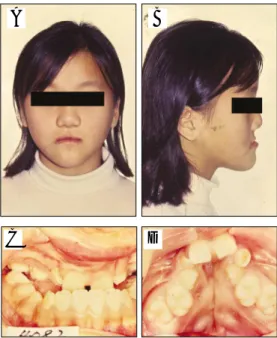
를 보였으며 좌우 비대칭의 정모를 보였다. 구내 임상소견 으로 전치부 반대교합, 협착된 상악궁, 구개의 양측성 골결 손부가 존재하였다(Fig. 1). /k/ 발음 시 과비음이 났다. 방 사선 사진 상 #12, 14, 15, 22, 24, 25, 35, 45이 결손되 었으며 두부규격방사선사진 계측에서 SNA 77.7�로 상악 의 후퇴양상을 보였으며 하악골은 81.4�로 정상 범주이었 다. 성장기 환자이기에 상악골을 전방견인하기로 치료계획 을 수립하고 face mask와 labiolingual appliance를 이용 해 1년간 상악골을 전방으로 견인하였다. 상악에 결손 치아 가 많아 성장 완료 시까지 기다린 뒤 악교정 수술을 시행하 기로 하고 유지 관리하였다. 5년 후 18세 1개월에 2차 교정 치료를 시작하여, 골결손부에 골이식을 시행하고, LeFort I 골절단술 후 RED를 이용한 골신연술을 시행하였다. 시행 직전 전치부 수평 피개량은 -14 mm 였으며 6일간의 잠복 기 후 첫 일주일은 1.5 mm/3회/하루의 비율로 견인하였고 그 이후는 1 mm/2회/하루의 비율로 견인하였다. 재발을 고려하여 수평피개량이 5 mm 가량 확보될 때까지 견인하 였다. 4주간의 골경화기를 거친 뒤 RED를 제거하고 face mask와 탄성 고무를 이용하여 5주간 유지하였다. 골신연 술 후 SNA가 72.7�에서 83.1�로 개선되었으며 ANB는 - 7.9�에서 4.1�로 개선되었다(Table 1). 측모는 오목한 측 모에서 볼록한 측모로 개선되었음을 임상사진을 통해 알 수 있었다(Fig. 2). 술 후 48개월까지 유지 관찰한 결과 SNA 는 81.5�로 약간의 재발을 보이나 전치 수평 피개가 적절하 고 교합도 비교적 양호하여 치료 결과가 잘 유지되고 있음 을 알 수 있다(Fig. 3, 4). 상악골을 상당한 양으로 전방 견 인 했음에도 불구하고 발음의 장애는 나타나지 않았다.
Fig. 1. 11-year-old female with BCLP at first visit;
extraoral view (A, B), intraoral view (C, D)
Table 1. Comparison of cephalometric values Initial Immediate Post-DO
after DO 48 months SNA(deg) 72.7� 83.1� 81.5�
SNB(deg) 80.6� 79.0� 77.9�
ANB(deg) -7.9� 4.1� 3.6�
SN-GoMe(deg) 31.1� 33.3� 32.1�
A
C D
B
증례 2
우측의 완전 편측성 구순구개열이 있는 22세 4개월의 남 성 환자로 #12 부위의 결손부로 인해 생긴 앞니 사이 빈 공 간을 주소로 교정과에 내원하였다. 1세 때 구순성형술, 3세
때 구개열수술, 20세 때 비성형술을 시행 받았으며 전신적 인 병력은 없었다. 구외 임상 소견으로 상악이 저성장 및 후 퇴되어 오목한 측모를 보였으며 비대칭적인 정모를 보였다.
구내 임상 소견으로 전치부의 수평피개량이 -5 mm로 반대 교합이었고 정출된 하악 전치로 인해 전치부 수직피개가 6mm로 과개교합이었다(Fig. 5). 발음 시 비음이 심하게 났다. 모형 분석 상 상악 치열궁에서 공간이 부족하였고 제 2소구치부위에 총생이 집중되어 있었다(상악 치열궁내 공 간부족량 -16.2 mm, #15, 25 배열 공간 부족량 -15.6 mm). 방사선 사진 상 #12 부위 골결손부가 관찰되었다.
두부규격방사선사진에서 SNA가 69.9�로 상악골이 후퇴양 상을 보였다.
Fig. 2. Preoperative state (A, B, G), Postoperative state (C, D, H), The RED device in place (E), Retention state using a facemask with elastic traction (F)
Fig. 4. Superimposition of preoperative state and retention state cephalogram;
corrected anterior crossbite by distraction osteogenesis of maxilla
Fig. 3. After 48 months from surgery, Well retained facial profile and occlusion; extraoral view (A, B), intraoral view (C, D, E)
A
A
C
E
D B
E F G H
B C D
는 점도 전치부 분절 내부고정골신장술을 선택하게 된 배경 이다. 하악골은 전치부의 정출이 심해 전치부를 교정적으로 압하 시 구치부 정출, 전치부 치근 흡수 등의 부작용이 예상 되어 전치부 분절골 절단술을 시행해 교합면을 평탄화 하기 로 하였다. 상악치열은 양측의 제 2소구치를 제외하고 lev- eling 시행하였으며 과정 중에 골결손부에 골이식을 시행하 였다. 하악치열은 전치부와 구치부 분절을 나누어 leveling 을 시행하였다. 상악에 급속구개확대스크류를 응용한 골신 연장치를 장착한 뒤 악교정 수술을 시행하였다(Fig. 6). 악 교정 수술 시행 직전 전치부 수평피개량은 -7 mm 였으며
였다. 부족한 공간은 술후 교정 단계에서 open coil spring 과 골고정원을 이용하여 #16, 17을 후방으로 이동해 확보 하였다. 골신연술 후 SNA는 70.6�에서 75.2�로, ANB는 -3.5�에서 0.5�로 개선되었다. 오목한 측모도 개선되었으나 하악의 전돌감은 약간 잔존해있다(Fig. 8, Table 2). 술후 34개월, 46개월 뒤 촬영한 두부규격방사선사진에서 SNA 는 각 74.9�, 74.6�로 비교적 안정적으로 유지되고 있음을 알 수 있다(Fig. 9). 상악체의 전방 분절만 견인하였기에 연 구 개 인 두 간 폐 쇄 부 전 (VPI, Velopharyngeal Incompetency)은 악화되지 않았다.
Fig. 5. 22-year-old male with UCLP(right) at first visit; extraoral view (A, B), intraoral view (C, D, E, F)
Fig. 6. Internal distraction device
A B
A B
C D
C D
E F
Ⅲ. 고 찰
대부분의 구순구개열자는 상악 열성장에 의한 중안면부 함몰 및 골격성 III급 부정교합의 형태를 보인다. 구순구개 열자의 상악골 발육저하는 조직의 결손과 기능장애라는 선 천적인 요소보다는 구순과 구개의 수술에 의한 반흔 조직의 영향으로 알려져 있다
1,2,11-14). Graber
11)와 Ross
12,13)는 상악 의 열성장에는 구개성형술의 시행시기와 방법이 가장 큰 영 향을 준다고 하였으며 최근에 이루어진 Alam 등
15)의 연구 에 의하면 구순구개열자의 치열궁 관계에 영향을 미치는 여 러 요소 중 구개성형술의 방법과 유전적 배경이 가장 중요 하다고 하였다.
성인 구순구개열자에서 악교정 수술이 필요한 비율에 대 하여 Ross
13)는 약 25%, Posnick
16)은 60% 정도로 보고하 였다. 특히 한국인의 두개안면구조는 서양인에 비해 골격성
Ⅲ급 부정교합이 되기 쉬운 유전적 배경이 있으므로 악교정 수술이 필요한 경우가 더욱 많은 것으로 추정된다. 그러나 Proffit와 Turvey
17)는 North Carolina대학에서 체계적으 로 잘 관리된 환자에서는 5 - 10% 정도에서만 악교정 수술 이 필요하다고 하여 적절한 치료 방침에 따른 체계적 관리 의 중요성을 강조하였다. 심한 상악골 열성장을 동반한 구 순구개열자의 안모와 교합을 개선하기 위하여 흔히 상악에
왔다. 그러나 구순구개열자에서는 반흔 조직과 혈류 공급 장애로 상악골의 전방이동량이 제한되고 술후 재발의 성향 이 높으며 수술 후 치유지연, 치아상실, 연조직과 골조직의 괴사, 감염과 같은 후유증이 발생할 수 있다
18,19).
이러한 문제를 극복하기 위하여 치유가골에 견인력을 가 하여 조직을 늘리는 골신연술 (또는 골신장술)이 사용되고 있다. 골신연술은 1905년 Codivilla
20)가 기형에 의한 짧은 대퇴골의 길이를 증가시키기 위해 처음 이용하였으나 그다 지 좋은 결과는 얻지 못하였다. 1950년대 후반부터 Ilijarov
21)는 많은 실험적 및 임상적 연구를 통해 골신연술 의 방법과 개념을 정립하여 골신연술의 속도와 리듬, 골막 과 골수, 혈행의 유지 및 견고한 고정의 중요성을 강조하였 다. 구강악안면외과 영역에서는 1972년 Snyder 등
22)이 개 에서 하악골의 신장을 시도하였고 1992년 McCarthy
23)가 임상적으로는 처음으로 반안면왜소증이 있는 어린이의 하 악골의 골신장을 시행하였다.
구순구개열자에서 골신연술을 적용한 사례로는 1997년 Polley와 Figueroa
24,25)가 심한 상악골 열성장이 있는 어린 이와 청소년에서 외부고정골신장기를 이용한 치료증례들을 처음 보고하였다. RED 시스템은 골을 신장시키는 도중에 도 견인 방향을 조절할 수 있고, 삽입과 제거가 용이한 장점 이 있지만 환자의 사회 생활에 지장을 주며 정신적으로 상
Fig. 8. After 46 months from surgery, Well retained facial profile and occlusion; extraoral view (A, B), intraoral view (C, D, E, F)
Fig. 9. Superimposition of preoperative and retention state cephalogram; anteriorly distract- ed anterior segment of maxilla, inferiorly intruded anterior segment of mandible
Table 2. Comparison of cephalometric values Initial Immediate Post-DO
after DO 46 months SNA(deg) 70.6� 75.2� 74.6�
SNB(deg) 74.1� 74.7� 74.7�
ANB(deg) -3.5� 0.5� 0.1�
SN - GoMe(deg) 28.2� 29.0� 28.8�
A B
C D
E F
에는 골신장량에 한계가 있고 정확한 방향으로 골신장을 하 기 어려우며 환자의 저작력에 의해 screw가 빠지거나 장치 가 파손이 될 수 있다. 이러한 단점들을 극복하기 위하여 계 속적으로 새로운 장치와 술식들이 개발되고 있다
31). 또한 최 근에는 치아 사이를 절단한 후 골신장술을 시행하여 측방 확장, 전상악골의 확장, 넓은 골 결손부를 감소시키는 등의 다양한 임상적 적용이 보고되고 있다
32-38).
증례 1은 성장이 종료된 성인 여성에서 외부고정골신장기 를 이용하여 치료하였다. 골신연술의 장점의 하나로 심리적 인 부담을 많이 가지는 어린이에서 어린 나이에도 시행할 수 있다는 것이나 이 환자는 굳이 조기 개선을 원하지 않았 고, 다수 치아의 선천 결손으로 골신연술을 위한 견고한 구 강내장치를 만들기 어려웠다. 상악 제 1대구치만을 이용하 여 구강내 장치를 만드는 경우 골신연술과 그 후의 보정을 위한 상악골전방견인 장치 적용 시에 치아의 과도한 동요로 효과적인 치료를 계속하기 어려울 수가 있다. 또한 어린 나 이에 골신연술을 통해 만족할 만한 결과를 얻었더라도 잔여 성장에 의해 상당량의 상, 하악골의 부조화가 초래될 수 있 다는 것도 부담이 되는 부분이다. 이 증례에서는 19 mm의 전방 견인이 이루어졌으며, 장기간에 걸친 평가에도 양호한 교합이 유지되었고 특히 발음도 악화되지 않았다. Guyette 등
39)은 골신연술 후 발음에 대한 영향을 평가하여 16.7%에 서 과비음이 현저히 증가한다고 보고하였으나 Harada 등
40)은 6명에서 평가한 결과이기는 하나 15 mm이하의 전방이 동에서는 구개범인두 기능에 현저한 영향을 주지는 않았다 고 보고하였다. 발음에 미치는 영향에 대해서는 더 많은 대 상과 보다 과학적인 방법으로 평가가 이루어져야 할 것으로 생각된다.
증례 2는 상악골을 분절한 후 상악골 급속확대에 이용되 는 Biederman type의 screw를 이용하여 상악골의 전방이 동과 치열궁 장경을 증가시켜 안모를 개선함과 동시에 공 간부족으로 구개측에 위치하던 상악 좌우측 제 2소구치를 배열하였다. 이 증례에서는 상악치열궁이 협착 되지 않아 치료 전에 구치부 폭경은 조화를 이루었고 견치 간 폭경도 전방분절의 전방이동 후 하악과 조화를 이룰 수 있었던 것 이 이러한 치료 계획을 설정한 이유였다. 구개 점막은 비교
있을 것이다 . 이상의 두 증례는 성인 구순구개열자에서도 중안면함몰과 교합을 개선하기 위하여 골신연술이 유용하 게 이용될 수 있음을 보여준다.
Ⅳ. 결 론
구순구개열이 있는 환자는 어린 나이에 시행한 구개와 구 순의 성형술로 인해 상악의 3차원적인 성장이 저해 받게 된 다. 이로 인해 중안면의 함몰, 전치부의 반대교합, 상악 치 궁의 협착, 불량한 치아배열 등이 흔히 관찰된다. 중안모의 함몰을 개선하기 위해 외과적 및 교정적 복합치료가 필요하 다. 골신연술은 전통적인 악교정 수술에 비하여 골조직의 위치개선과 함께 연조직도 신장시킬 수 있어 재발을 줄이고 상악골을 더 많이 전방 이동 시킬 수 있다. 또한 성장기 환 자에게도 사용할 수 있어 근래에 많이 사용되고 있다. 골신 열술에 이용되는 장치 중 내부고정골신장기는 각 환자의 상 태에 따라 다양하게 적용할 수 있다.
저자들은 각기 다른 골신장기를 이용하여 교합과 안모의 개선을 얻었고 장기간 결과가 안정적으로 유지된 두 가지 증례를 보고하였다.
References