www.jpis.org
pISSN 2093-2278 eISSN 2093-2286 Copyright © 2013 Korean Academy of PeriodontologyThis is an Open Access article distributed under the terms of the Creative Commons Attribution Non-Commercial License (http://creativecommons.org/licenses/by-nc/3.0/).
Evaluation of the correlation between insertion torque and primary stability of dental implants using a block bone test
Dorjpalam Bayarchimeg1, Hee Namgoong1, Byung Kook Kim2, Myung Duk Kim2, Sungtae Kim1, Tae-Il Kim1, Yang Jo Seol1, Yong Moo Lee1, Young Ku1, In-Chul Rhyu1, Eun Hee Lee3, Ki-Tae Koo1,*
1Department of Periodontology, Dental Research Institute, Seoul National University School of Dentistry, Seoul, Korea
2Implant R&D Center, Osstem Implant Co., Busan, Korea
3Dental Clinic, Healthcare System Gangnam Center, Seoul National University Hospital, Seoul, Korea
Purpose: Implant stability at the time of surgery is crucial for the long-term success of dental implants. Primary stability is considered of paramount importance to achieve osseointegration. The purpose of the present study was to investigate the correlation between the insertion torque and primary stability of dental implants using artificial bone blocks with different bone densities and compositions to mimic different circumstances that are encountered in routine daily clinical settings.
Methods: In order to validate the objectives, various sized holes were made in bone blocks with different bone densities (#10,
#20, #30, #40, and #50) using a surgical drill and insertion torque together with implant stability quotient (ISQ) values that were measured using the Osstell Mentor. The experimental groups under evaluation were subdivided into 5 subgroups ac- cording to the circumstances.
Results: In group 1, the mean insertion torque and ISQ values increased as the density of the bone blocks increased. For group 2, the mean insertion torque values decreased as the final drill size expanded, but this was not the case for the ISQ val- ues. The mean insertion torque values in group 3 increased with the thickness of the cortical bone, and the same was true for the ISQ values. For group 4, the mean insertion torque values increased as the cancellous bone density increased, but the cor- relation with the ISQ values was weak. Finally, in group 5, the mean insertion torque decreased as the final drill size increased, but the correlation with the ISQ value was weak.
Conclusions: Within the limitations of the study, it was concluded that primary stability does not simply depend on the in- sertion torque, but also on the bone quality.
Keywords: Bone density, Dental implants, Torque.
INTRODUCTION
Implant stability at the time of surgery is crucial for the long-term success of dental implants. Primary stability is considered of paramount importance to achieve osteointe- gration [1]. Primary implant stability can be defined as a func- tion of local bone quality and quantity, the geometry of an
implant, the placement and surgical technique used, and the precise fit in the bone. Thus, primary implant stability is con- sidered a significant parameter in achieving osseointegra- tion, and the orchestration of the elements mentioned is cru- cial for the long-term success of the implant [2,3].
Two main factors that influence primary stability of an im- plant during placement are the amount of bone-implant
Received: Dec. 17, 2012; Accepted: Jan. 18, 2013
*Correspondence: Ki-Tae Koo
Department of Periodontology, Dental Research Institute, Seoul National University School of Dentistry, 101 Daehak-ro, Jongno-gu, Seoul 110-744, Korea E-mail: [email protected], Tel: +82-2-2072-0108, Fax: +82-2-744-0051
tissue interface. Such stresses may be beneficial for enhanc- ing the primary stability of an implant, but they can reach a sufficiently high level to result in necrosis and local ischemia of the bone at the implant-tissue interface [4-6]. In the same respect, secondary stability can also be determined by the bone tissue response to the surgical trauma and the implant surface. The response is ultimately bone formation and re- modeling at the implant interface leading to increased fixa- tion and stability of the implant, although bone resorption resulting in implant failure can also occur during the initial healing period. In the literature, it is clear that surface rough- ness may result in more bone at the implant interface as well as a higher resistance to torque. Poor fixation may lead to micromovements during implant healing, which can poten- tially cause fibrous encapsulation and are associated with higher failure rates. Shorter healing periods are usually need- ed for implants with good primary stability. On the other hand, implants with poor stability need longer healing peri- ods to achieve sufficient gain in secondary stability. This sug- gests the possibility of determining the length of the healing period on an individual basis, making implant treatment saf- er, more effective, and less time-consuming in some cases [7].
Generally, clinicians evaluate primary stability using the percussion test or using their own perception during the in- sertion process. However, the lack of precision has motivated the development of different methods to objectively evaluate primary stability; in particular, peak insertion torque (IT) and resonance frequency analysis (RFA) are the most used global- ly. Clinically, RFA values or implant stability quotient (ISQ) values have been correlated with changes in implant stability during osseous healing. Thus, IT and ISQ values are thought to have a positive correlation [8,9]. However, the formula of higher IT torque translating into higher primary stability
bone varies significantly among patients. Therefore, the pur- pose of the present study was to investigate the correlation between IT and primary stability of dental implants using ar- tificial bone blocks with different bone densities and compo- sitions to mimic different circumstances that are encoun- tered in routine daily clinical settings.
MATERIALS AND METHODS
Bone specimens
The bone block of solid rigid polyurethane foam (Sawbones, Vashon, WA, USA) with various bone densities (cancellous bone: #10, #20, #30; cortical bone: #50; and homogeneous bone: #10, #20, #30, #40) were used in the present study (Fig.
1). Because the mean bone mineral density was 0.31 g/cm³ for the posterior maxilla and 0.55 g/cm³ for the anterior maxilla, polyurethane foam blocks with a bone density of 0.48 g/cm³ were chosen. Short fiber-filled epoxy sheets were used as a substitute for cortical bone. Because the mean cortical thick- ness for the mandible was 2.22±0.47 mm and the mean cor- tical bone thickness for the maxilla was 1.49±0.34 mm, the sheets with a corresponding thickness were selected [10]. The following five different cortical thicknesses were used: blocks without a cortical layer (only homogeneous bone), blocks with a 0.5 mm cortical thickness, blocks with a 1.0 mm corti- cal thickness, blocks with a 1.5 mm cortical thickness, and blocks with a 2.0 mm cortical thickness.
Experimental design
The experimental group under evaluation was subdivided into 5 subgroups according to the objectives. In group 1, the correlation between IT and implant stability according to the bone density was evaluated. Implants were placed in homo- geneous bone blocks with different bone densities (#10, #20,
#30, and #40) following osteotomy preparation with a final drill diameter of Ø3.6 mm. An increase in numerical size of the bone blocks represented an increase in the bone density.
Measurement of the IT and ISQ values were repeated 20 times for each bone density. In group 2, the correlation be- tween the IT and implant stability according to the size of the final drill diameter used was evaluated. Homogeneous bone blocks with a density of #20 were used.
The final drill diameters under evaluation were Ø2.7 mm, Ø3.0 mm, Ø3.3 mm, and Ø3.6 mm. The remaining process was identical to that for group 1. The objective of group 3 was to evaluate the correlation between the IT and implant sta- bility according to the thickness of the cortical bone. Using bone blocks with a cortical density of #50 and cancellous density of #20, the cortical bone was manipulated to have a Figure 1. Photographic presentation of the bone block with various
bone densities that were used in the present study.
final drill diameter was Ø3.6 mm and identical steps were performed.
The objective of group 4 was to evaluate the correlation be- tween the IT and implant stability according to the cancel- lous bone density by controlling the cortical bone thickness to 1.5 mm and using block bones with a uniform cortical density of #50. Only the density of the cancellous bone (#10,
#20, and #30) was different in the bone blocks for group 4.
In group 5, the correlation between the IT and implant sta- bility according to the size of the final drill diameter in the bone blocks with a uniform cortical thickness of 1.5 mm was evaluated. The density of the cortical bone (#50) and the can- cellous bone (#20) was controlled. The final drill diameters were Ø3.0 mm, Ø3.3 mm, Ø3.6 mm, and Ø3.8 mm.
Osteotomy preparation and fixture installation
All of the osteotomies were prepared with a gentle surgical technique using a surgical drill at a rotational speed of 800 rpm with opious external cooling. The drill was fixed in a stan-
the implant fixtures (Osstem Implant Co., Seoul, Korea) with a length of 11.5 mm and a diameter of 4.1 mm were placed in the prepared osteotomies. The diameter of the final drill was chosen and assigned according to the test protocol. The fol- lowing diameters were used: Ø2.7 mm, Ø3.0 mm, Ø3.3 mm, Ø3.6 mm, and Ø3.8 mm (Fig. 2).
IT and RF measurements
During installation, the peak IT was measured for all of the implants (Fig. 3). Following the final seating of the fixtures, the stability of each implant was measured in ISQ units us- ing the Osstell Mentor (Osstell, Göteborg, Sweden). The RF values were represented in the ISQ on a scale from 1 to 100 and were averaged for each implant (Fig. 4). Each measure- ment was performed up to 20 times [11-15].
Statistical analysis
The IT and ISQ values were summarized using means and standard deviations. One-way analysis of variance was used to compare the mean IT and ISQ values. The Pearson’s cor- relation coefficient was used to evaluate the correlation be- tween the IT and the ISQ at implant placement. A P-value of
<0.05 was considered statistically significant. Statistical anal- ysis was performed using the IBM SPSS ver. 20.0 (IBM Co., Armonk, NY, USA) [16-22].
RESULTS
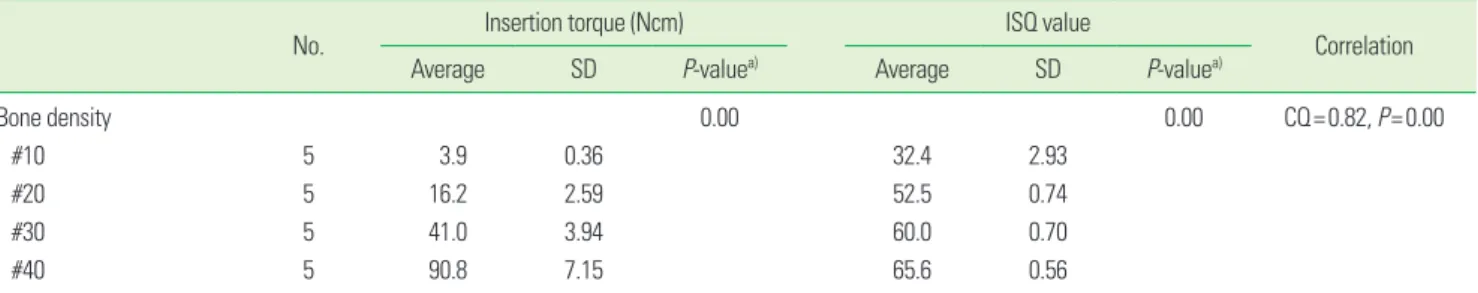
In group 1, the mean IT and ISQ value increased as the density of bone blocks increased, respectively (P=0.00 and P=0.00). The correlation between the two parameters was evaluated using Pearson’s correlation coefficient, and the re- sults suggested a positive correlation (CQ=0.82, P=0.00), which was statistically significant (Table 1, Fig. 5). For group 2,
Figure 4. Measurement of the implant stability quotient values us- ing the Ostell Mentor (Osstell).
Figure 3. Torque measurement at fixture placement.
Figure 2. Photographic presentation of the fixture type used in the present study (SS II fixture, Osstem Implant Co.).
No. Insertion torque (Ncm) ISQ value
Correlation
Average SD P-valuea) Average SD P-valuea)
Bone density 0.00 0.00 CQ=0.82, P=0.00
#10 5 3.9 0.36 32.4 2.93
#20 5 16.2 2.59 52.5 0.74
#30 5 41.0 3.94 60.0 0.70
#40 5 90.8 7.15 65.6 0.56
SD: standard deviation, ISQ: implant stability quotient, ANOVA: analysis of variance.
a)ANOVA.
Table 2. Correlation between the insertion torque and implant stability according to the final drill diameter.
No. Insertion torque (Ncm) ISQ value
Correlation
Average SD P-valuea) Average SD P-valuea)
Final drill diameter 0.00 0.01 CQ=-0.07, P=0.77
Ø2.7 mm 5 39.2 2.77 52.1 0.85
Ø3.0 mm 5 35.4 1.95 53.9 0.64
Ø3.3 mm 5 26.0 2.00 54.4 1.64
Ø3.6 mm 5 16.2 2.56 52.5 0.74
SD: standard deviation, ISQ: implant stability quotient, ANOVA: analysis of variance.
a)ANOVA.
Figure 5. A diagram presenting the correlation between the inser- tion torque (IT) and implant stability quotient (ISQ) values accord- ing to the various bone densities tested in the present study. The in- sertion torque increased according to the bone density, and the same was observed for the ISQ. The two variables appeared to have a strong positive correlation, which was statistically significant (cor- relation coefficient=0.82).
100 80 60 40 20
0 #10 #20 #30 #40
ISQ value IT (Ncm)
Figure 6. Diagram illustrating the relationship between the inser- tion torque and implant stability quotient (ISQ) according to the various drill sizes under evaluation. The insertion torque (IT) de- creased when the correlation with the ISQ value was weak and non- significant (correlation coefficient=0.07).
60 50 40 30 20 10
0 Ø2.7 Ø3.0 Ø3.3 Ø3.6
ISQ value IT (Ncm)
Table 3. Correlation between the insertion torque and implant stability according to the thickness of the cortical bone.
No. Insertion torque (Ncm) ISQ value
Correlation
Averge SD P-valuea) Average SD P-valuea)
Cortical bone thickness 0.00 0.00 CQ=0.84, P=0.00
0.5 mm 5 9.20 1.63 54.4 1.34
1.0 mm 5 12.0 1.58 55.7 1.68
1.5 mm 5 24.8 2.86 57.9 0.51
2.0 mm 5 25.4 3.97 57.9 0.45
SD: standard deviation, ISQ: implant stability quotient, ANOVA: analysis of variance.
a)ANOVA.
No. Insertion torque (Ncm) ISQ value
Correlation
Average SD P-valuea) Average SD P-valuea)
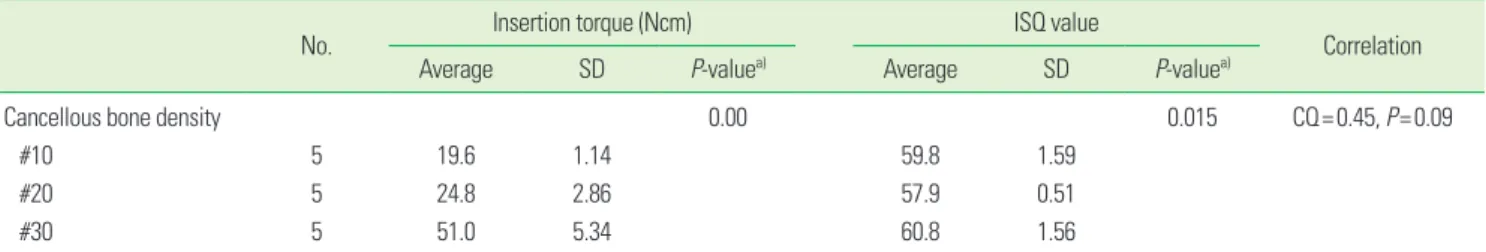
Cancellous bone density 0.00 0.015 CQ=0.45, P=0.09
#10 5 19.6 1.14 59.8 1.59
#20 5 24.8 2.86 57.9 0.51
#30 5 51.0 5.34 60.8 1.56
SD: standard deviation, ISQ: implant stability quotient, ANOVA: analysis of variance.
a)ANOVA.
the mean IT values decreased as the final drill size expanded, but this was not the case for the ISQ values. The mean ISQ values showed limited change as the drill size increased. The correlation coefficient between the IT and ISQ value was -0.07 with a P-value of 0.77, but it was considered nonsignifi- cant (Table 2, Fig. 6). The mean IT values in group 3 increased according to the thickness of the cortical bone, and the same was true for the ISQ values as well. It was also shown that the two parameters had a stronger linear relationship (CQ=0.84) with the values reaching statistical significance (P=0.00) (Ta- ble 3, Fig. 7). For group 4, the mean IT values increased as the cancellous bone density increased (P=0.00), but the correla- tion with the ISQ values was weak (CQ=0.45) and nonsignifi- Figure 8. Insertion torque (IT) increased according to the bone den-
sity, while the correlation with the implant stability quotient (ISQ) value was weak and non-significant (correlation coefficient=0.45).
70 60 50 40 30 20 10
0 #10 #20 #30
ISQ value IT (Ncm)
Table 5. Correlation between the insertion torque and implant stability according to the final drill diameter with cortical bone.
No. Insertion torque (Ncm) ISQ value
Correlation
Average SD P-valuea) Average SD P-valuea)
Final drill diameter 0.00 0.02 CQ=0.57, P=0.01
Ø3.0 mm 5 36.4 4.04 58.9 1.33
Ø3.3 mm 5 32.8 5.97 58.7 0.58
Ø3.6 mm 5 24.8 2.86 58.0 1.48
Ø3.8 mm 5 16.2 2.17 56.8 1.33
SD: standard deviation, ISQ: implant stability quotient, ANOVA: analysis of variance.
a)ANOVA.
Figure 7. The insertion torque (IT) increased according to the thick- ness of the cortical bone, as did the implant stability quotient (ISQ) value. The two variables had a stronger positive correlation (correla- tion coefficient=0.84) with statistically significant values.
70 60 50 40 30 20 10
0 0.5 mm 1.0 mm 1.5 mm 2.0 mm
ISQ value IT (Ncm)
Figure 9. Insertion torque (IT) decreased according to the size of the final drill, but the correlation (correlation coefficient=0.57) with the implant stability quotient (ISQ) value was weak and nonsignifi- cant.
70 60 50 40 30 20 10
0 Ø3.0 Ø3.3 Ø3.6 Ø3.8
ISQ value IT (Ncm)
Length (mm)
decreased according to the final drill size (P=0.00), but the correlation with the ISQ value was weak (CQ=0.57) and was non-significant (Table 5, Fig. 9).
DISCUSSION
The aim of the present study was to investigate the correla- tion between the IT and primary stability of dental implants through the IT test, and RFA analysis was performed using artificial bone blocks that simulated different circumstances that can ioccur in clinical practice. In general, primary im- plant stability is considered the most important factor in a successful implant treatment, and the distinct ranges of im- plant primary stability have been distinguished by the reso- nance frequency method [23-27]. Thus, RFA was used instead of the Periotest as a method to measure implant stability.
The results of the present study showed that the IT and ini- tial stability increased according to the increase in the bone density, resulting in a strong positive correlation. In other words, the initial stability was shown to be highly dependent on the bone density. The IT also increased according to the thickness of the cortical bone, and a slight increase was ob- served for initial stability. This shows that the volume of high dense cortical bone affects the initial stability and it corrobo- rates a recent study in which the same artificial bone model was used. In that study, an increase in the mean IT values was observed when the bone blocks with only trabecular bone (without a cortical layer) were compared to the groups with a cortical layer of 1 mm, 1 to 2 mm, and 2.5 mm [10].
The results from group 4 did not deviate much from group 2, which may suggest that the density of cancellous bone may have an impact on IT, but may have a limited effect on prima- ry stability. This may indicate that the thickness of cortical bone or the cortical outer layer functions as the primary de- terminant for primary stability. This was also reflected in the results of groups 2, 4, and 5, where the correlation between the two parameters was weak. Therefore, the only factors that showed a positive correlation between the IT and the ISQ val- ue were the bone density and thickness of the cortical bone.
In testing the impact of the final drill size, the IT decreased as the drill size expanded, but the initial stability showed lim- ited change. This showed that initial stability cannot be ac- quired by simply reducing the diameter of the final drill in attempts to increase the IT. Biologic and anatomical conse- quences such as the thickness of cortical bone seem to be significant factors affecting primary stability, and estimation of bone density and the optimal selection of drill size are im- portant.
Authors Byung Kook Kim and Myung Duk Kim are research- ers employed under Osstem Implant Co. (Seoul, Korea).
ACKNOWLEDGEMENTS
Our special thanks to Osstem Implant Co. (Seoul, Korea) for the technical support to conduct this study.
REFERENCES
1. Degidi M, Daprile G, Piattelli A, Iezzi G. Development of a new implant primary stability parameter: insertion torque revisited. Clin Implant Dent Relat Res 2011 Oct 18 [Epub].
http://dx.doi.org/10.1111/j.1708-8208.2011.00392.x.
2. Dilek O, Tezulas E, Dincel M. Required minimum primary stability and torque values for immediate loading of mini dental implants: an experimental study in nonviable bo- vine femoral bone. Oral Surg Oral Med Oral Pathol Oral Radiol Endod 2008;105:e20-7.
3. Stacchi C, Vercellotti T, Torelli L, Furlan F, Di Lenarda R.
Changes in implant stability using different site prepara- tion techniques: twist drills versus piezosurgery: a single- blinded, randomized, controlled clinical trial. Clin Implant Dent Relat Res 2011 Apr 19 [Epub]. http://dx.doi.org/10.1111/
j.1708-8208.2011.00341.x.
4. Nedir R, Bischof M, Szmukler-Moncler S, Bernard JP, Samson J. Predicting osseointegration by means of im- plant primary stability. Clin Oral Implants Res 2004;15:
520-8.
5. Isoda K, Ayukawa Y, Tsukiyama Y, Sogo M, Matsushita Y, Koyano K. Relationship between the bone density esti- mated by cone-beam computed tomography and the pri- mary stability of dental implants. Clin Oral Implants Res 2012;23:832-6.
6. Bilhan H, Geckili O, Mumcu E, Bozdag E, Sunbuloglu E, Kutay O. Influence of surgical technique, implant shape and diameter on the primary stability in cancellous bone.
J Oral Rehabil 2010;37:900-7.
7. Esposito M, Hirsch JM, Lekholm U, Thomsen P. Biologi- cal factors contributing to failures of osseointegrated oral implants. (II). Etiopathogenesis. Eur J Oral Sci 1998;106:
721-64.
8. Degidi M, Daprile G, Piattelli A. Primary stability determi- nation by means of insertion torque and RFA in a sample of 4,135 implants. Clin Implant Dent Relat Res 2010 Sep 17 [Epub]. http://dx.doi.org/10.1111/j.1708-8208.2010.00302.x.
9. Degidi M, Perrotti V, Strocchi R, Piattelli A, Iezzi G. Is in- sertion torque correlated to bone-implant contact percent-
morphometrical evaluation of 17 human-retrieved dental implants. Clin Oral Implants Res 2009;20:778-81.
10. Tabassum A, Meijer GJ, Wolke JG, Jansen JA. Influence of surgical technique and surface roughness on the primary stability of an implant in artificial bone with different cor- tical thickness: a laboratory study. Clin Oral Implants Res 2010;21:213-20.
11. Turkyilmaz I. A comparison between insertion torque and resonance frequency in the assessment of torque capacity and primary stability of Brånemark system implants. J Oral Rehabil 2006;33:754-9.
12. Kahraman S, Bal BT, Asar NV, Turkyilmaz I, Tozum TF.
Clinical study on the insertion torque and wireless reso- nance frequency analysis in the assessment of torque ca- pacity and stability of self-tapping dental implants. J Oral Rehabil 2009;36:755-61.
13. Roze J, Babu S, Saffarzadeh A, Gayet-Delacroix M, Hoorn- aert A, Layrolle P. Correlating implant stability to bone structure. Clin Oral Implants Res 2009;20:1140-5.
14. Huang HM, Lee SY, Yeh CY, Lin CT. Resonance frequency assessment of dental implant stability with various bone qualities: a numerical approach. Clin Oral Implants Res 2002;13:65-74.
15. Hong J, Lim YJ, Park SO. Quantitative biomechanical analysis of the influence of the cortical bone and implant length on primary stability. Clin Oral Implants Res 2012;
23:1193-7.
16. Dos Santos MV, Elias CN, Cavalcanti Lima JH. The effects of superficial roughness and design on the primary stabil- ity of dental implants. Clin Implant Dent Relat Res 2011;
13:215-23.
17. Bischof M, Nedir R, Szmukler-Moncler S, Bernard JP, Samson J. Implant stability measurement of delayed and immediately loaded implants during healing. Clin Oral Implants Res 2004;15:529-39.
18. Trisi P, De Benedittis S, Perfetti G, Berardi D. Primary sta- bility, insertion torque and bone density of cylindric im- plant ad modum Branemark: is there a relationship? An
19. Moon SH, Um HS, Lee JK, Chang BS, Lee MK. The effect of implant shape and bone preparation on primary stabil- ity. J Periodontal Implant Sci 2010;40:239-43.
20. Hsu JT, Fuh LJ, Tu MG, Li YF, Chen KT, Huang HL. The ef- fects of cortical bone thickness and trabecular bone strength on noninvasive measures of the implant primary stability using synthetic bone models. Clin Implant Dent Relat Res 2011 May 20 [Epub]. http://dx.doi.org/10.1111/
j.1708-8208.2011.00349.x.
21. Merheb J, Van Assche N, Coucke W, Jacobs R, Naert I, Qui- rynen M. Relationship between cortical bone thickness or computerized tomography-derived bone density values and implant stability. Clin Oral Implants Res 2010;21:612-7.
22. Khayat PG, Arnal HM, Tourbah BI, Sennerby L. Clinical outcome of dental implants placed with high insertion torques (up to 176 Ncm). Clin Implant Dent Relat Res 2011 May 20 [Epub]. http://dx.doi.org/10.1111/j.1708-8208.2011.
00351.x.
23. Molly L. Bone density and primary stability in implant therapy. Clin Oral Implants Res 2006;17 Suppl 2:124-35.
24. Sim CP, Lang NP. Factors influencing resonance frequen- cy analysis assessed by Osstell mentor during implant tis- sue integration: I. Instrument positioning, bone structure, implant length. Clin Oral Implants Res 2010;21:598-604.
25. Martinez H, Davarpanah M, Missika P, Celletti R, Lazzara R. Optimal implant stabilization in low density bone. Clin Oral Implants Res 2001;12:423-32.
26. Katsoulis J, Avrampou M, Spycher C, Stipic M, Enkling N, Mericske-Stern R. Comparison of implant stability by means of resonance frequency analysis for flapless and conventionally inserted implants. Clin Implant Dent Relat Res 2012;14:915-23.
27. Ostman PO, Hellman M, Sennerby L. Direct implant load- ing in the edentulous maxilla using a bone density-adapt- ed surgical protocol and primary implant stability criteria for inclusion. Clin Implant Dent Relat Res 2005;7 Suppl 1:S60-9.