Ⅰ. 서 론
Machined surface implant가 처음 도입된 이후 초기 골유 착을 촉진시키기 위해 임프란트 표면처리에 대한 연구가 지속적으로 시행되어 왔고 임상에 적용하면서 치유기간이 현저히 단축되고 있다. 최근 임상에서는 resorbable blast media (RBM) surface, sandblast large-grit acid-etching (SLA), anodized surface 등의 제품들이 많이 사용되고 있으며 많은 논문들에서 골유착 시기가 단축되며 조기 하중이 가능하 다고 언급되고 있다1-3.
한편 1990년대에 초기 골유착을 촉진시키기 위한 목적으 로 hydrosyapatite (HA) coated surface implant가 사용된 적이 있으나 실패율이 높다는 연구논문들이 발표되면서 많은 제품들이 시장에서 소멸되었다. 일부 학자들은 장기적으
로 볼 때 HA implant는 코팅층이 벗겨지거나 흡수되어 임 프란트와 골 사이에 공간이 생기며 이로 인해 역학적으로 불안정해지면서 골유착이 파괴될 가능성이 있다고 주장한 다. 또한 초기 골유착이 우수하더라도 HA 표면은 감염되 기 쉽고 일단 감염되면 코팅층이 흡수되면서 조기 탈락의 원인이 된다고 하였다4,5.
그러나 과거의 HA 코팅 임프란트들은 코팅 기술이 미숙 한 제품이 많았으며 제품에 따라 코팅의 질에 많은 차이가 있었다. 2005년 Trisi 등6은 10년간 기능한 HA 코팅 임프란 트의 부검검체에서 시편을 채취하여 조직학적 분석결과를 보고하였는데 HA 코팅층 소실은 25%를 넘지 않았고 bone- implant contact (BIC) ratio는 78.48%였음을 관찰하였다. 즉 적절한 보철치료가 진행되고 유지관리가 잘 시행되는 환 자들에서는 HA 코팅이 훼손되지 않고 장기간의 생존을 보 장할 수 있다고 하였다. 그 외에도 HA 코팅 임프란트의 좋 은 임상성적을 보고하는 문헌들이 많이 발표되었다7-9.
본 연구에서는 2가지 유형의 수산화인회석 코팅 임프란 트의 초기골유착 상태를 평가하기 위해 조직형태계측학적 분석을 통한 골-임프란트 접촉률(BIC ratio)을 측정하여 비 교하였다.
김 영 균
463-707 성남시 분당구 구미동300 분당서울대학교병원 치과 구강악안면외과 Young-Kyun Kim
Department of Oral and Maxillofacial Surgery, Section of Dentistry, Seoul National University Bundang Hospital
300 Gumi-dong, Bundang-gu, Seongnam, 463-707, Korea TEL: +82-31-787-7541 FAX: +82-31-787-4068 E-mail: [email protected]
수산화인회석 코팅 임프란트의 골치유과정에 관한 실험적 연구
엄태관1,2∙김종화3∙조인희2∙정창모4∙조용석5∙김영균3
1부산대학교 치의학전문대학원 치의학과, 2오스템임프란트 연구소,
3서울대학교 치의학대학원 분당서울대학교병원 치과 구강악안면외과,
4부산대학교 치의학전문대학원, 5앞선치과병원
Experimental study about the bony healing of hydroxyapatite coating implants
Tae-Gwan Eom1,2, Jong-Hwa Kim3, In-Hee Cho2, Chang-Mo Jeong4, Yong-Seok Cho5, Young-Kyun Kim3
1Interdisciplinary Program in Biomedical, Pusan National University, Busan, 2Implant R&D Center, OSSTEM IMPLANT Co., Ltd., Busan,
3Department of Oral and Maxillofacial Surgery, Section of Dentistry, Seoul National University Bundang Hospital, School of Dentistry, Seoul National University, Seongnam, 4Pusan National University, School of Dentistry, Busan, 5Apsun Dental Hospital, Seoul, Korea
Introduction: Hydroxyapatite(HA) coating implant can accelerate osseointegration, however, there are many controversies.
Materials and Methods: This study examined the early osseointegration of two types of hydroxyapatite coated implants. Twelve adult male minia- ture pigs (Medi Kinetics Micropigs, Medi Kinetics Co., Ltd., Busan, Korea) were used in this study. In the implants placed in the mandible, a histo- morphometric evaluation was performed to evaluate the bone-implant contact (BIC) ratio.
Results: The BIC ratio increased with time. TS III HA and Zimmer HA were not significantly different (P>0.05). At 8 weeks, the BIC of Zimmer HA was higher than TS III HA, but there was no significant difference (P>0.05).
Conclusion: HA coated implants will accelerate early osseointegration.
Key words:Bone-implant contact, Hydroxyapatite coated implant
[paper submitted 2011. 3. 11 / revised 2011. 7. 18 / accepted 2011. 7. 20]
Abstract (J Korean Assoc Oral Maxillofac Surg 2011;37:295-300)
Ⅱ. 연구 대상 및 방법
본 연구는 Medikinetics 연구소 동물실험윤리위원회의 허 가를 받은 후 시행되었다.(Medikinetics-IACUC: 100125- 001) Adult male miniature pigs (Medi Kinetics Micropigs, Medi Kinetics Co., Ltd., Busan, Korea) 12마리가 실험에 사 용되었다.
1. 전신마취
Atropine (0.05 mg/kg)을 정맥주사한 후 Zoletil (Virbac Laboratories, Carros, France)과 xylazine (Narcosyl, Intervet Korea, Seoul, Korea)을 근육 및 정맥주사하여 전신마취를 유도하였다. Isoflurane (Ifran, Hana Pharm Co., Ltd., Seoul, Korea) with oxygen, 1:1 (5-10 mL/kg/min)으로 전신마취를 유지하면서 수술을 진행하였다.
2. 임프란트 종류 및 수술
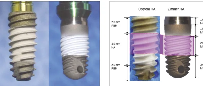
1군은 Osstem TS III HA, 2군은 Zimmer HA (MP-1 HA)로 배정하였다.(Fig. 1) 12 miniature pigs의 양측 대구치들을 발 치하고 4개월의 치유기간을 거친 후 2가지 종류의 임프란 트를 식립하였다. 구강내는 0.1% chlorhexidine 용액으로 소 독하고 구강외 피부는 베타딘과 에탄올로 닦은 후 소독된 소 공 으 로 덮 어 수 술 부 위 만 노 출 시 켰 다 . 수 술 부 위 에 1:100,000 에피네프린 함유 리도카인으로 침윤마취를 하여 국소적 지혈과 마취효과를 동시에 얻도록 하였다. 치조정 절개를 가한 후 점막골막피판을 형성하여 치조골을 노출 시켰다. 초기 드릴링을 시행한 후 2.0, 3.3, 3.5 mm 직경까지 드릴링을 시행한 후 직경 3.7 mm, 길이 8.5 mm 임프란트를
식립하였다. 덮개나사를 연결하고 봉합하여 수술을 종료 하였다. Dexamethasone-21-isonicotinate (Voren; Boehringer Ingelheim Korea Ltd, Seoul, Korea) 1 mL을 수술 후 마취회 복 전에 1회 근육 주사하였다. 진통 목적으로 Hyoscine-N- butylbromide (Buscopan Comp, Boehringer Ingelheim Korea Ltd.) 0.4 mg/kg를 수술 후 3일간 근주하였다. 술후 감염 방 지를 위해 Clemizole Penicillin G & Sodium Penicillin G (Antipen-SM, WooGene B&G Ltd, Seoul, Korea) 1 mL/10 kg 을 수술 후 2일 간격으로 3회 근주하였다.
3. 골-임프란트 접촉률 분석
수술 2주, 4주, 8주에 각 4마리씩 희생시켰으며 임프란트 와 주변 골조직을 포함한 시편을 채취하였다. 채취된 시편 은 10% phosphate buffered formalin 용액으로 고정하였다.
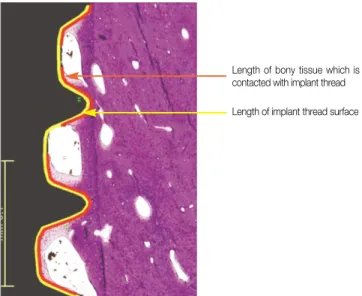
알콜을 이용하여 단계별 탈수를 한 후 레진(Technovit 7210 VLC, Heraeus Kulzer GmbH, Wehrheim, Germany)을 이용하 여 포매하였다. 포매된 block을 diamond band cutting system (Exakt CP, Exakt Apparatebau, Norderstedt, Germany)으로 협 설측 방향으로 절단하고 micro grinding system (Exakt 400CS, Exakt Apparatebau)을 이용하여 30-40 μm 두께로 연 마한 후 H&E stain을 하였다. 조직학적 관찰 및 조직계측학 적 분석을 위하여 광학현미경(BX51, Olympus Co., Tokyo, Japan)에 digital camera (DP71, Olympus Co.)를 부착하여 × 100 배율의 이미지를 얻은 다음 이미지분석 프로그램 (Image-Pro Plus, Media Cybernetics Inc., Maryland, SA)을 이 용하였다. 2가지 임프란트의 HA 코팅된 부위를 계측하기 위해 Ts III HA는 작은 나사산 2개, 큰 나사산 4개 부위, Zimmer 임프란트는 나사산 8개 부위를 기준으로 BIC를 측 정하였다.(Fig. 2) BIC(%)는 나사산과 접촉한 골조직의 길
Fig. 1.TS III HA and Zimmer HA. Fig. 2.Bone-implant contact measuring area.
Osstem HA Zimmer HA
2.0 mm RBM
4.0 mm HA
2.5 mm RBM
1.0 mm Machined 1.5 mm MTX
2.5 mm MP-1 HA
3.0 mm MTX
이와 나사산 표면의 전체 길이 비율을 사용하여 평가하였 다.(Fig. 3) 조직 슬라이드 제작 오류 등이 발생한 시편들은 분석대상에서 제외시켰으며 각 시기별 시편 수는 Table 1 (TS III HA군 21개, Zimmer군 22개)과 같았다.
나사산과 접촉한 골조직의 길이 BIC (%)= ×100
나사산 표면의 전체길이
4. 통계
SPSS version 12.0 (SPSS Inc., Chicago, IL, USA)을 이용하 여 Mann-Whitney test를 시행하였다. 유의성은 P<.05 로 하 였다.
Ⅲ. 결 과
1. 조직학적 소견
2주에 모든 군에서 기존에 존재하던 층판골(old lamellar bone)에서 새롭게 형성된 소주직골(newly formed trabecular woven bone)이 임프란트 나사산 쪽으로 자라 들어가고, 임 프란트 표면과 직접 접촉된 곳도 관찰되었다. 4주에는 신
생 소주골의 두께가 두꺼워지면서 골밀도가 증가되는 양 상을 보였다. 8주에는 신생골의 침착으로 골밀도가 더욱 증가하고 신생골에는 혈관구조와 골세포(osteocytes)들이 관찰되며, 골개조가 잘 이루어지고 있었다. 기간이 증가함 에 따라 모든 군에서 신생골의 형성이 증가하는 양상을 보 였다.(Figs. 4-9)
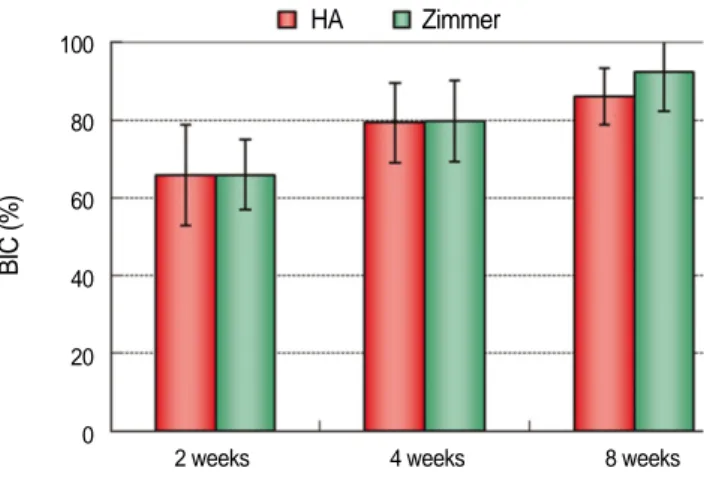
2. 골-임프란트 접촉률(Table 2, Fig. 10)
시간이 경과하면서 골-임플란트 접촉률은 점차 증가되었 다. 각 시기별 골-임프란트 접촉률은 TS III HA와 Zimmer HA 사이에 통계적으로 유의한 차이가 없었다.(P>0.05)
Fig. 3.Bone-implant contact measuring method.
Length of bony tissue which is contacted with implant thread Length of implant thread surface
Table 1.Specimens for bone-implant contact analysis
Period TS III HA Zimmer Total
2 weeks 6 8 14
4 weeks 7 6 13
8 weeks 8 8 16
Total 21 22 43
Fig. 4. Histological finding of TS III HA after 2 weeks. New bones grew from the cutting line of the host bone to con- tact the screw threads directly, which was superior to the two other surfaces.(H&E stain, ×100)
Fig. 5. Histological finding of TS III HA after 4 weeks. The thickness of the newly formed trabecular bones increased, and bone remodeling was ongoing.(H&E stain, ×100)
Ⅳ. 고 찰
임프란트 표면을 거칠게 하여 표면적을 증가시킴으로써 골유착 속도를 빠르게 하려는 생각에서 blasting and acid etching, RBM, SLA, Oxidation 등 다양한 방법들이 개발되 어 임상에 적용되고 있다. HA 코팅 임프란트는 오래 전부 터 임상에 적용되었으나 결과가 좋지 못하였고 학자들에 따라 상반된 의견들이 제시되었다.
Table 2.Bone-implant contact comparison between 2 groups
TS III HA Zimmer P value
2 weeks 65.74±12.89 65.88±8.94 0.796
4 weeks 79.24±10.18 79.77±10.46 1.000
8 weeks 85.97±7.29 92.22±9.86 0.064
P-values were calculated with Mann-Whitney test.
Fig. 7.Histological finding of Zimmer HA after 2 weeks was similar with that of TS III HA.
Fig. 8.Histological finding of Zimmer HA after 4 weeks was similar with that of TS III HA.
Fig. 6. Histological finding of TS III HA after 8 weeks.
Lamellar structures formed by the apposition of new bones were observed, and bone remodeling was ongoing.(H&E stain, ×100)
Fig. 9.Histological finding of Zimmer HA after 8 weeks was similar with that of TS III HA. Bone-implant contact ratio of Zimmer was higher than TS III HA, however, there was no statistically significant difference.
Fig. 10.Bone-implant contact ratio of TS III HA and Zimmer implant.
HA Zimmer
2 weeks 4 weeks 8 weeks 100
80
60
40
20
0
BIC (%)
초기 HA 코팅 제품들은 코팅하는 과정에서 충분한 결정 율을 유지하지 못해 실망스런 결과를 초래한 경우가 많았 다. Spline implant system은 HA를 plasma coating 한 후 압축, 수화열처리가공의 특수한 MP-1 과정을 거쳐 HA crys- tallinity를 97% 이상으로 높이면서 이전의 많은 문제점들 을 해결하였다10.
Kim 등11은 electron-beam evaporation process을 통해 500 um 두께의 thin calcium phosphate film을 SLA titanium sur- face에 침착시키는 방법을 소개하였다. Ca-P deposited SLA implant는 신생골 형성 능력이 현저하게 향상되었다고 언 급하였다. Rohner 등12은 Yorkshire pigs의 경골, 장골능, 견 갑골 부위에 4.1 mm 직경의 SLA 표면을 가진 ITI 임프란트 들을 식립하였으며 각각의 길이는 8, 10, 12 mm 였다. 골- 임프란트 접촉률은 3주 후 경골 35.2±11.5%, 견갑골 63.7
±6.8%, 장골능24.2±10.9%를 나타냈다. 6주 후에는 경골 44.4±10.9%, 견갑골73.8±8.2%, 장골능 44.2±10.6%로 증 가하였다. 12주 후에는 경골 46.8±12.6%, 견갑골 74.2±
6.5% , 장골능 52.5±11.2%를 나타냈다고 보고하였다. Choi 등13은 3가지 생체재료들을 사용하여 임프란트 주변의 3벽 성 골내결손부에 이식한 후 용해되는 것에 관한 실험적 연 구를 시행하였다. 이 연구에서 CaP nanocoating 임프란트들 이 사용되었으며 주변 결손부에는 무정형(amorphous calci- um phosphate, ACP), MBCP, FDBA가 이식되었다. 12주 후 조직형태계측학적 분석을 통해 골-임프란트 접촉률을 측 정한 결과 대조군에서는 32.12±12.11%, ACP 61.91±
24.56%, MBCP 63.57±21.57%, FDBA 48.65±12.7%를 보 였다고 발표하였다.
고전적인 HA 코팅 임프란트이 5년 성공율은 약 85%로 보고되었지만 Zimmer MP-1 HA의 5년 성공율은 97%로 매 우 높게 보고되었다14,15. 2010년 오스템사에서 개발된 TS III HA 임프란트는 상부의 일부(2 mm)는 RBM 표면이고 나머 지는 HA 코팅처리한 hybrid type을 채택하고 있다. 코팅 두 께는 TS III HA 20-70 um, Zimmer 20-150 um이며 두가지 제 품 모두 열프라즈마 방식의 코팅처리가 시행되었다. XRD 및 EDS 분석을 시행한 결과 Ca/P ratio는 Zimmer 1.65, TS III HA 1.69이고 결정화 정도는 Zimmer 96.3±0.6, TS III HA 98% 라고 자체 실험을 통해 발표되었다16.
HA 코팅 임프란트는 골표면과 접촉 시 우수한 생체적합 성을 보이고 골전도에 의한 주변 신생골의 성장을 유도하 면서 골성숙을 촉진시키는 장점을 가진다고 알려져 있다.
또한 임프란트와 골조직의 접촉강도를 현저히 증가시키기 때문에 골질이 불량한 부위에서 좋은 효과를 발휘할 수 있 고 골유착 기간이 단축되기 때문에 조기 부하를 시행할 수 있는 장점이 있다17-19. 임상적으로 골유착의 정도를 평가하 는 방법은 periotest, RFA, 타진, 촉진 및 치근단방사선사진 을 이용하는 방법 등이 있으나 정확하지 못한 것은 분명하
다20,21. 정확한 평가법은 기계적 분석법인 제거회전력
(removal torque value)과 조직형태계측학적 분석법인 골-임
프란트 접촉률이 있으나 동물실험을 통해서만 가능한 방 법이고 실제 임상에 적용할 수 없는 한계점이 있다19,22,23. 본 연구에서는 동물실험을 통해 골-임프란트 접촉률을 조직 형태계측학적으로 평가하였다.
본 연구에서 골-임프란트 접촉률은 시간이 경과하면서 증가하는 경향을 보였으며 2주, 4주 시점에는 2가지 실험 군이 매우 유사한 값을 보였다. 8주 군에서는 Zimmer 임프 란트의 골-임프란트 접촉률이 다소 높은 소견을 보였지만 통계적으로 유의성 있는 차이를 보이지 않았다. 본 연구 결 과들을 타 연구들과 직접적으로 비교하는 것은 어렵다. 왜 냐하면 실험 부위, 동물, 사용된 임프란트의 직경 및 길이, 표면 처리 등 실험조건이 다양하기 때문이다.
Ⅴ. 결 론
Miniature pigs 하악골에 식립된 2가지 HA 코팅 임프란트 의 골-임프란트 접촉률은 2주부터 높은 양상을 보였고 시 간이 경과하면서 증가하였다. 따라서 HA 코팅 임프란트는 골유착 기간을 단축시킬 가능성이 있다고 판단된다.
References
1. Jeong R, Marin C, Granato R, Suzuki M, Gil JN, Granjeiro JM, et al. Early bone healing around implant surfaces treated with variations in the resorbable blasting media method. A study in rabbits. Med Oral Patol Oral Cir Bucal 2010;15(1):e119-25.
2. Abdel-Haq J, Karabuda CZ, Arısan V, Mutlu Z, Ku¨rkçu¨M.
Osseointegration and stability of a modified sand-blasted acid- etched implant: an experimental pilot study in sheep. Clin Oral Implants 2011;22:265-74.
3. Zhu X, Ong JL, Kim S, Kim K. Surface characteristics and struc- ture of anodic oxide films containing Ca and P on a titanium im- plant material. J Biomed Mater Res 2002;60:333-8.
4. Whitehead RY, Lucas LC, Lacefield WR. The effect of dissolu- tion on plasma sprayed hydroxylapatite coatings on titanium.
Clin Mater 1993;12:31-9.
5. Wheeler SL. Eight-year clinical retrospective study of titanium plasma-sprayed and hydroxyapatite-coated cylinder implants. Int J Oral Maxillofac Implants 1996;11:340-50.
6. Trisi P, Keith DJ, Rocco S. Human histologic and histomorpho- metric analyses of hydroxyapatite-coated implants after 10 years of unction: a case report. Int J Oral Maxillofac Implants 2005;
20:124-30.
7. McGlumphy EA, Peterson LJ, Larsen PE, Jeffcoat MK.
Prospective study of 429 hydroxyapatite-coated cylindric om- niloc implants placed in 121 patients. Int J Oral Maxillofac Implants 2003;18:82-92.
8. Lee JJ, Rouhfar L, Beirne OR. Survival of hydroxyapatite-coated implants: a meta-analytic review. J Oral Maxillofac Surg 2000;
58:1372-9.
9. Morris HF, Ochi S. Hydroxyapatite-coated implants: a case for their use. J Oral Maxillofac Surg 1998;56:1303-11.
10. Chang YL, Lew D, Park JB, Keller JC. Biomechanical and mor- phometric analysis of hydroxyapatite-coated implants with vary- ing crystallinity. J Oral Maxillofac Surg. 1999;57:1096-108.
11. Kim H, Choi SH, Chung SM, Li LH, Lee IS. Enhanced bone forming ability of SLA-treated Ti coated with a calcium phos- phate thin film formed by e-beam evaporation. Biomed Mater 2010;5:044106.
12. Rohner D, Tay A, Chung SM, Hutmacher DW. Interface of un- loaded titanium implants in the iliac crest, fibula, and scapula: a histomorphometric and biomechanical study in the pig. Int J Oral Maxillofac Implants 2004;19:52-8.
13. Choi JY, Jung UW, Lee IS, Kim CS, Lee YK, Choi SH.
Resolution of surgically created three-wall intrabony defects in implants using three different biomaterials: an in vivo study. Clin Oral Implants Res 2011;22:343-8.
14. Thierer T, Davliakos JP, Keith JD Jr, Sanders JJ, Tarnow DP, Rivers JA. Five-year prospective clinical evaluation of highly crystalline HA MP-1-coated dental implants. J Oral Implantol 2008;34:39-46.
15. Artzi Z, Carmeli G, Kozlovsky A. A distinguishable observation between survival and success rate outcome of hydroxyapatite- coated implants in 5-10 years in function. Clin Oral Implants Res 2006;17:85-93.
16. Osstem Implant Co. TS III HA implant system clinical monitor- ing, 2010;1-36. Busan.
17. Cook SD, Key JF, Thomas KA, Jarcho M. Interface mechanics and histology of titanium and hydroxylapatite-coated titanium for dental implant applications. Int J Oral Maxillofac Implants 1987;2:15-22.
18. Block MS, Kent JN, Kay JF. Evaluation of hydroxylapatite-coat- ed titanium dental implants in dogs. J Oral Maxillofac Surg 1987;
45:601-7.
19. Baker D, London RM, O’Neal R. Rate of pull-out strength gain of dual-etched titanium implants: a comparative study in rabbits.
Int J Oral Maxillofac Implants 1999;14:722-8.
20. Friberg B, Sennerby L, Linden B, Grondahl K, Lekholm U.
Stability measurements of one-stage Bra�nemark implants during healing in mandibles. A clinical resonance frequency analysis study. Int J Oral Maxillofac Surg 1999;28:266-72.
21. Noguerol B, Muñoz R, Mesa F, de Dios Luna J, O’Valle F. Early implant failure. Prognostic capacity of Periotest: retrospective study of a large sample. Clin Oral Implants Res 2006;17:459-64.
22. Masuda T, Yliheikkila¨ PK, Felton DA, Cooper LF.
Generalizations regarding the process and phenomenon of os- seointegration. Part I. In vivo studies. Int J Oral Maxillofac Implants 1998;13:17-29.
23. Nkenke E, Fenner M, Vairaktaris EG, Neukam FW, Radespiel- Tro¨ger M. Immediate versus delayed loading of dental implants in the maxillae of minipigs. Part II: histomorphometric analysis.
Int J Oral Maxillofac Implants 2005;20:540-6.