83 대한안과학회지 2017년 제 58 권 제 1 호
J Korean Ophthalmol Soc 2017;58(1):83-86 ISSN 0378-6471 (Print)⋅ISSN 2092-9374 (Online)
https://doi.org/10.3341/jkos.2017.58.1.83
Case Report
윗눈꺼풀에 국한하여 발생한 신경집종 1예
A Case of Upper Eyelid Schwannoma
양 헌⋅노주헌
Heon Yang, MD, Joo Heon Roh, MD
성모안과병원
Sungmo Eye Hospital, Busan, Korea
Purpose: To report a rare case of upper eyelid schwannoma presenting as a chalazion.
Case summary: A 54-year-old male presented to our clinic with a slowly growing, painless recurred mass located in the middle area of the right upper eyelid margin. Surgical incision had been performed on a similar mass two year previous, although no his- tological analysis had been performed. On examination, a 4 × 3-mm-sized, firm, nonpigmented mass was palpable in the right upper eyelid, and no signs of neurofibromatosis were present elsewhere. The lesion was initially thought to be an eyelid mass, so we performed an excisional biopsy under local anesthesia. The lesion was easily isolated from the surrounding tissue and was excised completely. Histopathologically, the excised mass showed a compact arrangement of spindle cells forming pal- isades with Verocay bodies (Antoni A patterns). Immunohistochemistry revealed diffuse and strong S-100 protein positivity.
These findings resulted in the diagnosis of eyelid schwannoma.
Conclusions: Because of its rarity and solitary feature, eyelid schwannoma can be confused with chalazion. Thus, ophthalmolo- gists should consider schwannoma in the differential diagnosis of a slowly growing, painless recurred mass or a lesion with ma- lignant transformation after incomplete excision.
J Korean Ophthalmol Soc 2017;58(1):83-86 Keywords: Eyelid, Schwannoma
■Received: 2016. 7. 14. ■ Revised: 2016. 9. 8.
■Accepted: 2016. 12. 27.
■Address reprint requests to Joo Heon Roh, MD
Sungmo Eye Hospital, #409 Haeun-daero, Haeundae-gu, Busan 48064, Korea
Tel: 82-51-743-0775, Fax: 82-51-743-0776 E-mail: [email protected]
ⓒ2017 The Korean Ophthalmological Society
This is an Open Access article distributed under the terms of the Creative Commons Attribution Non-Commercial License (http://creativecommons.org/licenses/by-nc/3.0/) which permits unrestricted non-commercial use, distribution, and reproduction in any medium, provided the original work is properly cited.
눈꺼풀은 신체의 피부와 마찬가지로 다양한 조직을 포함 하고 있으며, 피부에 나타나는 종양이 눈꺼풀에도 발생할 수 있다.1,2 신경집종(schwannoma)은 말초신경집을 구성하 는 신경초세포(schwann cell)의 증식으로 생기는 양성종양 으로,3-6 척수와 교감신경, 경추신경, 미주신경에 주로 분포 하며, 눈에서는 대부분 안와에 발생하고 눈꺼풀에 발생하 는 신경집종은 매우 드물다.7
눈꺼풀의 신경집종은 현재까지 전 세계적으로 14건이 보 고되었으며,3-15 보통 콩다래끼(chalazion)와 유사한 임상양상 을 보인다.이전에 국내 보고된 1건의 사례는 종괴의 임상양 상이 멜라닌결핍모반(amelanocytic nevus)과 유사한 반면,11 저자들이 경험한 증례는 기존의 보고와 유사한 콩다래끼의 양상을 보여 이에 문헌고찰과 함께 보고하고자 한다.
증례보고
54세 남자가 우안 윗눈꺼풀의 재발된 종괴를 주소로 내 원하였다. 환자는 과거력상 2년 전 개인 안과에서 동일 부 위의 종괴를 콩다래끼로 진단 받고 절개술을 시행 받은 병 력이 있었다. 종괴는 절개 후 재발하여 2년 동안 통증 없이 크기가 천천히 커지는 양상을 보였다고 하였다. 병력 청취
84
- 대한안과학회지 2017년 제 58 권 제 1 호 -
Figure 1. Clinical photograph of the right eye. Preoperative
photograph shows a 4 × 3 mm-sized non-pigmented chala- zion-like mass on the right upper eyelid margin.A
B
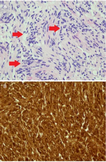
Figure 2. Light micrograph of excisional biopsy specimen.
(A) The Antoni type A area shows cellular proliferation of spindle cells arranged in a palisading fashion (red arrows). In this case, type B area is not identified (hematoxylin-eosin,
×400). (B) Immunostaining for S100 protein is strong and dif- fuse (S-100, ×400).
Figure 3. Clinical photograph of the right eye at 8 weeks after
excision. Postoperative photograph showed removed site was clear. No recurrence was observed.상 외상이나 가족력은 없었으며 신체검사상 신경섬유종증 소견은 보이지 않았다. 전안부 소견으로 우안 윗눈꺼풀 가
운데에 4 × 3 mm의 비색소성 단단한 눈꺼풀 종괴가 관찰 되었으며 다른 부위에서는 유사한 종괴가 발견되지 않았다 (Fig. 1). 촉진상 통증은 없었으며 움직이지 않는 양상을 보 였다. 위에 기술한 사항 외에 이상 소견은 없었으며, 세극 등 검사 및 안저 검사에서도 특이한 점을 찾을 수 없었다. 눈꺼풀 종괴의 정확한 진단을 위해 종괴의 절제 생검에 대한 설명을 하였으며, 환자의 동의하에 국소마취를 하고 눈꺼풀의 후방으로 접근하여 절제 생검을 시행하였다. 눈 꺼풀의 종괴는 경계가 분명하고 주변과 잘 박리되어 어려 움 없이 절제되었다.
조직병리검사상 Hematoxylin & eosin 염색하에서 과세 포성의 방추세포들이 울타리배열의 모습을 보이며 베로케 이 소체(Verocay body)를 구성하는 Antoni type A의 모습 을 보였으며, 면역조직 화학염색상에서는 S-100 단백질에 대해 미만성의 강한 양성 소견이 보였다(Fig. 2). 해당 소견 으로 눈꺼풀의 종괴가 신경집종임을 확인할 수 있었다. 종 괴 절제 8주 후 경과 관찰 시 절제된 부위는 깨끗하였고 재 발 소견은 보이지 않았다(Fig. 3).
고 찰
신경집종(schwannoma)은 매우 드문 질환으로서 신경초 세포(schwann cell)의 증식으로 발생하는 양성 종양이다.3-7 호발 양상은 대체로 단발성의 산발적 양상을 보이며, 다발 성은 신경섬유종증(neurofibromatosis)의 특징적인 소견이 다.7 다발성의 신경섬유종증이 있는 경우 드물게 악성으로 전환하며,3 눈꺼풀에서 신경집종의 악성전환은 아직 보고 된 바가 없다.13,14
눈에서는 주로 안와에 발생하고 전체 안와 종양의 1% 정 도를 차지하며,16 눈꺼풀에 발생하는 경우는 드물어 0.1%
85 - 양 헌⋅노주헌 : 윗눈꺼풀에 국한하여 발생한 신경집종 1예 -
정도 발견되는 것으로 알려져 있다.10 그 외에 보고된 눈의 호발 부위는 결막,17 포도막,18 공막19이다. 현재까지 눈꺼풀 에 발생한 신경집종은 세계적으로 14건이 보고되었고,3-15 그중 국내에서는 Lee et al11이 멜라닌결핍모반(amelanocytic nevus)과 유사한 1예를 보고하였다.
눈꺼풀에 발생하는 신경집종은 희소성과 산발적 특성으 로 인해 임상적으로 콩다래끼(chalazion)로 잘못 진단되는 경우가 많다.8 보고된 사례를 분석해 보면 14건 가운데 콩 다래끼(chalazion)와 유사한 경우가 12건 있었으며,3-9,12-15 멜라닌결핍모반(amelanocytic nevus)이나 봉입낭(inclusion cyst)과 유사한 사례가 2건 있었다.10,11 연령 분포는 소아에 서 발생한 경우가 2건,7,9 그 외는 성인에게 생긴 것으로 보 고되었다.3-6,8,10-15
호발 부위는 윗눈꺼풀이 9건,5,8-10 아래눈 꺼풀이 5건3,4,6,7,10-15
으로 나타났으며, 신경섬유종증을 가진 경우는 없었다.
신경집종의 진단은 조직병리학적으로 나타나는 안토니 A 형(Antoni type A), 안토니 B형(Antoni type B)의 특성과 면 역조직학적으로 보이는 S-100 단백질에 대한 양성 소견으 로 진단이 가능하다. 안토니 A형(Antoni type A)은 Hema- toxylin & eosin 염색하에서 방추세포들이 울타리배열의 빽 빽한 모습을 보이는 과세포성의 조직을 말하며, 안토니 B형 (Antoni type B)은 크고 둥근 투명세포들이 느슨한 모습을 보이는 저세포성의 조직이다.4 면역조직학적으로는 S-100 단백질에 대한 양성 소견이 강하게 나타나며 안토니 A형의 경우가 특징적이다.14 세포학적으로는 비정형성이 심하나 유사분열 양상은 드문 것으로 알려져 있다.13 본 증례는 병 리학적으로 안토니 A형의 양상을 보이고, 면역조직학적으 로 S-100 단백질에 미만성의 강한 양성 소견이 보여 신경집 종으로 진단하였다. 치료는 외과적 절제가 필요하며 재발을 막기 위해서 종괴를 완전히 제거해야 한다.7 불완전하게 절 제 시 재발되거나 악성 변형이 나타나기도 한다.6,7,11
이상에서 저자들이 경험한 1예를 바탕으로 고찰하여 볼 때, 절개술 등의 일반적인 치료에 반응하지 않는 비전형적 인 콩다래끼의 경우 감별진단 시 드물지만 신경집종의 가 능성도 고려해야 하며, 불완전한 절제 후 다시 느리게 자라 나거나 악성 변형이 의심되는 경우에는 조직검사를 시행할 것을 제안하는 바이다.
REFERENCES
1) Welch, RB, Duke JR. Lesions of the lids: a statistical note. Am J Ophthalmol 1958;45:415-26.
2) Allington HV, Allington JH. Eyelid tumors. Arch Dermatol 1968;97:50-65.
3) Sidiqui MA, Leslie T, Scott C, Mackenzie J. Eyelid schwannoma in a male adult. Clin Exp Ophthalmol 2005;33:412-3.
4) Chung YR, Moon S, Jang JW. Eyelid schwannoma in a Korean woman. Jpn J Ophthalmol 2007;51:231-2.
5) Baijal GC, Garg SK, Kanhere S, Monga S. Schwannoma of the eye-lid. Indian J Ophthalmol 1980;28:155-6.
6) Butt Z, Ironside JW. Superficial epitheloid schwannoma present- ing as a subcutaneous upper eyelid mass. Br J Ophthalmol 1994;78:586-8.
7) Gelincik I. Right upper eyelid schwannoma in a child: a case report. J Clin Exp Pathol 2012;2:1000116.
8) Shields JA, Guibor P. Neurilemoma of the eyelid resembling a re- current chalazion. Arch Ophthalmol 1984;102:1650.
9) Shields JA, Kiratli H, Shields CL, et al. Schwannoma of the eyelid in a child. J Pediatr Ophthalmol Strabismus 1994;31:332-3.
10) López-Tizón E, Mencía-Gutiérrez E, Gutiérrez-Díaz E, Ricoy JR.
Schwannoma of the eyelid: report of two cases. Dermatol Online J 2007;13:12.
11) Lee KW, Lee MJ, Kim NJ, et al. A case of eyelid schwannoma. J Korean Ophthalmol Soc 2009;50:290-3.
12) Yuichi O, Tatsuo Y. A case of schwannoma in the upper eyelid. J Eye Ophthalmic Plast Reconstr Surg 2009;25:50-2.
13) Kumar S, Kumar S, Kulshrestha R. Cystic schwannoma of eyelid in an Indian male: a rare presentation. Orbit 2008;27:407-9.
14) Singh S, Saraf S, Goswami D, Singh S. Case report of isolated schwannoma‑a rare eyelid tumor. US Ophthalmic Review, 2014;
7:143-5.
15) Magdum RM, Paranjpe R, Kotecha M, Patil P. Solitary eyelid schwannoma. Med J DY Patil Univ 2014;7:502-4.
16) Rootman J, Goldberg C, Robertson W. Primary orbital schwan- nomas. Br J Ophthalmol 1982;66:194-204.
17) Le Marc’hadour F, Romanet JP, Fdili A, et al. Schwannoma of the bulbar conjunctiva. Arch Ophthalmol 1996;114:1258-60.
18) Shields JA, Font RL, Eagle RC Jr, et al. Melanotic schwannoma of the choroid: immunohistochemistry and electron microscopic observations. Ophthalmology 1994;101:843-9.
19) Graham CM, McCartney AC, Buckley RJ. Intrascleral neuril- emmoma. Br J Ophthalmol 1989;73:378-81.
86
= 국문초록 =
윗눈꺼풀에 국한하여 발생한 신경집종 1예
목적: 눈꺼풀에 국한하여 발생하는 신경집종은 매우 드문 질환이다. 저자들은 콩다래끼와 유사한 형태로 발생한 증례가 있어 이를 보고하는 바이다.
증례요약: 54세 남자가 재발된 우안 윗눈꺼풀의 종괴를 주소로 내원하였다. 과거력상 환자는 2년 전 개인 안과에서 콩다래끼로 진단 받고 절개술을 시행 받은 병력이 있으며, 종괴는 절개 후 재발하여 2년 동안 통증 없이 크기가 천천히 커지는 양상을 보였다. 내원 당시 우안 윗눈꺼풀 가운데에 4 × 3 mm의 단단한 비색소성 눈꺼풀 종괴가 관찰되었으며, 신경섬유종증과 같은 전신질환은 없었다.
진단을 위해 국소마취하에 종괴를 절제 생검하였으며, 종괴는 경계가 분명하였고 주변과 잘 박리되었다. 절제된 종괴는 조직병리검사 상 방추세포들이 울타리 배열의 베로케이 소체를 구성하는 모습을 보였고, 면역조직화학염색상 S-100 단백질에 대하여 미만성의 강 한 양성 소견을 보였다. 이러한 소견으로 눈꺼풀에 발생한 신경집종으로 진단하였다.
결론: 눈꺼풀에 발생하는 신경집종은 매우 드물지만 콩다래끼와 유사한 양상을 보이는 경우가 있어 간과하기 쉽다. 따라서 본 증례와 같이 불완전한 절제 후 다시 느리게 자라나거나 악성 변형이 의심되는 종괴는 감별진단 시 신경집종도 고려할 필요가 있다.
<대한안과학회지 2017;58(1):83-86>
- 대한안과학회지 2017년 제 58 권 제 1 호 -