ISSN 0378-6471 (Print)⋅ISSN 2092-9374 (Online)
https://doi.org/10.3341/jkos.2017.58.7.879
Case Report
합성경막을 이용한 아메드밸브삽입술 후 발생한 공막융해와 동반된 튜브 짓무름
Tube Erosion with Scleral Melting after Ahmed Valve Implantation Using a Synthetic Dural Substitute
안현민1,2⋅박종운2
Hyun Min Ahn, MD1,2, Jong Woon Park, MD, PhD2
연세대학교 의과대학 안과학교실1, 국민건강보험 일산병원 안과2 Department of Ophthalmology, Yonsei University College of Medicine1, Seoul, Korea Department of Ophthalmology, National Health Insurance Service Ilsan Hospital2, Goyang, Korea
Purpose: The objective of this case report was to present tube erosion of Ahmed valve implantation using a synthetic dura sub- stitute (Neuro-Patch®, B. Braun, Boulogne, France).
Case summary: Tube erosion was caused by dissolution of the conjunctiva and partial-thickness scleral tunnel in 5 patients who received Ahmed valve implantation using a synthetic dura substitute for glaucoma treatment 2 to 4 months after the operation.
Furthermore, the patients required re-operation for preventing secondary complications such as endophthalmitis.
Conclusions: This case series using a synthetic dura substitute in Ahmed valve implantation demonstrated the risk of tube ero- sion with scleral tunnel melting and following secondary complications even with a partial-thickness scleral tunnel method.
J Korean Ophthalmol Soc 2017;58(7):879-884
Keywords: Ahmed valve complication, Neuro-patch, Synthetic dura, Tube erosion
■Received: 2017. 2. 9. ■ Revised: 2017. 4. 17.
■Accepted: 2017. 6. 23.
■Address reprint requests to Jong Woon Park, MD, PhD Department of Ophthalmology, National Health Insurance Service Ilsan Hospital, #100 Ilsan-ro, Ilsandong-gu, Goyang 10444, Korea
Tel: 82-31-900-0590, Fax: 82-31-900-0049 E-mail: [email protected]
*Conflicts of Interest: The authors have no conflicts to disclose.
ⓒ2017 The Korean Ophthalmological Society
This is an Open Access article distributed under the terms of the Creative Commons Attribution Non-Commercial License (http://creativecommons.org/licenses/by-nc/3.0/) which permits unrestricted non-commercial use, distribution, and reproduction in any medium, provided the original work is properly cited.
녹내장 방수유출 삽입 장치 중 하나인 아메드밸브는 녹 내장 여과 수술에 있어 매우 유용한 장치로 알려져 있다.1 아메드밸브(Ahmed glaucoma valve®; New World Medical Inc., Rancho Cucamonga, CA, USA)를 포함한 방수 유출 장치의 튜브는 대개의 경우 전방에 위치하게 삽입되며, 이 때 튜브를 고정시키고 노출을 방지하기 위해 다양한 방법
이 사용될 수 있다. 흔하게 사용되는 방법 중 하나로 부분 층 공막 터널 혹은 절편을 만들어 튜브를 공막 사이 공간에 위치시키는 방법이 있다.2 이외에도 기증 공막, 심막, 경막 과 같은 조직뿐만 아니라 콜라겐 매트릭스와 같은 다양한 합성 재료들이 이식물로 사용될 수 있으며, 공막 터널 혹은 절편과 이식물을 동시에 사용하기도 한다.3,4 본 증례에서 사 용한 합성경막(Neuro-Patch®; B. Braun, Boulogne, France)은 폴리에스터 우레탄 제재로 뇌수술 시 흔하게 사용하는 합 성재료로 1995년 유럽에서 경막결손이나 뇌척수 감압술 시 사용되도록 허가되었다(Fig. 1).5
아메드밸브삽입술의 합병증으로는 수술 후 맥락막삼출 을 동반 혹은 동반하지 않은 저안압이 가장 높으며, 전방출 혈, 유리체출혈, 밸브 폐쇄, 밸브 위치이상, 관 미란, 관에 의한 각막 손상, 방수 누출, 안내염 등이 있다.6,7 이 중 관이 결막이나 각막의 짓무름을 발생시키는 경우는 2-7% 정도
Table 1. The history of the patients with Ahmed valve implantation using a synthetic dura substitute
Case Age Sex R/L Systemic disease Previous ocular disease Previous ocular surgery Glaucoma type
1 44 M R DM PDR None NVG
2 55 M L Dyslipidemia None None POAG
3 65 F R DM, CRF, CAOD PDR Cataract OP NVG
L PDR Cataract OP NVG
4 67 M L HBV carrier IOL dislocation Vitrectomy, Cataract OP Secondary
5 56 F R DM, CRF PDR None NVG
L PDR None NVG
R = right; L = left; M = male; F = male; DM = diabetic mellitus; PDR = proliferative diabetic retinopathy; NVG = neovascular glaucoma; POAG = primary open-angle glaucoma; CRF = chronic renal failure; CAOD = coronary artery occlusive disease; OP = operation; HBV = hepatitis B-viral; IOL = intraocular lens.
Figure 1. Image of Neuro-patch
® (B. Braun, Boulogne, France).3 × 3 mm Neuro-patchy was placed on scleral flap and covered with conjunctival flap.
로 보고되고 있다. 관이 노출될 경우 이차적인 안내염 등의 감염이 발생할 수 있다.8,9 본 증례보고에서는 합성경막을 부분층 공막터널 상부에 이식한 아메드밸브삽입술을 시행 받은 환자 중 튜브 짓무름이 발생하여 치료를 요하게 된 다 섯 증례를 보고하고자 한다(Table 1, 2).
증례보고
아메드 벨브 삽입술 방법
각막의 윤부 12시 방향에 6-0 vicryl로 견인 봉합을 시행 한 뒤 상이측에 원개 기저 결막절개를 실시하였다. 결막과 테논낭을 공막으로부터 박리하여 아메드밸브 공간을 확보 하였다. 각막의상이측으로 3 × 3 mm 부분층 공막터널을 만들었다. 평형염액을 이용하여 아메드밸브 작동상태를 확 인(priming)하였다. 밸브를 각막 윤부로부터 8 mm 떨어진 곳에 8-0 nylon을 이용하여 공막에 고정하였다. 튜브의 사 면을 위로 향하게 하여 전방에 약 2 mm 들어가는 크기로 자른 후 윤부에서 약 1 mm 위치에 부분층 공막터널 밑으
로 23 G 바늘을 이용하여 홍채면과 평행하게 천자한 이후 튜브를 위치시켰다. 이후 8-0 nylon을 이용하여 공막편 모 서리를 공막에 고정하였다. 공막편 위로 3 × 3 mm 크기의 합성경막을 위치시킨 이후, 견인 봉합 시 사용한 각막윤부 의 6-0 vicryl을 이용하여 결막봉합을 시행하였다.
증례 1
당뇨 과거력의 44세 남성으로, 안과적으로 양안 신생혈 관 녹내장이 동반된 증식성 당뇨망막병증으로 치료받고 있었 다. 브린졸라미드-티몰롤 복합제(Elazop®, Alcon, Fort Worth, TX, USA) 및 브리모니딘(Alphagan®, Allergan, Irvine, CA, USA) 점안제를 투여하였으나 우안 안압 42로 지속적으로 상승된 소견을 보여 2회의 유리체내 베바시주맙(Avastin®, Genentech, CA, USA,) 주사를 시행하였으나 일시적인 안 압 하강만 있었을 뿐, 지속적으로 안압 상승 소견을 보여 합성경막을 사용한 아메드밸브삽입술을 시행하였다. 수술 후 우안 안압 12로 안정적으로 유지되었으나, 수술 2개월 째 밸브 튜브가 튜브 짓무름으로 인해 결막 밖으로 노출되 어 합성경막을 제거하고 10-0 nylon으로 튜브를 고정, 손실 된 결막 부위에 콜라젠 글라이코아미노글라이칸 제재를 이 용하여 복원술을 시행하게 되었다.
증례 2
이상지혈증 외 특이 내과력이 없는 55세 남성으로, 안과 적으로 양안 원발성 개방각 녹내장을 주소로 치료받고 있 었다. 도졸라미드-티몰롤 복합제(Cosopt®, Merck & Co, Whitehouse Station, NJ, USA), 브리모니딘, 라타노프로스 트(Xalatan®, Bausch & Lomb, Tampa, FL, USA) 점안액을 사용하였으나 안압 30 mmHg 이상으로 조절되지 않아 좌 안 마이토마이신을 사용한 섬유주절제술을 시행하였다. 섬 유주절제술 7개월 후 도졸라미드-티몰롤 복합제 및 라타노 프로스트 점안제와 아세타졸라마이드 경구약을 사용하였 으나 좌안 안압 33 mmHg로 다시 조절되지 않아 합성경
Table 2. The treatment and complications of the patients with Ahmed valve implantation using a synthetic dura substitute
Case PreoperativeIOP (mmHg) Preoperative treatment
Immediate post- operative IOP
(mmHg)
Post-operative IOP at 1 month
(mmHg)
Complication type
Interval until complication
Treatment for complication
1 42 Glaucoma medications,
Intravitreal Avastin #2
6 12 Tube exposure 2 months Revision
2 33 Trabeculectomy,
Glaucoma medications
8 15 Iris touching,
Tube exposure
3 months Revision
3 56
Right eye
Glaucoma medications, Intravitreal Avastin #2
9 14 Valve exposure 4 months Removal
42 Left eye
Glaucoma medications, Intravitreal Avastin #2
13 15 Tube exposure 4 months Revision
4 42 Glaucoma medications,
Anti-inflammatory drugs
8 12 Synthetic Dura
exposure
3 months Revision
5 32 Right eye
Glaucoma medications, Intravitreal Avastin #1
9 10 Tube exposure 2 months None (Patient
refuse) 28
Left eye
Glaucoma medications, Intravitreal Avastin #1
9 10 Tube exposure 2 months None (Patient
refuse) IOP = intraocular pressure calculated with Goldmann applanation tonometer (mmHg).
A B
C D
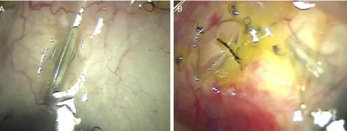
Figure 2. Images of tube erosions with scleral melting in Ahmed valve implantation using a synthetic dura substitute (case 4). (A)
One month after the operation, synthetic dura substitute was shown through the thinning conjunctival in the left eye. (B) The con- junctival thinning was aggravated for 2 weeks. (C) 3 months after the operation, Ahmed valve tube was exposed with melting sclera and conjunctiva and synthetic dura substitute was difficult to be recognized from the original shape. (D) Ahmed valve revision was performed with collagen glycoaminoglycan substitute.막을 사용한 아메드밸브삽입술을 시행하였다. 수술 후 좌 안 안압 15 mmHg 정도로 잘 유지되었으나, 수술 1개월째, 삽입한 합성결막 바로 위쪽의 결막이 얇아지기 시작하다가
3개월째 결막과 공막 및 합성경막이 융해되면서 튜브 일부 가 밖으로 노출되었으며, 더불어 밸브 튜브 끝이 홍채와 닿 으면서 염증이 발생하게 되었다. 때문에 합성 경막을 제거
A B
Figure 3. Four months after Ahmed valve implantation using a synthetic dura substitute in case 3. (A) Ahmed valve tube was exposed
with conjunctival buttonhole in the left eye. (B) Ahmed valve body was exposed through the conjunctival buttonhole in right eye.하면서 10-0 nylon으로 튜브를 고정하였고, 손실된 결막 부 위에 콜라젠 글라이코아미노글라이칸 제재를 이용하여 아 메드밸브복원술을 시행하였다(Fig. 2).
증례 3
당뇨와 만성신부전, 관상동맥협착에 의해 중재술을 시행 받은 과거력이 있는 65세 여성으로, 안과적으로 양안 신생 혈관 녹내장이 동반된 증식성 당뇨망막병증으로 치료받고 있던 분이었다. 양안 모두 백내장 수술 과거력이 있었으며, 양안 유리체내 베바시주맙 주사술을 2회 시행 받았던 환자 로, 우안 시력 안전 수동 변별, 좌안 시력 0.2였으며 도졸라 미드-티몰롤 복합제, 브리모니딘, 라타노프로스트 점안액을 사용하였으나 지속적으로 양안 안압 40 mmHg 이상으로 조절되지 않아 양안 합성경막을 이용한 아메드밸브삽입술 을 시행 받았다. 수술 후 양안 안압하강제를 사용하지 않고 안압 15 mmHg 정도로 유지되었다. 수술 후 4개월째, 우안 아메드밸브 본체가 결막 결손 부위로 돌출되었으며, 좌안 또한 밸브 튜브가 노출되었다(Fig. 3). 우안은 합성경막 및 아메드밸브제거술을 시행하였으며, 좌안의 경우 합성경막 만 제거한 이후 결막 봉합을 통한 수복을 시행하였다.
증례 4
B형 간염 보균자인 67세 남성으로, 안과적으로 양안 백 내장 수술 및 좌안 인공수정체 이탈로 인한 유리체 절제술 과 이차 인공수정체 삽입술을 시행 받은 과거력이 있는 환 자였다. 좌안 유리체 절제술 후 이차성 녹내장으로 6개월간 치료 받던 환자로, 브린졸라미드-티몰롤 복합제 및 브리모 니딘 점안제 안압하강제 및 염증 치료를 위해 리멕솔론 점 안액을 사용하였으나 좌안 안압 40 mmHg로 상승되어 좌
안 합성경막을 이용한 아메드밸브삽입술을 시행하였다. 수 술 후 좌안 안압 12 mmHg로 잘 유지되었다. 하지만 수술 후 3개월째 결막이 얇아지는 소견과 함께 삽입되었던 합성 경막이 노출되었으며, 결국 합성경막을 제거하게 되었고, 콜라젠 글라이코아미노글라이칸 제재로 아메드밸브복원술 을 시행하였다.
증례 5
당뇨 및 만성신부전으로 투석을 하고 있는 56세 여자 환 자로, 양안 신생혈관 녹내장을 동반한 증식성 당뇨망막병 증으로 타 병원에서 치료를 받고 있다가 양안 도졸라미드- 티몰롤 복합제, 브리모니딘, 라타노프로스트 점안제를 사용 하였으나 양안 시력 0.02, 안압 30 mmHg 정도로 수술적 치 료를 위해 전원된 환자였다. 수술 2주 전 양안 유리체내 베 바시주맙 주사술을 시행하였으나 안압은 하강하지 않았으 며, 결국 합성경막을 이용한 아메드밸브삽입술을 시행하였 다. 수술 후 안압하강제 사용 없이 양안 안압 10 mmHg로 안정적으로 유지되었으나 수술 2개월째 양안 밸브 튜브가 결막 밖으로 노출되는 소견이 발견되었다. 환자 및 보호자 가 적극적 치료를 원하지 않아 항생제 안약을 사용하면서 외래 경과 관찰 중이다.
고 찰
녹내장 밸브 삽입술은 난치성 녹내장 수술에 있어 유용 한 방법으로 알려져 있으나 몇몇 합병증이 보고되고 있다. 대표적인 합병증으로 밸브 튜브 바로 위쪽으로 결막이 얇 아지면서 구멍(buttonhole)이 발생하는 튜브 짓무름이 있 다.10 이러한 합병증은 방수누출(leakage)뿐만 아니라 안내
염(endophthalmitis)과 같은 감염을 유발시킬 수 있는 것으 로 알려져 있다.11 이러한 튜브 짓무름을 막기 위하여 공막 터널이나 절편을 만드는 방법 또는 이식물을 이용한 방법 등이 알려져 있다.2 일반적으로 이식물을 사용하지 않고 공 막 터널 혹은 절편만을 이용하는 방법이 수술적 용이성 및 비용 문제로 인하여 선호되지만, 안내 튜브 회전에 의한 각 막 내피의 손상과 절편을 만드는 과정의 위험성뿐만 아니
라,12,13 공막과 결막에도 튜브 짓무름을 발생시키기 때문에
튜브 노출에 의한 이차적 합병증이 발생할 수 있다고 알려 졌다.14 때문에 보완적 방법으로 기증 공막, 심막, 경막과 같은 조직뿐만 아니라 콜라겐 글라이코아미노글라이칸 등 의 다양한 합성 재료가 이용된다.4
본 증례에서는 합성경막(Neuro-Patch®, B. Braun)을 사용 하여 공막 터널을 보완하였다. 합성경막은 비흡수성 소섬 유의 폴리에스터 우레탄 재제로 뇌수술 후 발생한 경막결 손 수복을 위해 빈번하게 사용되는 제재이다.15 합성경막은 폴리에스터 우레탄 재제의 특성상 물리적인 힘의 부하에 강할 뿐 아니라 다양한 세포로 분화할 수 있는 간엽세포 분 화, 증식 및 세포 간의 결합을 유도하여 조직과 강하게 유 착된다는 장점에 의해 경막성형술이나 다양한 뼈의 재건술 및 유방암에서의 조직재건에 사용된다.16-18 생체 조직을 직 접 이용하지 않는 점에서 이득이 있을 뿐 아니라 다양한 치 료 영역에서 그 효과 및 이용 가능성이 밝혀진 점에서 녹내 장 밸브 삽입술에서도 충분히 효과가 있을 것으로 기대하 였다. 그러나 본 증례에서는 수술 2-4개월 후, 공막 터널 융 해와 합성경막과 결막을 뚫고 아메드밸브 튜브 노출이 발 생하였으며, 결과적으로 이차적인 합병증을 방지하기 위해 합성결막을 제거하는 재수술을 요하게 되었다.
합병증의 원인으로는 다양한 것들을 생각해 볼 수 있다.
먼저 일반적으로 시행되는 녹내장 밸브 삽입술 후 발생한 튜브 짓무름 현상의 원인을 본 증례에서도 추정해 볼 수 있 다. 당뇨 과거력과 같은 환자 인자와 봉합으로 인한 미세혈 류의 압박은 조직 허혈을 발생시키고 세포괴사를 유발시킬 수 있다는 보고가 있다.19 더불어 길어진 수술 시간과 많아 진 수술 재료들은 조직 변화가 튜브 짓무름을 발생시킨다 는 보고도 있다.13 이에 빗대어 볼 때, 본 증례에서도 합성 경막을 추가적으로 삽입하는 과정에서 길어진 수술시간과 늘어난 재료 또한 이러한 문제를 촉진시킬 수 있을 것으로 본다. 더하여 본 증례에서 사용된 합성경막 재료에 의한 원 인을 고려해야 된다. 비록 합성경막의 안과적 사용에 대해 보고된 논문은 없지만, 합성경막을 사용한 신경외과 수술 에서 조직 감염 및 염증에 의한 괴사로 합병증이 발생하였 다는 몇몇 연구들이 보고되었다.5,20 이와 관련해 본 증례 환 자에서 신경외과에서 보고된 바와 같이 합성경막과 직접
접촉된 부위에서 시작된 결막 두께 및 혈관 변화로 확인할 수 있는 염증에 의한 괴사 조직의 발생은, 합성경막 조직자 체에 의한 염증성 변화로, 튜브 짓무름 및 공막과 결막의 융해가 발생했을 가능성을 생각해 봐야 할 것이다. 또한 직 접적으로 실험해 보지는 않았으나, 상대적으로 단단하고 탄성이 강한 합성경막의 경우, 조직 특성상 경막재건술이 나 뼈의 재건술보다 압박을 통한 허혈이 결막과 공막에서 상대적으로 더 잘 유발될 수 있을 가능성 또한 생각해 볼 수 있다. 결론적으로, 본 증례를 통하여 아메드밸브삽입술 에서 합성경막을 이용하는 경우 부분층 공막터널을 같이 만든다 하여도 결막과 공막 융해가 동반된 튜브 짓무름이 발생할 수 있는 위험성을 확인할 수 있었다.
REFERENCES
1) Lim K, Allan BD, Lloyd AW, et al. Glaucoma drainage devices;
past, present, and future. Br J Ophthalmol 1998;82:1083-9.
2) Rosenberg LF, Krupin T. Implants in glaucoma surgery. The Glaucomas 1996;3:1783-807.
3) Smith MF, Doyle JW, Ticrney Jr JW. A comparison of glaucoma drainage implant tube coverage. J Glaucoma 2002;11:143-7.
4) Rosentreter A, Mellein AC, Konen WW, Dietlein TS. Capsule ex- cision and Ologen implantation for revision after glaucoma drain- age device surgery. Graefes Arch Clin Exp Ophthalmol 2010;
248:1319-24.
5) Malliti M, Page P, Gury C, et al. Comparison of deep wound in- fection rates using a synthetic dural substitute (neuro-patch) or per- icranium graft for dural closure: a clinical review of 1 year.
Neurosurgery 2004;54:599-603; discussion 603-4.
6) Dubey S, Sharma V, Agrawal A, et al. Safety and efficacy of Ahmed glaucoma valve implantation in refractory glaucomas in Northern Indian eyes. Saudi J Ophthalmol 2015;29:103-8.
7) Kim SW, Kim YH, Yun IS, Ahn JH. Surgical treatment for tube erosion after Ahmed valve implantation. J Korean Ophthalmol Soc 2016;57:453-60.
8) Huang MC, Netland PA, Coleman AL, et al. Intermediate-term clinical experience with the Ahmed Glaucoma Valve implant. Am J Ophthalmol 1999;127:27-33.
9) Wilson MR, Mendis U, Paliwal A, Haynatzka V. Long-term fol- low-up of primary glaucoma surgery with Ahmed glaucoma valve implant versus trabeculectomy. Am J Ophthalmol 2003;136:464- 70.
10) Heuer DK, Budenz D, Coleman A. Aqueous shunt tube erosion. J Glaucoma 2001;10:493-6.
11) Francis BA, DiLoreto DA, Chong LP, Rao N. Late-onset bacterial endophthalmitis following glaucoma drainage implantation.
Ophthalmic Surg Lasers Imaging 2003;34:128-30.
12) Melamed S, Fiore PM. Molteno implant surgery in refractory glaucoma. Surv Ophthalmol 1990;34:441-8.
13) Trubnik V, Zangalli C, Moster MR, et al. Evaluation of risk factors for glaucoma drainage device-related erosions: a retrospective case-control study. J Glaucoma 2015;24:498-502.
14) Rosentreter A, Schild AM, Dinslage S, Dietlein TS. Biodegradable
= 국문초록 =
합성경막을 이용한 아메드밸브삽입술 후 발생한 공막융해와 동반된 튜브 짓무름
목적: 합성경막을 이용한 아메드밸브삽입술에서 튜브 짓무름이 발생한 다섯 증례를 보고하고자 한다.
증례요약: 안압 조절을 위해 합성경막을 이용한 아메드밸브삽입술을 시행한 5명의 녹내장 환자에서 2-4개월 후 결막 및 부분층 공막 터널 융해와 더불어 튜브 짓무름이 발생하였으며, 안내염 등의 이차적인 합병증을 막기 위해 재수술이 필요하게 되었다.
결론: 아메드밸브삽입술에서 합성경막을 이용하는 경우 부분층 공막터널을 병행하여 수술하는 경우에도 공막터널의 융해와 동반된 튜브 짓무름이 발생하여 이에 따른 이차적인 합병증이 발생할 수 있는 위험성이 있다.
<대한안과학회지 2017;58(7):879-884>
implant for tissue repair after glaucoma drainage device surgery. J Glaucoma 2012;21:76-8.
15) Gudmundsson G, Søgaard I. Complications to the use of vicryl-col- lagen dural substitute. Acta Neurochir (Wien) 1995;132:145-7.
16) Raul JS, Godard J, Arbez-Gindre F, Czorny A. Use of polyester urethane (Neuro-Patch) as a dural substitute. Prospective study of 70 cases. Neurochirurgie 2003;49(2-3 Pt 1):83-9.
17) Cardwell RD, Kluge JA, Thayer PS, et al. Static and cyclic me- chanical loading of mesenchymal stem cells on elastomeric, elec- trospun polyurethane meshes. J Biomechl Eng 2015;137. doi:
10.1115/1.4030404. Epub 2015 Jun 3.
18) Zanetta M, Quirici N, Demarosi F, et al. Ability of polyurethane foams to support cell proliferation and the differentiation of MSCs into osteoblasts. Acta Biomater 2009;5:1126-36.
19) Huddleston SM, Feldman RM, Budenz DL, et al. Aqueous shunt exposure: a retrospective review of repair outcome. J Glaucoma 2013;22:433-8.
20) El Majdoub F, Löhr M, Maarouf M, et al. Transmigration of fi- brino-purulent inflammation and malignant cells into an artificial dura substitute (Neuro-Patch): report of two cases. Acta Neurochir (Wien) 2009;151:833-5.