ISSN 0378-6471 (Print)⋅ISSN 2092-9374 (Online)
http://dx.doi.org/10.3341/jkos.2015.56.5.784
Case Report
안내후방콘택트렌즈삽입술 7년 후 발생한 급격한 내피세포 감소 1예
Sudden Loss of Endothelial Cell Density 7 Years after Receiving an Implantable Contact Lens: A Case Report
강동완⋅엄영섭⋅임재원⋅강수연⋅김효명⋅송종석
Dong Wan Kang, MD, Young Sub Eom, MD, Jay Won Rhim, MD, Su Yeon Kang, MD, Hyo Myung Kim, MD, PhD, Jong Suk Song, MD, PhD
고려대학교 의과대학 구로병원 안과학교실
Department of Ophthalmology, Korea University Guro Hospital, Korea University College of Medicine, Seoul, Korea
Purpose: To report a case of decreased endothelial cell density 7 years after posterior chamber phakic intraocular lens implantation.
Case summary: A 45-year-old man with high myopia combined with astigmatism was treated with Toric implantable Collamer Lens (ICL) implantation. The patient’s best corrected visual acuity was 0.7 in both eyes before the operation. After the treatment, his uncorrected visual acuity was 0.9 and corrected visual acuity was 1.0 in both eyes, indicating an improvement in visual function. Preoperative endothelial cell density measured 3,063 cells/mm2 in the right eye and 3,126 cells/mm2 in the left eye. At 5 years postoperatively, measurements were 2,897 cells/mm2 in the right eye and 2,974 cells/mm2 in the left, showing little change. However, a 6-year postoperative measurement of 2,198 cells/mm2 in the right eye and 2,803 cells/mm2 in the left showed a slight decrease in endothelial cell density in the right eye, and a follow-up measurement one year later displayed a rap- id decline to 1,272 cells/mm2 in the right eye and 2,852 cells/mm2 in the left eye. The Toric ICL lens was removed from the right eye and phacoemulsification and posterior chamber intraocular lens implantation was performed. Two-month postoperative en- dothelial cell density was 1,257 cells/mm2 and endothelial cell damage from the operation itself was minimal.
Conclusions: ICL implantation may cause complications related to corneal endothelial cells as well as glaucoma. Patients should receive regular follow-up examinations for endothelial cell density.
J Korean Ophthalmol Soc 2015;56(5):784-788
Key Words: Endothelial cell loss, ICL implantation, Implantable contact lens
■Received: 2014. 11. 28. ■ Revised: 2015. 1. 8.
■Accepted: 2015. 4. 1.
■Address reprint requests to Jong Suk Song, MD, PhD
Department of Ophthalmology, Korea University Guro Hospital,
#148 Gurodong-ro, Guro-gu, Seoul 152-703, Korea Tel: 82-2-2626-3178, Fax: 82-2-2626-1261 E-mail: [email protected]
* This study was presented as an e-poster at the 112th Annual Meeting of the Korean Ophthalmological Society 2014.
* This study was supported in part by Alumni of department of ophthalmology, Korea University College of Medicine in 2015.
ⓒ2015 The Korean Ophthalmological Society
This is an Open Access article distributed under the terms of the Creative Commons Attribution Non-Commercial License (http://creativecommons.org/licenses/by-nc/3.0/) which permits unrestricted non-commercial use, distribution, and reproduction in any medium, provided the original work is properly cited.
현재 행해지고 있는 굴절이상을 교정하는 방법으로는 엑 시머레이저를 이용한 방법과 안내렌즈 삽입술로 나누어 볼 수 있다. 레이저를 이용한 방법으로는 굴절교정레이저각막 절제술(photorefractive keratectomy, PRK), 레이저각막절삭 성형술(laser in-situ keratomileusis, LASIK), 레이저각막상 피절삭성형술(laser epithelial keratomileusis, LASEK) 등이 있으며 야간 눈부심, 달무리, 각막혼탁, 각막확장증 등의 부 작용이 있고 각막조직의 양에 제한이 있어 고도근시 환자 의 굴절교정에 있어서는 제한이 있다.1-3 안내렌즈삽입술은 고도근시 환자의 교정에 제한이 적고 굴절력의 예측이 가 능하고 조절기능이 유지될 수 있으며 교환 및 제거가 가능 하다는 장점이 있다.4,5 현재 임상에서 사용되고 있는 유수
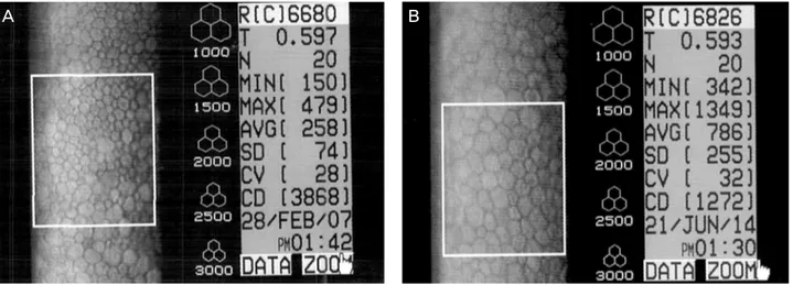
Figure 1. Corneal endothelial cell density of the right eye. (A) Before ICL implantation operation. (B) Seven years after ICL im-
plantation operation. R = right; T = thickness; N = number; SD = standard deviation; CV = coefficient of variation; CD = cell density; ICL = implantable contact lens.정체용 안내렌즈로는 고정 위치나 방법에 따라 눈안삽입콘 택트렌즈(Implantable contact lens, ICL)와 같은 후방렌즈, Phakic6, Nuvita 렌즈와 같은 전방각지지렌즈, 알티산렌즈 와 같은 홍체고정안내렌즈로 나눌 수 있다. 그 중 ICL과 같 은 안내후방콘택트렌즈는 홍채와 수정체 사이에 위치함에 따라 백내장을 유발할 가능성이 있고 동공차단 녹내장이나 악성녹내장 발생 등이 가능하다는 단점이 있으나, 전방에 위 치하는 전방각지지렌즈나 홍채고정렌즈에 비해 각막 내피와 의 거리가 멀어 내피세포 손상을 일으킬 위험이나 전방 내 구조의 손상이 적다는 장점이 있어 널리 사용되고 있다.6-9
각막내피세포는 손상을 받게 되면 재생되지 않고 내피세 포의 거대화, 이동, 육각형세포비율의 감소 등의 복구과정 을 거치며 각막내피세포의 밀도가 감소하게 된다.10 정상인 에서 각막내피세포의 밀도는 2,500 cells/mm2 정도로 알려 졌으며 외부 손상 등에 의해 그 밀도가 500 cells/mm2 이내 로 감소하면 각막 부종과 부전이 발생하게 된다.11 ICL은 타 렌즈보다 각막내피세포에 미치는 영향은 적은 것으로 알려졌으나 내피세포의 변화와 감소에 대한 보고와 논의가 국내외로 계속 진행되고 있다.12-20
본 증례는 본원에서 ICL 삽입술을 받은 환자에서 6년째 까지 정상내피세포 밀도를 보이다가 7년째 급격한 내피세 포 감소가 발생한 환자가 있어 이를 보고하고자 하며, 나아 가 ICL과 같은 안구 내 수술 시행 후 정기적인 각막내피세 포의 변화를 측정하는 것의 중요성에 대해서 이야기해 보 고자 한다.
증례보고
45세 남자 환자가 시력교정을 목적으로 외래 방문하였
다. 환자는 고도근시로 25년간 소프트렌즈를 착용하고 있 었다. 환자는 특별한 외상이나 각막질환, 포도막염 등 안과 적인 과거력은 없었다. 초진 시 시행한 굴절력 검사상 최대 교정시력은 우안 0.7 (-14.0 Dsph=-2.0 Dcyl×30 A), 좌안 0.8 (-13.0 Dsph=-5.0 Dcyl×175 A)이었고 세극등현미경검 사 및 안저검사상 특이소견은 보이지 않았다. 각막내피세 포검사를 위하여 비접촉성 경면현미경(SP-2000P: Konan, Tokyo, Japan)을 이용, 25개의 cell dotting 후 기계가 자동 으로 계산하는 방식의 center method로 시행하였고, 수술 전 시행한 각막내피세포밀도는 우안 3,063 cells/mm2, 좌안 3,126 cells/mm2로 측정되었다(Fig. 1A). 다소의 polymegath- ism은 보였으나 endothelial cell density나 polymorphism은 정상범위였고, 특별한 내피세포의 이상은 관찰되지 않았다.
환자와의 상의 후 시력교정을 위해 Toric implantable contact lens (Toric ICL; STAAR Surgical AG, Nidau, Switzerland) 삽입술을 시행하기로 하였고, 먼저 동공폐쇄녹내장의 예방 을 위해 수술 전 Nd-YAG 레이저를 이용하여 주변부홍채 레이저절개술을 시행하였다.
수술은 0.5% Proparacaine hydrochloride (Alcaine, Purrs, Belgium)로 점안마취를 시행 후 12시와 6시 방향에 전방 출입구를 만들고 전방에 점탄물질 1.4% NaHA (Healon GV, Abbott, Abbott Park, IL, USA)를 한 뒤 3.0 mm의 이측 투명각막절개창을 통해 제조사의 삽입장치(STAAR ICL in- jector system)를 이용하여 ICL을 삽입하였다. 전방 점탄물 질을 제거한 후 Carbachol 0.01% (Miostat, Alcon, ForthWorth, TX, USA)를 전방에 주입하여 동공을 축소시켰고, 투명각막 절개창은 10-0 Nylon을 이용하여 봉합하였다. 수술 중 각막 내피와 수정체, 홍채 등에 닿지 않도록 조작에 주의를 기울 였다. 수술 후 1달 동안 0.5% Levofloxacin (Cravit, Santen,
A B
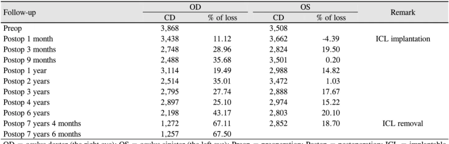
Table 1. Changes in corneal endothelial cell density (CD, cells/mm
2)Follow-up OD OS
Remark
CD % of loss CD % of loss
Preop 3,868 3,508
Postop 1 month 3,438 11.12 3,662 -4.39 ICL implantation
Postop 3 months 2,748 28.96 2,824 19.50
Postop 9 months 2,488 35.68 3,501 0.20
Postop 1 year 3,114 19.49 2,988 14.82
Postop 2 years 2,514 35.01 3,472 1.03
Postop 3 years 2,795 27.74 2,888 17.67
Postop 4 years 2,897 25.10 2,974 15.22
Postop 6 years 2,198 43.17 2,803 20.10
Postop 7 years 4 months 1,272 67.11 2,852 18.70 ICL removal
Postop 7 years 6 months 1,257 67.50
OD = oculus dexter (the right eye); OS = oculus sinister (the left eye); Preop = preoperation; Postop = postoperation; ICL = implantable contact lens.
Figure 2. Changes in corneal endothelial cell density. OD =
oculus dexter (the right eye); OS = oculus sinister (the left eye).Osaka, Japan)과 1% Prednisolone acetate (Pred-Forte, Allergan, CA, USA)를 하루 4회 점안하였다.
수술 후 1개월째 나안시력은 우안 1.0, 좌안 0.9로 측정되 었고 ICL 렌즈의 vaulting은 우안 1½ Corneal thickness (CT), 좌안 1 CT였다. 각막내피세포밀도는 1개월째 우안 3,438 cells/mm2, 좌안 3,662 cells/mm2로 측정되었고, 6개 월째는 우안 2,488 cells/mm2, 좌안 3,501 cells/mm2로 우안 에서 조금 감소한 모습을 보였다. 1년 간격으로 각막내피세 포밀도를 측정하였고 수술 후 4년째의 각막내피세포밀도는 우안 2,897 cells/mm2, 좌안 2,974 cells/mm2로 별 변화가 없 었다. 6년째에는 우안이 2,198 cells/mm2 좌안이 2,803 cells/mm2로 정상범위의 각막내피세포 밀도이긴 하지만 우 안에 내피세포의 감소가 관찰되었다
1년 뒤 수술 후 7년째 각막내피세포밀도를 측정하였을 때 우안 1,272 cells/mm2, 좌안 2,852 cells/mm2로 우안의 내 피세포밀도가 급격히 감소한 것이 관찰되었다(Fig. 1B). 환 자가 호소하는 특별한 증상은 없었고, 나안시력은 우안 0.9, 좌안 0.8이었으며 ICL 렌즈의 Vaulting은 양안 모두 1 CT 였다. 각막내피세포의 손상을 막기 위해 ICL을 제거하기로 하였고, 부동시를 해결하기 위해 초음파유화술과 인공수정
체 삽입술을 동시에 시행하였다. 수술 중 각막내피세포를 보 호하기 위해 분산성 점탄물질인 Viscoat (Alcon Laboratories, Inc., Forth Worth, TX, USA)를 사용하여 ICL을 제거한 후, 초음파유화술 및 인공수정체 삽입술을 시행한 뒤 10-0 Nylon으로 절개창을 봉합하였다.
수술 후 두 달째 측정한 각막내피세포는 우안 1,257 cells /mm2로 수술로 인한 각막내피세포의 손상은 심하지 않았 다(Table 1, Fig. 2). 수술 후 나안시력은 0.9였으며 정기적 으로 각막내피세포검사를 시행하며 경과관찰 중이다.
고 찰
ICL과 같은 유수정체용 후방렌즈는 1980년대 Fyodorov et al18,19이 후방내 실리콘 인공수정체를 삽입하기 시작한 이후 널리 시술되어 왔고, 국내에서는 1997년 최초로 소개 된 후 현재 활발히 시행되고 있는 굴절 교정술이다.14 시력 회복이 빠르며 빛번짐이 적고 술후 조절이 유지되며 추후 렌즈를 교체하거나 제거할 수 있는 가역적인 수술방법이라 는 장점이 있다.20 처음에는 실리콘 재질로 되어 있어 백내 장에 발생빈도가 많았지만 현재 사용되고 있는 ICL은 친수 성 Collamer 재질로 되어 있어 합병증을 줄이게 되었고,19 본 증례에서도 높은 생체적합성을 가지는 Collamer 재질의 Visian Toric ICL 렌즈를 사용하였다. 최근에는 Cental flow 를 가지고 있는 ICL도 출시되어 동공폐쇄녹내장의 발생 위 험을 줄이기 위하여 따로 주변부홍체절개술을 시행할 필요 가 없어지는 등 합병증의 위험을 줄이는 데 있어 많은 발전 이 이루어지고 있다.
각막내피세포밀도의 감소는 안내렌즈삽입술 후 발생가 능한 중요한 합병증 중 하나로, 인간의 각막내피세포는 생 체 내에서 증식에 제한이 있어 손상을 받게 되면 재생되지 않고 남아있는 각막내피세포의 거대화, 이동, 로젯형성, 육
각형세포비율의 감소 등의 과정을 거치며 각막내피세포의 밀도가 감소하게 된다.10 후방렌즈는 전방에 위치하는 전방 각지지렌즈나 알티산렌즈 등과 비교하여 백내장, 동공차단 녹내장 및 악성녹내장의 발생 빈도가 더 높은 반면 내피세 포 손상은 적다고 알려졌다4
ICL 삽입 후 각막내피세포의 변화에 대해 국내외로 많은 논문들에서 보고되고 있다.12-17,21 대부분의 논문에서 경과 관찰기간 동안 내피세포의 유의한 변화가 없거나 2년 이내 에 내피세포의 유의한 감소가 있으나 그 이후로는 통계적 으로 유의한 감소가 없는 안정된 상태를 보였다고 보고하 였다. 단기적인 연구로는 Dejaco-Ruhswurm et al4의 연구나 Choi et al21의 연구에서 ICL 삽입 후 1년째에 각막내피세포 의 유의한 감소를 보였지만 그 이후로는 특별한 변화가 없 는 안정된 양상을 보였다는 보고를 하고 있고, Choi et al21 은 3년간의 경과관찰을 통하여 술 전과 비교하였을 때 통 계적으로 유의한 각막내피세포의 변화는 보이지 않았다고 보고하였다. 중장기적인 연구에서는 Han and Lee22는 술 후 5년 동안 전반적으로 내피세포가 감소하는 양상을 보였으나 통계적으로 유의하지 않았다는 보고를 하였고, Igarashi et al23은 ICL 삽입 후 8년간의 기간 동안 추적관찰을 통하여 최대 22.8%의 각막내피세포의 감소를 보고하였다. 이와 같 이 술 후 내피세포감소에 대해서 많은 연구가 있었지만 본 증례에서와 같이 약간의 감소를 보이나 정상범위 안에 내피 세포밀도를 보이다가 술 후 7년이 지나서 1,500 cells/mm2 미 만의 감소를 보인 보고는 없었다.
유수정체인공수정체 삽입 후 각막내피세포의 감소에 영 향을 주는 요인은 다양한 것으로 알려졌다. 수술 중 수술 기구나 수정체에 의한 각막내피의 직접적 손상에 의해 주 로 발생할 수 있고, 임상전단계의 염증반응의 독성 효과 또 한 내피세포의 감소를 초래할 수 있다.4,24 외상에 의한 감소 는 수술 후 비교적 조기경과 시 관찰된다는 점에서 본 증례 와는 거리가 있다고 할 수 있겠다.24 술 후 지속적인 각막내 피세포 감소의 원인으로는 ICL 자체로 인해 발생한 임상전 단계의 염증반응에 의한 것으로 생각되고 있으며 이는 홍 채와 ICL의 직접적인 접촉이나 혈액-방수장벽의 장애로 추 측되고 있다.4,24 본 증례보고에서는 각막내피세포의 갑작스 런 급격한 감소에 있어 환자가 호소한 외상이나 감염 등의 이벤트는 없었지만, 1년이라는 경과관찰 기간 동안 환자가 인지하지 못한 전방 내 염증반응을 초래할 수 있는 질환에 이환되어 있을 가능성을 생각해 볼 수 있었고, 경과관찰 기 간 동안 환자의 안압, 전방 내 염증, 각막의 상태 등이 모두 특별한 이상소견을 보이지 않았으므로 지속적인 임상전단 계의 염증이 갑자기 심화되어 내피세포에 어떠한 영향을 주었을 가능성에 대해서 고려해 볼 수 있었다.
비록 본 증례에서 급격한 내피세포의 감소에 있어서의 정확한 원인을 규명하지는 못하였지만 이에 대한 연구의 필요성과, 임상적으로 안정되어 있는 환자일지라도 지속적 인 경과관찰의 필요성과 중요성을 이야기했다는 점에 본 증례에 의의를 둘 수 있겠다. 갑작스러운 각막내피세포의 감소를 예방하기 위해서 연 1-2회 정도의 정기적인 경과관 찰과 공초점현미경이나 비접촉성 경면현미경을 이용한 각 막내피세포의 측정이 행해져야 하며, 내피세포의 감소가 일어났을 경우 유발요인에 따라 빠른 약물치료나 ICL 제거 술 등 수술적 중재가 필요할 것이다. 정기적인 경과관찰을 하지 않고 지내왔더라면 ICL 제거술을 시행할 수도 없을 정도의 각막내피세포의 감소가 일어나거나 더 나아가 각막 부전이 발생하여 돌이킬 수 없는 결과를 초래할 수도 있었 을 것이다. 술 후 비교적 안정적인 모습을 보이더라도 꼭 정기적 각막내피세포 변화를 측정해야 함의 중요성을 다시 한 번 일깨워준 증례라 할 수 있겠다.
REFERENCES
1) Heitzmann J, Binder PS, Kassar BS, Nordan LT. The correction of high myopia using the excimer laser. Arch Ophthalmol 1993;111:
1627-34.
2) Geggel HS, Talley AR. Delayed onset keratectasia following laser in situ keratomileusis. J Cataract Refract Surg 1999;25:582-6.
3) Holladay JT, Dudeja DR, Chang J. Functional vision and corneal changes after laser in situ keratomileusis determined by contrast sensitivity, glare testing, and corneal topography. J Cataract Refract Surg 1999;25:663-9.
4) Dejaco-Ruhswurm I, Scholz U, Pieh S, et al. Long-term endothe- lial changes in phakic eyes with posterior chamber intraocular lenses. J Cataract Refract Surg 2002;28:1589-93.
5) Javitt JC. Clear-lens extraction for high myopia. Is this an idea whose time has come? Arch Ophthalmol 1994;112:321-3.
6) Sanders DR, Doney K, Poco M; ICL in Treatment of Myopia Study Group. United States Food and Drug Administration clinical trial of the Implantable Collamer Lens (ICL) for moderate to high my- opia: three-year follow-up. Ophthalmology 2004;111:1683-92.
7) Sarikkola AU, Sen HN, Uusitalo RJ, Laatikainen L. Traumatic cat- aract and other adverse events with the implantable contact lens. J Cataract Refract Surg 2005;31:511-24.
8) Smallman DS, Probst L, Rafuse PE. Pupillary block glaucoma sec- ondary to posterior chamber phakic intraocular lens implantation for high myopia. J Cataract Refract Surg 2004;30:905-7.
9) Kodjikian L, Gain P, Donate D, et al. Malignant glaucoma induced by a phakic posterior chamber intraocular lens for myopia. J Cataract Refract Surg 2002;28:2217-21.
10) Bourne RR, Minassian DC, Dart JK, et al. Effect of cataract sur- gery on the corneal endothelium: modern phacoemulsification compared with extracapsular cataract surgery. Ophthalmology 2004;111:679-85.
11) Jacobs PM, Cheng H, Price NC, et al. Endothelial cell loss after cataract surgery-the problem of interpretation. Trans Ophthalmol
= 국문초록 =
안내후방콘택트렌즈삽입술 7년 후 발생한 급격한 내피세포 감소 1예
목적: 안내후방콘택트렌즈(implantable Collamer Lens, ICL) 삽입술을 받은 환자에서 6년째까지 정상내피세포 밀도를 보이다가 7년째 급격한 내피세포 감소가 발생한 환자가 있어 이를 보고하고자 한다.
증례요약: 남자 45세, 난시를 동반한 고도근시 환자에서 Toric ICL 삽입술을 시행하였다. 환자는 수술 전 최대교정시력이 양안 모두 0.7이었으며, 수술 후에는 나안시력이 양안 모두 0.9, 교정시력이 양안 모두 1.0으로 시력향상을 보였다. 수술 전 각막내피세포 밀도는 우안 3,063 cells/mm2, 좌안 3,126 cells/mm2였고 수술 후 5년째의 각막내피세포 밀도는 우안 2,897 cells/mm2 좌안 2,974 cells/mm2 로 별 변화가 없었으나 6년째에는 우안 2,198 cells/mm2 좌안 2,803 cells/mm2로 내피세포의 감소가 관찰되었다. 1년 후 다시 측정한 각막내피세포 밀도는 우안 1,272 cells/mm2 좌안 2,852 cells/mm2로 우안의 내피세포밀도가 급격히 감소한 것이 관찰되었고, 우안의 Toric ICL 렌즈를 제거하고 수정체유화술 및 인공수정체 삽입술을 시행하였다. 수술 후 두 달째 측정한 각막내피세포 밀도는 1,257 cells/mm2로 수술로 인한 각막내피세포의 손상은 심하지 않았다.
결론: ICL과 같은 유수정체인공수정체를 삽입한 환자에서는 수술 후 백내장은 물론 각막내피세포에 영향을 줄 수 있어 정기적이고 규칙적인 내피세포밀도 측정을 시행해야 한다.
<대한안과학회지 2015;56(5):784-788 >
Soc U K 1982;102 (pt 2):291-3.
12) Han SY, Lee KH. Long term effect of ICL implantation to treat high myopia. J Korean Ophthalmol Soc 2007;48:465-72.
13) Chun YS, Lee JH, Lee JM, et al. Outcomes after implantable con- tact lens for moderate to high myopia. J Korean Ophthalmol Soc 2004;45:480-9.
14) Lee SY, Cheon HJ, Baek TM, Lee KH. Implantable contact lens to correct high myopia (clinical study with 24 months follow-up). J Korean Ophthalmol Soc 2000;41:1515-22.
15) Pesando PM, Ghiringhello MP, Di Meglio G, Fanton G. Posterior chamber phakic intraocular lens (ICL) for hyperopia: ten-year fol- low-up. J Cataract Refract Surg 2007;33:1579-84.
16) Sanders DR, Doney K, Poco M; ICL in Treatment of Myopia Study Group. United States Food and Drug Administration clinical trial of the Implantable Collamer Lens (ICL) for moderate to high my- opia: three-year follow-up. Ophthalmology 2004;111:1683-92.
17) Edelhauser HF, Sanders DR, Azar R, Lamielle H; ICL in Treatment of Myopia Study Group. Corneal endothelial assessment after ICL implantation. J Cataract Refract Surg 2004;30:576-83.
18) Fyodorov SN, Zuev VK, Tumanyan ER, Larionov Y. Analysis of long term clinical and functional results of intraocular correction of
high myopia. Ophthalmosurg (Moscow) 1990;2:3-6.
19) Fyodorov SN, Zuyev VK, Aznabayev BM. Intraocular correction of high myopia with negative posterior chamber lens. Ophthalmosurg 1991;3:57-8.
20) Jiménez-Alfaro I, Gómez-Tellería G, Bueno JL, Puy P. Contrast sensitivity after posterior chamber phakic intraocular lens im- plantation for high myopia. J Refract Surg 2001;17:641-5.
21) Choi WS, Lee HY, Seo SG, Her J. Clinical outcomes of implant- able contact lens and iris-fixed intraocular lens for correction of myopia. J Korean Ophthalmol Soc 2008;49:1406-14.
22) Han SY, Lee KH. Long term effect of ICL implantation to treat high myopia. J Korean Ophthalmol Soc 2007;48:465-72.
23) Igarashi A, Shimizu K, Kamiya K. Eight-year follow-up of posteri- or chamber phakic intraocular lens implantation for moderate to high myopia. Am J Ophthalmol 2014;157:532-9.e1.
24) Jiménez-Alfaro I, Benítez del Castillo JM, García-Feijoó J, et al.
Safety of posterior chamber phakic intraocular lenses for the cor- rection of high myopia: anterior segment changes after posterior chamber phakic intraocular lens implantation. Ophthalmology 2001;108:90-9.