Introduction
The treatment for unicompartmental osteoarthritis (OA) in young patients is still a controversial issue. The possible treatment methods of this disease are nonoperative or operative treatment; operative treatment can be divided into arthroscopy, high tibial osteotomy (HTO), and unicompartmental arthroplasty (UKA)13). However, the treatment outcomes of these surgical treatment modalities for unicompartmental OA have become subjects of controversy.
The ideal indication for HTO is young (<65 years old), active patients affected by symptomatic mild to moderate varus knee (around 10°) with medial compartment involvement (less than grade III Ahlback classification), good knee range of motion, no severe knee instability, and mild meniscus injury46). On the other hand, UKA is ideal if the patients are >60 years old and not obese and have low activity demand and unicompartmental femoral or tibial condyle avascular necrosis7,8). However, only a small proportion of patients can be considered eligible for either HTO or UKA; they are the socalled borderline patients9,10). The indications for both HTO and UKA include ages 55 to 65 years, moderate activity, mild varus alignment, less than grade I knee instability, and Ahlback grades I to II arthrosis.
In cases of advanced medial compartment arthritis accompa
nied by kissing lesions, HTO is known to be a relative contrain
dication, and UKA is preferred instead11). However, in relatively young patients, UKA or total knee arthroplasty (TKA) can be disastrous when complications, such as component loosening, failure, and infection occur7). Furthermore, revisions of UKA and TKA in younger patients have shown poor outcomes12). On the
High Tibial Osteotomy versus Unicompartmental Knee Arthroplasty for Medial Compartment Arthrosis with Kissing Lesions in Relatively Young Patients
Seung Min Ryu, MD, Jae Woo Park, MD, Ho Dong Na, MD, and Oog Jin Shon, MD
Department of Orthopedic Surgery, Yeungnam University Medical Center, Daegu, Korea
Purpose: The purpose of this study is to compare the clinical and radiographic outcomes of high tibial osteotomy (HTO) and unicompartmental arthroplasty (UKA) in advanced medial compartment arthritis accompanied by kissing lesions in relatively young patients.
Materials and Methods: Fortyfive patients were divided into the HTO (n=23) and UKA (n=22) groups. Clinically, we evaluated the Lysholm knee scoring scale, visual analogue scale, Hospital for Special Surgery, and Western Ontario and McMaster Universities Osteoarthritis index scores preoperatively, 6 and 12 months postoperatively, and at the final followup. Radiographically, we measured the femoraltibial angle and mechanical axis deviation preoperatively and at the final followup.
Results: All clinical outcomes gradually improved in both groups from the postoperative period to the final followup. At the final followup, all clinical outcomes were slightly better in the UKA group than in the HTO group; however, differences were not statistically significant.
Conclusions: HTO is comparable to UKA in terms of clinical outcomes. Thus, the results of this study suggest that HTO might be a good alternative treatment to UKA for medial unicompartmental arthritis accompanied by kissing lesions in relatively young patients.
Keywords: Knee, Osteoarthritis, Osteotomy, Arthroplasty, Unicompartmental pISSN 2234-0726 · eISSN 2234-2451
Knee Surgery & Related Research
Received February 3, 2017; Revised (1st) March 11, 2017;
(2nd) May 4, 2017; (3rd) June 20, 2017;
Accepted June 26, 2017
Correspondence to: Oog Jin Shon, MD
Department of Orthopedic Surgery, Yeungnam University Medical Center, 170 Hyeonchungro, Namgu, Daegu 42415, Korea Tel: +82536203640, Fax: +82536284020
Email: maestro[email protected]
17
This is an Open Access article distributed under the terms of the Creative Commons Attribution NonCommercial License (http://creativecommons.org/licenses/bync/4.0/) which permits unrestricted noncommercial use, distribution, and reproduction in any medium, provided the original work is properly cited.
Copyright © 2018 KOREAN KNEE SOCIETY www.jksrr.org
other hand, some studies have reported that HTO combined with arthroscopic microfracture in young patients with unicompart
mental arthritis with kissing lesions can be effective in alleviating pain and in preserving the joint in a shortterm followup1315).
However, to the best of our knowledge, no previous study com
paring the outcomes of HTO and UKA in young patients with unicompartmental arthritis with kissing lesions has been reported.
Hence, the purpose of this study was to compare the clinical and radiographic outcomes between HTO and UKA in relatively young patients with unicompartmental arthritis with kissing lesions.
Materials and Methods
1. Patient Selection
This study was approved by the Institutional Review Board of our hospital. From January 2007 to January 2012, all patients who were treated with HTO or UKA and had medial compart
mental arthritis with kissing lesions were reviewed in this study.
The kissing lesions affected both the contacting femoral and tibial condyles; these were diagnosed when the femoral condyle and tibial condyle were in contact on 45° flexion weightbearing posterioranterior radiographs (Fig. 1). Intraoperatively, these were classified as Outerbridge grade IV lesions that appeared to be in contact with each other in the femoral condyle and tibial condyle. Of 60 patients in total, seven patients whose followup were <2 years, three who had lateral meniscus lesions or lateral compartment arthritis, three who had insufficiencies in the an
terior or posterior cruciate ligaments, and two who had severe patellofemoral OA were excluded. No patient who had a defor
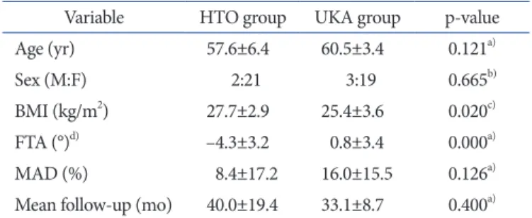
mity or history of trauma in any limb was included to evaluate the accurate alignment of the lower limbs. Ultimately, 45 patients were divided into the HTO group (n=23) and UKA group (n=22) for a comparative analysis (Table 1). Age, sex, and mean follow
up period were not statistically significantly different; however, body mass index (BMI) showed a statistically significant differ
ence between the two groups (p=0.020). The patients were asked to decide whether to undergo either HTO or UKA after a discus
sion with the surgeon about the prognosis, rehabilitation pro
gram, and their expectation of the level of physical activity after surgery. Based on the evaluation of the patient’s compliance, their ability to reduce weight, and hygiene, we finally decide proper treatment. HTO was recommended for patients with severe la
bor, such as farming, and UKA was recommended for sedentary patients in urban areas.
2. Surgical Technique
In the HTO group, a longitudinal skin incision was created at the medial side of the tibial tuberosity. The superficial medial col
lateral ligament was completely released below the osteotomy site.
Biplanar medial open wedge osteotomy was performed using the ChronOS vivify spacer (DePuy Synthes, Solothurn, Switzerland) and TomoFix plate (DePuy Synthes). The target correction angle was measured at the point where the mechanical axis of the lower limb passed through the Fujisawa point, which was 62.5% from the medial tibial articular margin16).
In the UKA group, a standard medial parapatellar arthrotomy was performed. UKA was performed without extension to the
Fig. 1. A 54yearold female who had medial compartmental arthritis combined with a kissing lesion. The kissing lesion was diagnosed when the femoral condyle and tibial condyle were seen in contact on the 45°
flexion weight bearing posterioranterior radiograph.
Table 1. Patient Demographics
Variable HTO group UKA group pvalue
Age (yr) 57.6±6.4 60.5±3.4 0.121a)
Sex (M:F) 2:21 3:19 0.665b)
BMI (kg/m2) 27.7±2.9 25.4±3.6 0.020c)
FTA (°)d) –4.3±3.2 0.8±3.4 0.000a)
MAD (%) 8.4±17.2 16.0±15.5 0.126a)
Mean followup (mo) 40.0±19.4 33.1±8.7 0.400a) Values are presented as mean±standard deviation.
HTO: high tibial osteotomy, UKA: unicompartmental arthroplasty, BMI:
body mass index, FTA: femoraltibial angle, MAD: mechanical axis deviation.
a)MannWhitney Utest.
b)Fisher exact test.
c)Student ttest.
d)Varus is defined as minus; valgus is defined as plus.
vastus medialis obliquus and without patella eversion. The sigma unicompartmental knee prostheses (DePuy, Warsaw, IN, USA) were applied in all patients in the UKA group.
3. Evaluation Tools
Clinically evaluation was performed using the Lysholm knee scoring scale17), visual analogue scale18), Hospital for Special Sur
gery (HSS)19), and Western Ontario and McMaster Universities Osteoarthritis index scores20) preoperatively, 6 and 12 months postoperatively, and at the final followup.
We radiographically measured the femoraltibial angle (FTA) and mechanical axis deviation (MAD) preoperatively and at the final followup (Fig. 2). Negative FTA and MAD values were given to the knees in varus alignment.
To determine the intra and interobserver reliabilities of the ra
diographic outcomes, two observers (SMR and HDN) evaluated 20 randomly selected radiographs regardless of HTO or UKA that were included in this series. The two observers were blinded to patient information and independently measured the preoper
ative and postoperative FTA and MAD values of the radiographs
and repeated the measurements 3 weeks later. The intra and in
terobserver reliabilities were analyzed using intraclass correlation coefficients (ICCs) with 95% confidence intervals.
We also compared the complications after surgery between the two procedures, such as fixation failure, intraarticular fracture, infection, and component loosening or breakage.
4. Statistical Analysis
Power calculations for sample size were performed using the G*Power program21). The sample size was based on the results reported by Ivarsson and Gillquist22). The mean Lysholm knee scoring scale scores of the HTO and UKA groups were 78 and 91, respectively, and their corresponding standard deviations were 19 and 11, respectively. With a power of 0.8, 20 cases for each group were needed for this study.
All statistical analyses were performed using the IBM SPSS ver.
23.0 (IBM Co., Armonk, NY, USA), and a pvalue of <0.05 was considered statistically significant. The MannWhitney Utest, Fisher exact test, and Student ttest were used to show statistical differences between the two groups. Wilcoxon signed rank test was also used to compare radiographic values between preopera
tive and final followup assessments.
Results
1. Clinical Outcomes
The clinical outcomes are shown in Table 2. All clinical out
comes gradually improved in both groups from the postopera
tive period to the final followup. At the postoperative 6 months, Lysholm knee scoring scale and HSS score showed better result in UKA group, and difference was statistically significant (p=0.032 and p=0.041). However, all clinical outcomes between the two groups showed no significant differences statistically at the post
operative 1 year and the final followup (p>0.05).
2. Radiographic Outcomes
The radiographic outcomes are shown in Tables 1 and 3. The mean FTA was –4.3° and 0.8° in the HTO and UKA groups, re
spectively, before surgery, and the difference was statistically sig
nificant (p=0.000). At the final followup, the mean FTA was 7.8°
and 5.6° in the two groups, respectively. The mean MAD was 8.4%
and 16.0%, respectively, before surgery, and the difference was not statistically significant (p=0.126). However, at the final fol
lowup, the mean MAD was 56.7% and 44.5%, respectively. The differences from the preoperative assessment to the final follow
up in both groups were all statistically significant (all p=0.000).
A B
Fig. 2. (A) Femoraltibial angle was defined as the angle between the anatomical axis of the femur and the tibia in the anteroposterior (AP) view of the knee joint. (B) For the assessment of mechanical axis devia
tion (MAD), a line was drawn from the center of the ankle joint to the head of the femur through that of the knee joint on the weight bearing fulllimb AP radiograph. MAD was calculated as the horizontal distance from this line to the medial edge of the tibial plateau divided by the total width of the tibial plateau.
Both intra and interobserver reliability testing results were 0.8 (Table 4). The ICC values of MAD were greater than those of FTA for both intra and interobserver reliabilities.
3. Complications
No specific complications were observed during the 1year shortterm followup period in both groups.
Discussion
This study was conducted to compare the results of HTO and UKA for medial unicompartmental knee arthritis accompanied by kissing lesions in relatively young patients. There were no significant differences in the clinical outcomes between the two treatment methods at the final followup. However, the radio
graphic outcomes showed that HTO tended to correct a greater angle than UKA.
Numerous studies reported the clinical outcomes of HTO and UKA in unicompartmental arthritis23,24). Although these studies did not compare such conditions accompanied by kissing lesions,
the differences in the clinical outcomes were not statistically sig
nificant between the two procedures. Similarly, the clinical out
comes of this study showed no statistically significant difference between HTO and UKA in the cases accompanied by kissing lesions at the final followup.
On the other hand, there are several reports that clinical out
come of UKA is better in unicompartmental arthritis less than 1 year follow up after surgery. Ivarsson and Gillquist22) reported that UKA group showed better results than HTO group at 6 months postoperatively. And Borjesson et al.25) also reported that timedistance variables of gait showed clinically significant dif
ferences in favor of the UKA group at 3 months postoperatively.
Our results also showed that UKA group showed better results than HTO group at 6 months postoperatively in Lysholm and HSS score with statistically significance.
Only a few studies have directly compared the radiographic outcomes of HTO and UKA in unicompartmental arthritis26,27). In general, reported radiographic outcomes showed no statisti
cally significant difference between HTO and UKA. However, the mean MADs of HTO and UKA of this study were 56.7% and 44.5%, respectively, and HTO was closer to the Fujisawa point16). We speculate that the differences in the radiographic data were Table 3. Radiographic Outcomes of the HTO Group and UKA Group
Group Preoperative Final followup pvalueb) FTA (°)a)
HTO –4.3±3.2 7.8±2.7 0.000
UKA 0.8±3.4 5.6±1.9 0.000
MAD (%)a)
HTO 8.4±17.2 56.7±12.2 0.000
UKA 16.0±15.5 44.5±13.8 0.000
Values are presented as mean±standard deviation.
HTO: high tibial osteotomy, UKA: unicompartmental arthroplasty, FTA:
femoraltibial angle, MAD: mechanical axis deviation.
a)Varus is defined as minus; valgus is defined as plus.
b)Wilcoxon signed rank test.
Table 4. Intraobserver and Interobserver Reliabilities of the Radiographic Evaluations
FTA (°) MAD (%)
Intraobserver reliability 0.946 (0.844–0.985) 0.977 (0.956–0.985) Interobserver reliability 0.892 (0.687–0.971) 0.901 (0.728–0.968) Values are presented as intraclass correlation coefficients (95% confi
dence interval).
FTA: femoraltibial angle, MAD: mechanical axis deviation.
Table 2. Clinical Outcomes of the HTO Group and UKA Group Group Preoperative POD
6 mo POD
1 yr Final followup Lysholm score
HTO 50.2±15.7 81.4±14.6 86.6±13.9 87.4±12.0 UKA 51.4±7.7 85.2±9.3 88.2±9.9 89.2±10.2
pvaluea) 0.641 0.032 0.937 0.445
VAS score
HTO 6.5±1.2 3.8±1.5 3.2±1.5 2.2±1.2
UKA 6.4±1.0 3.2±1.4 3.1±1.8 1.5±1.7
pvaluea) 0.194 0.889 0.482 0.303
HSS score
HTO 55.6±13.1 80.1±7.3 85.3±6.6 85.1±8.3
UKA 55.0±9.0 84.1±8.3 86.3±7.3 86.6±7.9
pvaluea) 0.520 0.041 1.000 0.625
WOMAC score
HTO 57.0±22.9 28.2±17.2 18.6±19.1 16.5±17.5 UKA 63.2±18.3 24.0±18.3 16.5±18.8 14.9±16.9
pvaluea) 0.453 0.716 0.751 0.169
Values are presented as mean±standard deviation.
HTO: high tibial osteotomy, UKA: unicompartmental arthroplasty, POD: postoperative day, Lysholm: Lysholm knee scoring scale, VAS:
visual analogue scale, HSS: Hospital for Special Surgery, WOMAC:
Western Ontario and McMaster Universities Osteoarthritis index.
a)MannWhitney Utest.
because of the surgeon’s own target angle, which was different between the two surgical methods.
HTO was once known as a relative contradiction for patients with advanced arthritis accompanied by a kissing lesion, and ar
throplasty is considered to offer more favorable outcome in most cases combined with kissing lesions11). However, there are nu
merous patients to which knee arthroplasty can be hardly applied owing to the risk of infection and the difficulty in management.
Although there was no major complication in the UKA group in our study, if complications occur, such as component loosen
ing or infection, it is disastrous for young patients as revision operations are more difficult. Extended life expectancy has made patients more unwilling to undergo knee revision arthroplasty, and a longterm followup has found that UKA is not effective in every case2830). In our study, although all clinical outcomes were slightly better in the UKA group than in the HTO group, there was no statistically significant difference. Therefore, we suggest that HTO might be a good alternative treatment for medial uni
compartmental arthritis with kissing lesions in relatively young, borderline patients considering the risk of arthroplasty.
The main limitations of this study are that it was a retrospec
tive study, and the number of cases was relatively small. Fur
thermore, the duration of followup was relatively short. Since the differences in both preoperative BMI and FTA between the two groups were statistically significant, it is presumed that an accurate comparison between the two groups could not be evalu
ated. Moreover, as the clinical outcomes between the two groups showed nonparametric values, we could not perform a repeated measures analysis of variance (ANOVA) analysis; thus, we could not evaluate the interaction between the time and treatment method. However, to the best of our knowledge, this is the first study to compare the clinical and radiographic outcomes of HTO and UKA in medial compartmental arthritis in relatively young patients with kissing lesions.
Conclusions
HTO was comparable to UKA in terms of clinical outcomes and complications in unicompartmental arthritis accompanied by kissing lesions in relatively young, borderline patients. Thus, the results of this study suggest that HTO might be a good alter
native treatment to UKA for medial unicompartmental arthritis with kissing lesions in this type of patient population.
Conflict of Interest
No potential conflict of interest relevant to this article was re
ported.
References
1. Billings A, Scott DF, Camargo MP, Hofmann AA. High tibial osteotomy with a calibrated osteotomy guide, rigid internal fixation, and early motion: longterm followup. J Bone Joint Surg Am. 2000;82:709.
2. Murphy SB. Tibial osteotomy for genu varum. Indications, preoperative planning, and technique. Orthop Clin North Am. 1994;25:47782.
3. Weale AE, Lee AS, MacEachern AG. High tibial osteotomy using a dynamic axial external fixator. Clin Orthop Relat Res. 2001;(382):15467.
4. Naudie D, Bourne RB, Rorabeck CH, Bourne TJ. The Install Award: survivorship of the high tibial valgus osteotomy: a 10 to 22year followup study. Clin Orthop Relat Res. 1999;
(367):1827.
5. Preston S, Howard J, Naudie D, Somerville L, McAuley J.
Total knee arthroplasty after high tibial osteotomy: no dif
ferences between medial and lateral osteotomy approaches.
Clin Orthop Relat Res. 2014;472:10510.
6. Naudie DD, Amendola A, Fowler PJ. Opening wedge high tibial osteotomy for symptomatic hyperextensionvarus thrust. Am J Sports Med. 2004;32:6070.
7. Kozinn SC, Scott R. Unicondylar knee arthroplasty. J Bone Joint Surg Am. 1989;71:14550.
8. Borus T, Thornhill T. Unicompartmental knee arthroplasty.
J Am Acad Orthop Surg. 2008;16:918.
9. Dettoni F, Bonasia DE, Castoldi F, Bruzzone M, Blonna D, Rossi R. High tibial osteotomy versus unicompartmental knee arthroplasty for medial compartment arthrosis of the knee: a review of the literature. Iowa Orthop J. 2010;30:131
40.
10. Rossi R, Bonasia DE, Amendola A. The role of high tibial osteotomy in the varus knee. J Am Acad Orthop Surg. 2011;
19:5909.
11. Bonasia DE, Dettoni F, Sito G, Blonna D, Marmotti A, Bruz
zone M, Castoldi F, Rossi R. Medial opening wedge high tib
ial osteotomy for medial compartment overload/arthritis in the varus knee: prognostic factors. Am J Sports Med. 2014;
42:6908.
12. Hang JR, Stanford TE, Graves SE, Davidson DC, de Steiger
RN, Miller LN. Outcome of revision of unicompartmental knee replacement. Acta Orthop. 2010;81:958.
13. Pascale W, Luraghi S, Perico L, Pascale V. Do microfractures improve high tibial osteotomy outcome? Orthopedics. 2011;
34:e2515.
14. van den Bekerom MP, Patt TW, Rutten S, Raven EE, van de Vis HM, Albers GH. Arthroscopic debridement for grade III and IV chondromalacia of the knee in patients older than 60 years. J Knee Surg. 2007;20:2716.
15. Shon OJ, Park SJ, Shim BJ, Lee DY. Comparative study of clinical and radiographic outcomes of high tibial osteotomy in patients with kissing lesions and nonkissing lesions. Knee Surg Relat Res. 2017;29:28894.
16. Fujisawa Y, Masuhara K, Shiomi S. The effect of high tibial osteotomy on osteoarthritis of the knee: an arthroscopic study of 54 knee joints. Orthop Clin North Am. 1979;10:585
608.
17. Lysholm J, Gillquist J. Evaluation of knee ligament surgery results with special emphasis on use of a scoring scale. Am J Sports Med. 1982;10:1504.
18. Carlsson AM. Assessment of chronic pain. I. Aspects of the reliability and validity of the visual analogue scale. Pain.
1983;16:87101.
19. Insall JN, Dorr LD, Scott RD, Scott WN. Rationale of the Knee Society clinical rating system. Clin Orthop Relat Res.
1989;(248):134.
20. Bellamy N, Buchanan WW, Goldsmith CH, Campbell J, Stitt LW. Validation study of WOMAC: a health status instrument for measuring clinically important patient relevant outcomes to antirheumatic drug therapy in patients with osteoarthritis of the hip or knee. J Rheumatol. 1988;15:183340.
21. Erdfelder E, Faul F, Buchner A. GPOWER: a general power analysis program. Behav Res Methods Instrum Comput.
1996;28:111.
22. Ivarsson I, Gillquist J. Rehabilitation after high tibial oste
otomy and unicompartmental arthroplasty: a comparative study. Clin Orthop Relat Res. 1991;(266):13944.
23. Nwachukwu BU, McCormick FM, Schairer WW, Frank RM, Provencher MT, Roche MW. Unicompartmental knee ar
throplasty versus high tibial osteotomy: United States prac
tice patterns for the surgical treatment of unicompartmental arthritis. J Arthroplasty. 2014;29:15869.
24. Yim JH, Song EK, Seo HY, Kim MS, Seon JK. Comparison of high tibial osteotomy and unicompartmental knee arthro
plasty at a minimum followup of 3 years. J Arthroplasty.
2013;28:2437.
25. Borjesson M, Weidenhielm L, Mattsson E, Olsson E. Gait and clinical measurements in patients with knee osteoarthri
tis after surgery: a prospective 5year followup study. Knee.
2005;12:1217.
26. Takeuchi R, Umemoto Y, Aratake M, Bito H, Saito I, Kum
agai K, Sasaki Y, Akamatsu Y, Ishikawa H, Koshino T, Saito T. A mid term comparison of open wedge high tibial oste
otomy vs unicompartmental knee arthroplasty for medial compartment osteoarthritis of the knee. J Orthop Surg Res.
2010;5:65.
27. StukenborgColsman C, Wirth CJ, Lazovic D, Wefer A. High tibial osteotomy versus unicompartmental joint replacement in unicompartmental knee joint osteoarthritis: 710year followup prospective randomised study. Knee. 2001;8:187
94.
28. WDahl A, Robertsson O, Lidgren L. Surgery for knee os
teoarthritis in younger patients. Acta Orthop. 2010;81:1614.
29. Warme BA, Aalderink K, Amendola A. Is there a role for high tibial osteotomies in the athlete? Sports Health. 2011;3:
5969.
30. Mancuso F, Dodd CA, Murray DW, Pandit H. Medial uni
compartmental knee arthroplasty in the ACLdeficient knee.
J Orthop Traumatol. 2016;17:26775.