• 검색 결과가 없습니다.

Navigated versus Conventional Technique in High Tibial Osteotomy: A Meta-Analysis Focusing on Weight Bearing Effect

N/A
N/A
Protected

Academic year: 2021

Share "Navigated versus Conventional Technique in High Tibial Osteotomy: A Meta-Analysis Focusing on Weight Bearing Effect"

Copied!
18
0
0

로드 중.... (전체 텍스트 보기)

전체 글

(1)

Introduction

High tibial osteotomy (HTO) is an established surgical treat­

ment option for younger patients with medial compartment knee

osteoarthritis (OA) with varus deformity that shifts the load on the knee joint by changing lower limb alignment

1)

. Optimal limb alignment is a paramount factor for satisfactory surgical results of HTO

2)

because poorly corrected alignment has been reported as one of the important causes of unsatisfactory clinical outcome after HTO

3­5)

.

Navigation was introduced in HTO to improve the accuracy of alignment correction
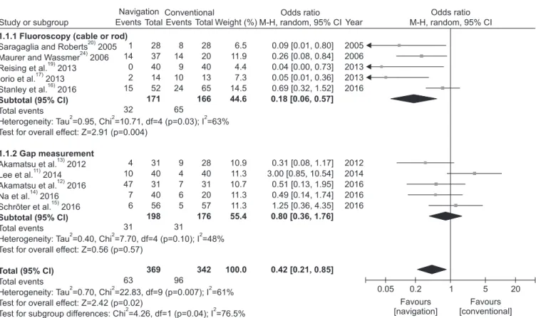
6,7)

, but it is still unclear whether navigated HTO is superior to the conventional technique regarding the achievement of the target coronal alignment. Recent systematic reviews or meta­analyses reported that the use of navigation in HTO could improve the precision of coronal alignment correc­

tion

8­10)

. However, there is conflicting evidence showing less outli­

er in the conventional HTO using the weight bearing scanogram technique

11)

or comparable accuracy in coronal alignment

12­16)

. Conventional HTO can be divided into different categories based on the detailed surgical technique, particularly by the method for

Navigated versus Conventional Technique in

High Tibial Osteotomy: A Meta­Analysis Focusing on Weight Bearing Effect

Kyung Wook Nha, MD, PhD 1, *, Young­Soo Shin, MD, PhD 2, *, Hyuk Min Kwon, MD 3 , Jae Ang Sim, MD, PhD 3 , and Young Gon Na, MD 3

1

Department of Orthopedic Surgery, Inje University Ilsan Paik Hospital, Goyang;

2

Department of Orthopedic Surgery, College of Medicine, Hallym University Hospital, Chuncheon;

3

Department of Orthopedic Surgery, Gachon University Gil Medical Center, Incheon, Korea

Purpose: We aimed to determine whether navigated opening wedge high tibial osteotomy (HTO) is superior to the conventional technique in terms of accuracy of the coronal and sagittal alignment correction, functional outcome, and operative time.

Methods: Studies comparing navigated and conventional HTO were included in this meta­analysis. We compared the incidence of radiological outliers in coronal alignment and tibial slope maintenance, mean differences in functional outcome scales, and operative time. Subgroup analyses were performed on coronal alignment accuracy based on the intraoperative method of alignment confirmation: fluoroscopy vs. gap measurement method.

Results: Twelve studies were included: there were 434 knees in the navigated HTO studies and 405 knees in the conventional HTO studies. The risk of outlier was lower in navigated HTO than in conventional HTO; however, the difference was not significant when navigated HTO was compared with conventional HTO performed using the gap measurement method. Tibial slope maintenance was comparable or better in navigated HTO.

No difference was found in the American Knee Society function and Lysholm scores. Navigated HTO necessitated a longer operative time of approximately 10 minutes.

Conclusions: The use of navigation in HTO can improve accuracy in both coronal and sagittal alignments, but its clinical benefit is unclear.

Keywords: Knee, Osteoarthritis, Tibia, Osteotomy, Computer-assisted surgery, Meta-analysis pISSN 2234-0726 · eISSN 2234-2451

Knee Surgery & Related Research

Received November 20, 2017; Revised (1st) August 28, 2018;

(2nd) October 8, 2018; (3rd) December 1, 2018;

Accepted February 8, 2019

Correspondence to: Young Gon Na, MD

Department of Orthopedic Surgery, Gachon University Gil Medical Center, 21 Namdong­daero 774beon­gil, Namdong­gu, Incheon 21565, Korea

Tel: +82­32­460­3384, Fax: +82­32­461­3214 E­mail: [email protected]

*These authors contributed equally to this study.

81

This is an Open Access article distributed under the terms of the Creative Commons Attribution Non­Commercial License (http://creativecommons.org/licenses/by­nc/4.0/) which permits unrestricted non­commercial use, distribution, and reproduction in any medium, provided the original work is properly cited.

Copyright © 2019 KOREAN KNEE SOCIETY www.jksrr.org

(2)

intraoperative confirmation of the correction amount. Fluoros­

copy method is used to confirm the corrected alignment by using a cable or metal rod under fluoroscopy

17­20)

. Gap measurement, which is preoperatively planned on a weight bearing scanogram, is sometime preferred

11­15)

. The former method relies on the in­

formation obtained in the supine position without loading on the joint, which is the same in navigated HTO

11)

. Although a recent study reported a linear relationship between mechanical axis deviation measured in intraoperative post­osteotomy fluoros­

copy and that in postoperative standing radiography, the greater discrepancy can be associated with the higher joint line conver­

gence angle

21)

. Lee et al.

22)

reported an unsatisfactory correlation of coronal alignment measurement between navigation and postoperative standing radiography, suggesting the measurement in supine position without weight bearing effect as the inherent limitation of navigation. Specogna et al.

23)

reported the difference of the coronal alignment in the supine position and that in the standing position influenced by the weight bearing effect. Previ­

ous systematic reviews and meta­analyses did not take much consideration into the weight bearing effect in the comparison of the navigation and conventional techniques

8­10)

.

Even if navigated HTO can improve the accuracy of the align­

ment correction, whether the improved accuracy will lead to better clinical outcomes is still controversial

8,10)

. Compared with the conventional technique, the navigation technique requires additional procedures, such as tracker fixation and registration;

hence, the longer operative time can be a disadvantage of navigat­

ed HTO

13,17,24)

. However, the operative time was not investigated in the previous meta­analysis

8)

. In addition, theoretically, tracker fixation can cause specific navigation­related complications, such as screw site fracture or infection and neurovascular injury

25­27)

.

This meta­analysis aimed to determine whether navigated opening wedge HTO is superior to the conventional technique in terms of the accuracy of the coronal and sagittal alignment cor­

rection, functional outcome, operative time, and complication rate. Regarding the coronal alignment accuracy, we specifically focused on the weight bearing effect associated with the intra­

operative surgical technique of conventional HTO. We hypoth­

esized that the use of navigation would improve the accuracy of alignment correction but it would not offer clinical benefit superior to that of the conventional technique in medial opening wedge HTO.

Materials and Methods

This meta­analysis was conducted according the guidelines of

the preferred reporting items for systematic reviews and meta­

analysis (PRISMA) statement (http://www.prisma­statement.

org).

1. Data and Literature Sources

This study was based on Cochrane Review Methods

28)

. Multiple comprehensive databases, including MEDLINE, EMBASE, Web of Science, SCOPUS, and the Cochrane Library, were searched for studies that compared the outcome of the navigated and conventional HTO. The language was restricted to English, and the publication date was limited from January 1, 2000 to August 15, 2016. The following keywords were used in the title, abstract, MeSH (Medical Subject Headings), and keyword fields: knee or tibia, OA, genu varum, osteotomy, computer­assisted, navigation, fluoroscopy, cable method, weight bearing, load bearing, axial load, axial force, and standing. Different search protocols were used for each database, and the detailed search strategy is de­

scribed in Appendix 1. After the initial electronic search, relevant articles and their bibliographies were manually searched. The identified articles were assessed individually for inclusion.

2. Study Selection

Study inclusion was decided independently by two reviewers on the basis of the predefined selection criteria. Titles and abstracts were read; the full article was evaluated if suitability could not be determined. Studies were included in the meta­analysis if they (1) compared radiologic and/or clinical outcomes in patients who underwent opening wedge HTO using either the navigation or conventional technique (either randomized controlled trials or retrospective studies); (2) included data on at least one of the radiographic outcome parameters regarding the accuracy of the coronal alignment. Studies were excluded if they met the follow­

ing exclusion criteria: (1) closing wedge HTO or dome osteoto­

my; (2) double­level osteotomy; (3) combined procedure; (4) not comparative studies (case series or case report); (5) systematic review, meta­analysis, or proceeding; (6) not involving humans (i.e., cadaveric or animal study); or (7) not written in English. In­

stitutional Review Board approval or informed consent (written/

oral) from the participants was not needed because all analyses were based on previously published studies.

3. Data Extraction

Two investigators independently recorded data from each study

by using a predefined data extraction form. The extracted data

were then cross­checked by the two investigators. Any unresolved

disagreement between the two reviewers was reviewed by a third

(3)

investigator. Recorded variables were as follows: study charac­

teristics (author, country, publication year, study period, study design, and level of evidence), patient demographics (number of knees and patients, age, and sex), surgical technique (type of navigation, fixation device, gap filling method and material, pre­

operative planning method, intraoperative alignment confirma­

tion, and follow­up period), radiographic parameters (correction target, acceptable range, postoperative coronal and sagittal align­

ments, and incidence of outlier), functional outcomes (American Knee Society [AKS] knee and function score, Lysholm score, and other scores), and operative time.

4. Assessment of Methodological Quality

Two investigators independently assessed the methodologi­

cal quality of each study by using the Newcastle–Ottawa Scale, as recommended by the Cochrane Non­randomized Studies Methods Working Group

29)

. The Newcastle–Ottawa Scale’s star system includes three domains: selection of the study groups, comparability of the groups, and ascertainment of the exposure or outcome of interest for case­control or cohort studies. Each assessed study could be awarded a maximum of one star for each numbered item within the selection (four items) and outcome (three items) domain. A maximum of two stars could be awarded for comparability (one item). Studies with scores higher than six points were considered high quality. Any unresolved disagree­

ments between investigators were resolved by consensus or by consultation with a third investigator.

5. Data Synthesis and Analysis

The main outcomes of the meta­analysis were the propor­

tion of the coronal and sagittal alignment outliers after HTO, functional outcomes, and operative time. Random­effects meta­

analyses were performed to pool these outcomes across included studies. The continuous variables were assessed using the mean difference (MD) with 95% confidence interval (CI), whereas dichotomous data were assessed using the odds ratio (OR) and its 95% CI. Statistical significance was considered if p<0.05.

Heterogeneity was determined using the I

2

statistics, which es­

timates the proportion of between­study inconsistencies due to actual differences between studies, rather than differences due to a random error. The value of I

2

ranged from 0% (complete consistency) to 100% (complete inconsistency). The I

2

statistics with values of 25%, 50%, and 75% indicated low, moderate, and high heterogeneity, respectively. For the coronal alignment out­

lier, we performed subgroup analysis based on the intraoperative alignment confirmation method, to explore a potential source of

heterogeneity. Forest plots were used to graphically present the results of individual studies and the respective pooled estimate of effect size. All statistical analyses were performed using RevMan ver. 5.3 (The Cochrane Collaboration, Copenhagen, Denmark).

Results

1. Identification of Studies

Fig. 1 shows the details of study identification, inclusion, and exclusion. An electronic search yielded 387 studies in MEDLINE, 525 in EMBASE, 481 in SCOPUS, 348 in Web of Science, and 22 in the Cochrane Library. One additional publication was identi­

fied through manual searching. After removal of 986 duplica­

tions, 777 studies remained; of these, 723 were excluded after review of the abstracts and full­text articles, and an additional 43 studies were excluded for various reasons according to the exclusion criteria. Finally, 12 studies were included in this meta­

analysis.

2. Study Characteristics and Patient Populations

The 12 included studies involved 434 knees and 405 knees that underwent navigated HTO and conventional opening wedge HTO, respectively (Table 1). Three studies were the randomized controlled trials, one was a prospective comparative study, and the other eight studies were retrospective comparative studies.

Ten studies reported the proportion of the outlier in coronal alignment after HTO, and only two studies reported those in sagittal alignment (Table 2). Five studies reported functional out­

come with AKS knee and function score, Lysholm score, Hospital for Special Surgery (HSS) score, and modified Cincinnati Rating System Questionnaire (Table 3). Information regarding the op­

erative time was reported in seven studies.

3. Methodological Quality Assessment

The quality of the 12 studies included in the meta­analysis is summarized in Table 4. All the included studies were of high quality (Newcastle–Ottawa Scale >6). All 12 studies included in this meta­analysis had a low risk of selection bias and compared demographic data of patients undergoing opening wedge HTO.

The follow­up of <3 months for radiographic evaluation was considered to have a higher risk of bias because radiographic measurements of coronal alignment may change over time

30)

. 4. Coronal Alignment

The target correction angle in coronal alignment was presented

with different radiographic parameters in each study: hip–knee–

(4)

ankle angle (HKA) in seven studies

14,15,17,18,20,24,31)

, femorotibial angle (FTA) in two studies, which were reports of the same au­

thors

12,13)

, and weight bearing line coordinate (WBL) in seven studies (Table 2)

11,13,14,16,18,19,31)

. The target was similar but not per­

fectly identical across the studies. Among the 12 studies, 5 studies reported a better accuracy of coronal alignment correction in the navigated HTO group

17­20,24)

, whereas 6 studies found no differ­

ence between the navigated and conventional HTO groups

12­16,31)

, and 1 study reported a better outcome in the conventional HTO group

11)

. In the pooled results of aggregate analysis for compari­

son of navigated and conventional HTO regarding accuracy of coronal alignment, using the outlier proportion as the outcome parameter, which was reported in 10 studies, navigated HTO showed a lower risk of outlier than conventional HTO (OR, 0.42;

95% CI, 0.21–0.85) (Fig. 2). We performed a subgroup analysis based on the method of intraoperative alignment confirmation.

The risk of outlier was lower in navigated HTO than that in conventional HTO using fluoroscopy to confirm intraoperative alignment (OR, 0.18; 95% CI, 0.06–0.57). However, no significant difference was found between navigated HTO and conventional HTO using intraoperative gap measurement method (OR, 0.80;

95% CI, 0.36–1.76). We also performed a subgroup analysis based on the type of fixation device, locking plates vs. non­

locking plates (Fig. 3). Navigated HTO showed less risk of outlier

than conventional HTO did when the locking plates were used (OR, 0.42; 95% CI, 0.22–0.81), whereas the difference was not significant when non­locking devices were used (OR, 0.38; 95%

CI, 0.08–1.91).

5. Sagittal Alignment

Of the 12 studies, eight reported the postoperative tibial slope and/or its change before and after HTO

11­15,17,18,31)

. Among the eight studies, five reported better accuracy of sagittal align­

ment maintenance after navigated HTO

12­14,17,31)

, whereas three found no difference between the navigated and conventional

HTO

11,15,18)

. Pooled analysis was performed using only two stud­

ies that reported the outlier proportion after HTO

12,17)

. Navigated HTO showed less risk of outlier than the conventional HTO did (OR, 0.06; 95% CI, 0.01–0.62) (Fig. 4). However, the limited number of studies included in the pooled analysis made it dif­

ficult to determine the comparative risk of outlier in the sagittal alignment between the navigated and conventional HTO.

6. Functional Outcome

Among the 12 studies included in the current meta­analysis, only 5 studies reported functional outcome with different mea­

sures: AKS knee score in three studies

12,13,17)

, AKS function score in two studies

12,13)

, Lysholm score in four studies

12,13,18,31)

, HSS Fig. 1. Preferred Reporting Items for Sys­

temic Reviews and Meta­analyses (PRIS­

MA) flow diagram for selection of studies.

(5)

Ta bl e 1. D et aile d C ha rac ter ist ics o f I nc lude d S tudies St ud y Co unt ry St ud y desig n (le ve l o f ev i den ce)

St ud y per io d (y r)

N o. o f kn ees (p at ien ts) Sex (M/F) Ag e (y r) Typ e o f n av iga tio n Fixa tio n de vice Ga p f illin g Pre op pl an nin g me tho d

In tra­ op era tiv e alig n m en t co nf ir­ m at io n

F/U Co rr ec tio n t ar get Radio­ gra phic eva lu a­ tion

Fun ct­ io na l eva lu­ at io n

HK A (°) FT A (°) WB L (%) Sa ra ga gli a an d Ro ber ts

20)

2005

Fra nce Ret ros pe c­ tive , co m pa ra­ tiv e s tud y (III)

N: 2001– 2002 C: 1997– 2000

N: 28 (N/A) C: 28 (N/A)

N/A N: 54 (35–71) C: 55 (27–70)

Or th oP ilo t (B . Bra un A es cu lap , Tu ttlin gen, G er m an y) AO T ­p lat e (S yn th es, So lo th ur n, Sw itzer lan d) TC P we dge D ug da le (2–6° va lgu s o r 182–186° of HK A)

N: n av i­ ga tio n C: f luo ro­ sco py (c ab le)

3 mo N/A 4 N/A N/A M aur er a nd W as s m er

24)

2006

Ge rm any Ret ro­ sp ec tiv e, co m pa ra­ tiv e s tud y (III) 2003– 2006 N: 44 (N/A) C: 23 (N/A) 33 F M os tly 50–70 Or th oP ilo t (HT O ver . 1.3; B . Bra un A es cu lap , Tu ttlin gen, G er m an y) To m oFix (S yn th es, W es t Ch es ter , P A, USA) N ot c lea r D ug da le N: n av i­ ga tio n C: n ot cle ar

Bef or e di s­ ch arge

N/A 3 N/A N/A Kim et a l.

18)

2009 Ko re a Ret ros pe c­ tive , co m pa ra­ tiv e s tud y (III)

N: 2005– 2007 C: 2004– 2005

N: 47 (45) C: 43 (40) N: 43/4 C: 38/5 N: 54±4.8 C: 53.5±5.8 Or th oP ilo t (B . Bra un A es cu lap , Tu ttlin gen, G er m an y)

N: d ua l o pen we dg e p lat es (A es cu lap , Se ou l, K or ea) C: m odif ied Pudd u pl at e (T aes an So l., S eo ul , Ko re a) N: a llog en ou s ch ip b on e C: a ut og en ou s trico rtic al bon e

D ug da le N: n av i­ ga tio n C: f luo ro­ sco py (c ab le)

1 y r 1 y r 3–5 N/A 62 Aka m ats u et al.

13)

2012 Ja pa n Ret ros pe c­ tive , co m pa ra­ tiv e s tud y (III)

N: 2006– 2009 C: 2003– 2006

N: 31 (26) C: 28 (22) N: 5/21 C: 11/11 N: 62±9 C: 57±9 Or th oP ilo t (HT O ver . 1.3; B . Bra un A es cu lap , Tu ttlin gen, G er m an y) To m oFix (S yn th es, So lo th ur n, Sw itzer lan d)

β­ Tr ica lci um ph os ph at e we dg es (O lym pu s Ter um o Bio m at er ia ls, To ky o, Ja pa n)

N: D ug da le C: M ini aci N: n av i­ ga tio n (a m oun t of ch ange on p lan­ nin g) C: ga p m ea sur e­ m ent

12 m o 12 m o N/A 170 62

(6)

Ta bl e 1. C ont inu ed St ud y Co unt ry St ud y desig n (le ve l o f ev i den ce)

St ud y per io d (y r)

N o. o f kn ees (p at ien ts) Sex (M/F) Ag e (y r) Typ e o f n av iga tio n Fixa tio n de vice Ga p f illin g Pre op pl an nin g me tho d

In tra­ op era tiv e alig n m en t co nf ir­ m at io n

F/U Co rr ec tio n t ar get Radio­ gra phic eva lu a­ tion

Fun ct­ io na l eva lu­ at io n

HK A (°) FT A (°) WB L (%) Io rio et a l.

17)

2013 Ita ly RCT (I) N/A N: 14 (13) C: 13 (11) N: 7/6 C: 7/4 N: 56.5±6.2 (40–62) C: m edi an 54. 8 (38–67)

Kin em at ics ­ ba se d im ag e­ fre e na viga tio n sys tem (Or th oP ilo t; B . Bra un A es cu lap , Tu ttlin gen, G er m an y) wi th HT O s of twa re ver . 1.4 (3D O pen ­ we dg e; B . B ra un Aes cu lap) Posi tio n HT O Pl at e (B . B ra un Aes cu lap)

D eh ydra te d eq uin e w edg e (O sto pl an t, Bio te ck, I ta ly) D ug da le N: n av i­ ga tio n C: f luo ro­ sco py (c ab le)

3 mo M edi an of 39 mo (ra ng e, 12–72 mo )

2–6 N/A N/A Rei sin g et al.

19)

2013 G er m any Ret ro sp ec­ tive m atch ed gro up co m pa ra­ tiv e s tud y (III)

N: 2005– 2009 C: 2005– 2009

N: 40 (40) C: 40 (40) N: 32/8 C: 32/8 N: 43.6±11.4 C: 43.6±11.5 O rth op ilot

TM

(A es cu lap C o., Tu ttlin gen, G er m an y; S of twa re: Or th op ilo t­s of twa re fo r HT O V 1.5)

To m oFix (S yn th es, So lo th ur n, Sw itzer lan d) N ot c lea r D on e b ut no t c lea r N: n av i­ ga tio n C: f luo ro­ sco py (m et al ro d)

2–45 day N/A N/A N/A 62 Le e et a l.

11)

2014 Ko re a Pro ­ sp ec tiv e co m­ pa ris on stud y (II)

N/A N: 40 (40) C: 40 (38) N: 9/31 C: 8/30 N: 51.9 (40–64) C: 54.4 (48–63) O rth op ilot

HT O 1.4 (B . B ra un A es cu lap , Tu ttlin gen, G er m an y) na viga tio n sys tem

N: p lat e w ith in ter lo ck in g scr ews C: A O T ­p lat e (S yn th es, So lo th ur n, Sw itzer lan d) N: a llog en ou s ch ip b on e C: a ut og en ou s trico rtic al bon e

N: M ini aci C: D ug da le (WBS pr ein ted on t he re al­ size d pa per)

N: n av i­ ga tio n C: ga p m ea sur e­ m en t+ flu or o­ sco py (c ab le)

8 w k N/A N/A N/A 62 Ri beir o et al.

31)

2014 Brazi l Ret ro sp ec­ tiv e, co m­ pa ra tiv e stud y (III)

2004– 2012 N: 18 (18) C: 20 (20) N: 17/1 C: 12/8 N: 46.6 C: 48.4 HT O v er . 1.5 Or th oP ilo t

sys tem (A es cu lap , Tu ttlin gen, G er m an y) N: HT O

p lat e (A es cu lap) C: A nt ho ny

pl at e (F ra nce Blo c S.A)

N: B ios orb

(β ­T ric alci um ph os ph at e we dg e, O tis, Fra nce) C: a ut og en ou s trico rtic al bon e N: n o C: D ug da le N: n av i­ ga tio n C: ga p m ea sur e­ m ent

12 m o 12 m o 3–6 N/A 65–70

(7)

Ta bl e 1. C ont inu ed St ud y Co unt ry St ud y desig n (le ve l o f ev i den ce)

St ud y per io d (y r)

N o. o f kn ees (p at ien ts) Sex (M/F) Ag e (y r) Typ e o f n av iga tio n Fixa tio n de vice Ga p f illin g Pre op pl an nin g me tho d

In tra­ op era tiv e alig n m en t co nf ir­ m at io n

F/U Co rr ec tio n t ar get Radio­ gra phic eva lu a­ tion

Fun ct­ io na l eva lu­ at io n

HK A (°) FT A (°) WB L (%) Aka m ats u et al.

12)

2016 Ja pa n RCT (I) 2010– 2012 N: 31 (31) C: 31 (31) N: 8/23 C: 8/23 N: 63.6±8.4 C: 66.3±8.4 CT ­b as ed Or th oM ap 3D n av iga tio n (Or th oM ap3D , St ry ker , K ala m azo o, MI, USA) a nd im ag e­ fre e k ne e N av iga tio n s of twa re in t he S try ker N av iga tio n S ys tem (S try ker)

To m oFix (S yn th es, So lo th ur n, Sw itzer lan d) 2 b ­T CP we dg es (O lym pu s Ter um o Bio m at er ia ls, To ky o, J ap an) N: n av i­ ga tio n C: M ini aci

N: n av i­ ga tio n C: ga p m ea sur e­ m ent

24 m o (CT : 3 mo )

24 m o N/A FT A 170 N/A N a et a l.

14)

2016 Ko re a Ret ro­ sp ec tiv e, co m­ pa ra tiv e stud y (III)

N: 2012– 2013 C: 2010– 2012

N: 40 (34) C: 20 (17) N: 7/33 C: 9/11 N: 55.4±5.8 (43–73) C: 50±9.5 (29–60) Or th oP ilo t (B . Bra un A es cu lap , Tu ttlin gen, G er m an y) To m oFix (S yn th es, So lo th ur n, Sw itzer lan d) Al log en ou s ch ip b on e D ug da le N: n av i­ ga tio n C: ga p m ea sur e­ m ent

>3 m o >3 m o 3 N/A 62 Sc hr öt er et al.

15)

2016 G er m any RCT (I) N/A N: 56 (56) C: 57 (57) N: 47/9 C: 36/21 N: 45±8 C: 47±8 Or th oP ilo t (B . Bra un A es cu lap , Tu ttlin gen, G er m an y)

To m oFix (S yn th es, So lo th ur n, Sw itzer lan d)

No D ug da le (m edi­ CAD)

N: n av i­ ga tio n C: ga p m ea sur e­ m ent

6 w k 6 w k 2–3 N/A N/A St an ley et al.

16)

2016 Au stra lia Ret ro­ sp ec tiv e, co m pa ra­ tiv e s tud y (III)

2007– 2013 N: 52 (50) C: 65 (61) N: 39/11 C: 53/8 49.2 (30–64) (B ra in lab A G, Fe ld kir ch en, Ba yer n, G er m an y)

N: Pudd u pl at e (A rthr ex, N ap les, FL, USA) C: D yn af ix vs. Os te oto m y Sys tem (EB I­ Bio m et, Pa rsi pp an y, NJ , USA) Ei th er a llog ra ft bon e or sy nt het ic b on e su bs tit ut e.

D ug da le (58%) N: n av i­ ga tio n C: f luo ro­ sc opy

12 m o N/A N/A N/A 58 M: m ale , F : f em ale , P re op: P re op era tiv e, F/U: f ol lo w­ up , HK A: hi p–k ne e–a nk le a ng le, FT A: f em or ot ib ia l a ng le, WB L: w eig ht b ea rin g lin e co or din at e, N: n av iga tio n, C: co nv en tio na l, N/A: n ot ava ila ble , T CP : t ric alci um p hos ph at e, HT O: hig h t ib ia l os te ot om y, R CT : ra ndo mize d co nt ro lle d t ria l, WBS: w eig ht b ea rin g s ca nog ra m, CT : co m pu te d t om og ra ph y.

(8)

Ta bl e 2. R adiog ra phic O ut co m es St ud y Ta rg et (accep ta ble ra ng e) Pos to p a lig nm en t (co ro na l) Pos to p alig nm en t (s ag itt al) O ut lier % (co ro na l) O ut lier % (s ag itt al) Con clu sion HK A (°) FT A (°) WB L (%)

Ti bi al slop e ch ange (°)

HK A (°) FT A (°) WB L (%) Tib ia l sl op e Un der ­ cor re ct ion O ve r­ cor re ct ion To ta l Tib ia l sl op e ch ange Sa ra ga gli a an d R ob er ts

20)

2005

4 (2–6) N/A N/A N/A N: 3.5±1.26 C: 4.0±2.28 (p=0.0015) N/A N/A N/A N/A N/A HK A; N: 1/28 (4) C: 8/28 (29) (p=0.0248)

N/A Co ro na l: N>C M aur er an d W as sm er

24)

2006

3 (2–5) N/A N/A N/A N/A N/A N/A N/A HK A; N: 13/37 (35) C: 13/20 (65) HK A; N: 1/37 (3) C: 1/20 (5) HK A; N: 14/37 (38) C: 14/20 (70) (p=0.016)

N/A Co ro na l: N>C Kim et a l.

18)

2009 3–5 (N/A) N/A 62 N/A N: 3.9±1.0 C: 2.7±2.2 (p=0.001)

N/A N: 62.3±2.9 C: 58.7±6.6 (p=0.001) N: 10.0±2.3 C: 10.8±2.2 (p=0.116)

N/A N/A N/A N/A Co ro na l: N>C Sa gi tta l: N=C Aka m ats u et a l.

13)

2012 N/A 170 (167–173) 62 0 (N/A) N/A N: 169.3±2.0 C: 170.9±3.4 (p=0.037)

N/A Pos to p; N: 9.5±4.1 C: 13.2±4.0 (p=0.001) Slo pe c ha ng e; N: 0.6±2.1 C: 3.5±3.6 (p=0.001) FT A>173; N: 0/31 (0) C: 5/28 (18) (p=0.014)

FT A<167; N: 4/31 (13) C: 4/28 (14) FT A; N: 4/31 (13) C: 9/28 (32) (n.s.)

N/A Cor on al: N=C (les s un der co rr ec tio n in N g ro up) Sa gi tta l: N>C Io rio et a l.

17)

2013 2­ 6 N/A N/A 0 (±2) N/A N/A N/A Slo pe c ha ng e; N: N/A C: +2.8±1.6 (p=0.0021)

HK A; N: 1/14 (7) C: 8/13 (62) HK A; N: 1/14 (7) C: 2/13 (15) HK A; N: 2/14 (14) C: 10/13 (77) (p=0.0392) N: 0 (0) C: 10 (76) (p<0.003)

Co ro na l: N>C Sa gi tta l: N>C Rei sin g et a l.

19)

2013 N/A N/A 62 (50–70) N/A N/A N: 171.1±1.5 C: 170.9±2.5 (n.s.)

N: 58.8±6.1 C: 58.6±8.1 (n.s.) N/A WB L; N: 0/40 (0) C: 5/40 (12.5) WB L; N: 0/40 (0) C: 4/40 (10) WB L; N: 0/40 (0) C: 9/40 (22.5) (p<0.05)

N/A Co ro na l: N>C Le e et a l.

11)

2014 N/A N/A 62 (50–70) N/A N/A N/A N: 56.8±9.7 C: 63.5±7.8 (p=0.001)

Pos to p; N: 11.5±2.7 C: 8.2±2.1 (p=0.453)

N/A N/A WB L; N: 10/40 (25) C: 4/40 (10) (p=0.034)

N/A Co ro na l: N<C Sa gi tta l: N/D

(9)

Ta bl e 2. C ont inu ed St ud y Ta rg et (accep ta ble ra ng e) Pos to p a lig nm en t (co ro na l) Pos to p alig nm en t (s ag itt al) O ut lier % (co ro na l) O ut lier % (s ag itt al) Con clu sion HK A (°) FT A (°) WB L (%)

Ti bi al slop e Ch ange (°)

HK A (°) FT A (°) WB L (%) Tib ia l sl op e Un der ­ cor re ct ion O ve r­ cor re ct ion To ta l Tib ia l sl op e ch ange Ri beir o et a l.

31)

2014 3–6 N/A 65–70 (N/A) N/A N: 3.06±1.76 C: 3.35±3.27 (p=0.733)

N/A N/A Pos to p; N: 10.11±3.18 C: 13.35±3.75 (p=0.014) Slo pe c ha ng e; N: +1.94 C: +4.4 (N/A)

N/A N/A N/A N/A Co ro na l: N/D Sa gi tta l: N>C Aka m ats u et a l.

12)

2016 N/A 170 (165–175) N/A 0 (±2.5) N/A N: 168.5±2.9 C: 168.1±3.7 (p=0.633)

N: 68.6±18.3 C: 73.5±15.0 (p=0.259)

Pos to p; N: 12.0±4.0 C: 15.1±4.8 (p=0.009) Slo pe c ha ng e; N: –0.2±1.5 C: +1.6±2.9 (p=0.005) N/A N/A FT A; N: 4/31 (12.9) C: 7/31 (22.6) (p=0.319)

N: 4/31 (12.9) C: 16/31 (51.6) (p=0.001)

Co ro na l: N/D Sa gi tta l: N>C N a et a l.

14)

2016 3 (2–6) N/A 62 (55–70) N/A N: 3.5±1.9 C: 3.3±2.1 (p=0.755)

N/A N: 64.5±8.6 C: 62.9±9.1 (p=0.509)

Pos to p; N: 8.0±4.9 C: 11.1±4.1 (p=0.021) Slo pe c ha ng e; N: +0.3±4.7 C: +3.0±3.4 (p=0.024)

HK A; N: 3/40 (8) C: 4/20 (20) (p=0.208) WB L; N: 1/40 (3) C: 3/20 (15) (p=0.103)

HK A; N: 4/40 (10) C: 2/20 (10) (p=1.000) WB L; N: 7/40 (18) C: 3/20 (15) (p=1.000)

HK A; N: 7/40 (18%) C: 6/20 (30%) (p=0.326) WB L; N: 8/40 (20) C: 6/20 (30) (p=0.519)

N/A Co ro na l: N/D Sa gi tta l: N>C

(10)

Ta bl e 2. C ont inu ed St ud y Ta rg et (accep ta ble ra ng e) Pos to p a lig nm en t (co ro na l) Pos to p alig nm en t (s ag itt al) O ut lier % (co ro na l) O ut lier % (s ag itt al) Con clu sion HK A (°) FT A (°) WB L (%)

Ti bi al slop e Ch ange (°)

HK A (°) FT A (°) WB L (%) Tib ia l sl op e Un der ­ cor re ct ion O ve r­ cor re ct ion To ta l Tib ia l sl op e ch ange Sc hr öt er et al.

15)

2016 In di vi su alize d, typ ica lly 2–3 fo r m odera te OA

N/A N/A N/A N: 1.8±2.1 C: 1.7±2.2 (n.s.) N/A N/A Slo pe c ha ng e; N: 2.1±3.9 C: 2.7±3.9 (p<0.001) N/A N/A HK A; N: 6/56 (11) C: 5/57(8)

N/A Co ro na l: N/D Sa gi tta l: N/D St an ley et a l.

16)

2016 N/A N/A 58 (5 er ro r: 53–63; 10 er ro r: 48–68)

N/A N: 2.1 (–5.0 to 11.0) C: 1.1 (–9.1 to 7.6) (p=0.08) N/A N: 20 (–7 t o 43) C: 16 (–19 t o 45) (p=0.28)

N/A N/A N/A WB L; 1) 53–63 N: 25/52 (48.1) C: 40/65 (61.5) (p=0.13) 2) 48–68 N: 15/52 (28.8) C: 24/65 (36.9) (p=0.10) WB L<20%; 1) 53–63 N: 10/19 (52.6) C: 21/31 (67.7) (p=0.15) 2) 48–68 N: 7/19 (36.8) C: 12/31 (38.7) (p=0.02)

N/A Co ro na l: N/D (N>C in l ar ge def or mi ty su bg ro up) Pos to p: p os to pera tiv e, HK A: hi p–k ne e–a nk le a ng le, FT A, f em or ot ib ia l a ng le, WB L: w eig ht b ea rin g lin e co or din at e, N/A: n ot a va ila ble , N: n av iga tio n, C: co nv en tio na l, N>C: n av iga te d g ro up sh ows b et ter r es ul t, n.s.: n o sig nif ica nt dif fer en ce b et we en g ro ups, N/D: n o dif fer en ce , N<C: co nv en tio na l g ro up s ho ws b et ter r es ul t, O A: os te oa rthr iti s.

(11)

Ta bl e 3. O pera tiv e T im e a nd F un ct io na l O ut co m es St ud y O pera tiv e t im e (min) AKS ­k ne e AKS ­fun ct io n Ly sho lm O th er s co re Con clu sion O pera tiv e t im e Fun ct io n Kim et a l.

18)

2009 N: 78.8±3.9 C: 77.8±3.5 (p=0.516) N/A N/A Pre op ; N: 56±6 C: 55±5 (p=0.263) Pos to p; N: 85±6 C: 83±5 (p=0.047) HSS s co re: Pre op ; N: 58±5 C: 57±5 (p=0.465) Pos to p; N: 84±8 C: 79±7 (p=0.009)

N/D N>C Aka m ats u et a l.

13)

2012 N: 112±21 C: 96±10 (p<0.001) Pre op ; N: 52±11 C: 49±12 (n.s.) Pos to p; N: 88±8 C: 84±8 (n.s.)

Pre op ; N: 60±16 C: 56±14 (n.s.) Pos to p; N: 97±7 C: 96±8 (n.s.) Pre op ; N: 53±11 C: 53±12 (n.s.) Pos to p; N: 95±5 C: 96±6 (n.s.)

N/A Lo ng er in n av iga tio n N/D Io rio et a l.

17)

2013 C: 23 min s ho rter (p<0.001) Pre op ; N: 51.4±9.9 C: 54.7±11.8 (n.s.) Pos to p; N: 85.1±7.3 C: 79.4±6.2 (n.s.)

N/A N/A M odif ied Cin cinn at i Ra tin g S ys tem Q ues tio nn air e: Pre op ; N: 46.5±7.2 C: 46.1±7.9 (n.s.) Pos to p; N: 84.6±8 C: 67.7±13.3 (n.s.)

Lo ng er in n av iga tio n (23 min) N/D Rei sin g et a l.

19)

2013 N: 141 (90–223) min C: 141 (82–246) min (n.s.) N/A N/A N/A N/A N/D N/A Ri beir o et a l.

31)

2014 N/A N/A N/A Pre op ; N: 46.83±16.86 C: 40.85±15.46 (p=0.357) Pos to p; N: 91.94±11.67 C: 87.6±11.12 (p=0.033)

N/A N/A N>C

(12)

score in one study

18)

, and modified Cincinnati Rating System Questionnaire in one study

17)

. Among the five studies that report­

ed the functional outcome, two reported better functional out­

come in navigated HTO

18,31)

, whereas others found no difference between navigated and conventional HTO

12,13,17)

. The results of the aggregate analysis based on each functional outcome measure are shown in Fig. 5. Navigated HTO showed better AKS knee score than conventional HTO did, but no difference was found in the AKS function and Lysholm scores. However, comparison of the functional outcome between navigated and conventional HTO was limited by the paucity of studies reporting functional outcome. In addition, two studies that provided the majority of the functional outcome data were conducted by Akamatsu et al.

12,13)

, which might have substantially affected the results of the pooled analysis.

7. Operative Time

Of the 12 studies, 7 reported the operative time of navigated and conventional HTO

12­15,17­19)

. Four studies reported longer operative time in the navigated HTO group than that in the conventional HTO group

12,13,15,17)

, whereas three studies reported comparable operative time between the two groups

14,18,19)

. Pooled analysis was available for five studies that reported both mean values and standard deviations of the operative time

12­15,18)

: the operative time was longer as much as 9.91 minutes (95% CI, 3.60–16.22) in the navigated HTO group than that in the conven­

tional HTO group (Fig. 6).

8. Complications

Only three studies compared the complication rate between the Ta bl e 3. C ont inu ed St ud y O pera tiv e t im e (min) AKS ­k ne e AKS ­fun ct io n Ly sho lm O th er s co re Con clu sion O pera tiv e t im e Fun ct io n Aka m ats u et a l.

12)

2016 N: 100±11.2 C: 83.3±15.5 (p<0.001) N: 87.5±5.8 C: 84.9± 0.0 (p=0.231) N: 99.4±2.8 C: 97.9±4.0 (p=0.106)

N: 90.4±7.3 C: 87.3±10.1 (p=0.181)

N/A N<C N/D N a et a l.

14)

2016 N: 41.3±5.6 C: 39.2±4.9 (p=0.232; s kin in cisio n–s cr ew f ixa tio n)

N/A N/A N/A N/A N/D N/A Sc hr öt er et a l.

15)

2016 N: 97±25 (w ith A/S) C: 84±17 (w ith A/S) (p=0.016) N: 84±17 (w ith ou t A/S) C: 66±17 (w ith ou t A/S) (p=0.015) N/A N/A N/A Lo ng er in n av iga tio n N/A AKS: A m er ica n K ne e So ciet y, N: na viga tio n, C: co nv en tio na l, N/A: n ot ava ila ble , P re op: pr eo pera tiv e, P os to p: p os to pera tiv e, HSS: H os pi ta l f or Sp eci al S ur ger y, N/D: no dif fer en ce , N>C: na viga te d g ro up s ho ws b et ter r es ul t, n.s.: n o sig nif ica nt dif fer en ce b et we en g ro ups, N<C: co nv en tio na l g ro up s ho ws b et ter r es ul t, A/S: a rthr os co pic s ur ger y.

Table 4. Newcastle–Ottawa Quality Assessment Scale (Cohort Study)

Study Selection

(****) Comparability

(**) Outcome (***) Saragaglia and Roberts

20)

2005 **** * **

Maurer and Wassmer

24)

2006 **** * *

Kim et al.

18)

2009 **** ** ***

Akamatsu et al.

13)

2012 **** ** ***

Iorio et al.

17)

2013 **** ** ***

Reising et al.

19)

2013 **** ** **

Lee et al.

11)

2014 **** * **

Ribeiro et al.

31)

2014 **** ** ***

Akamatsu et al.

12)

2016 **** ** ***

Na et al.

14)

2016 **** ** **

Schröter et al.

15)

2016 **** ** **

Stanley et al.

16)

2016 **** * ***

(13)

navigation and conventional groups

13,17,18)

. Kim et al.

18)

observed two delayed unions and one varus collapse of the 47 cases of nav­

igated HTO (6.3%), whereas two cases of delayed union was ob­

served among 43 cases of conventional HTO (4.7%). They found that all these complications were related to the breakage of the lateral tibial cortex during opening of the osteotomy site

18)

. Aka­

matsu et al.

13)

reported lateral unstable knees but found no dif­

ference between the groups: 2 knees with lateral cortex breakage and 3 knees with lateral tibial plateau fracture in the navigated HTO group (16.1%), whereas 4 knees with lateral cortex break­

age in the conventional group (14.3%). Iorio et al.

17)

reported only two cases of broken screws in the conventional HTO group (15.4%). Three studies reported no major complication in their series

12,16,24)

. The other studies did not report any results regard­

ing complications

14,19,20,31)

, or complicated patients were excluded from the studies

11,15)

. The paucity of the studies reporting com­

plications hindered quantitative comparison of the complication rates between the navigation and conventional methods.

Discussion

The most important findings of this meta­analysis are that navi­

gated HTO improved the accuracy of alignment correction in the coronal plane and maintained the tibial slope better than con­

ventional HTO did but there was no difference in the functional outcome. Furthermore, with use of gap measurement for intra­

operative alignment confirmation in the conventional procedure, the accuracy of the coronal alignment correction of conventional HTO was comparable to that of navigated HTO that required a longer operative time. These findings imply there is lack of evi­

dence to support the routine use of navigation in medial opening wedge HTO.

In this meta­analysis, compared with the conventional method, navigated HTO showed superior accuracy in coronal alignment correction based on the assessment of the outlier risk. Our find­

ings were consistent with those of the previous meta­analysis and systematic reviews that compared conventional and navigated HTO and concluded that the use of navigation improves the accuracy of coronal alignment correction

8­10)

. However, interest­

Fig. 2. Results of aggregate analysis for comparison of navigated and conventional high tibial osteotomy regarding the accuracy of coronal alignment, including subgroup analysis by the method of intraoperative alignment control: fluoroscopy vs. gap measurement. Numbers for ‘‘Events’’ refer to out­

lier; numbers for ‘‘Total’’ refer to total evaluated patients; ‘‘Weight’’ is calculated as 1/(within­study variation+between­study variation). M­H: Mantel–

Haenszel estimation method, CI: confidence interval, df: degree of freedom.

(14)

ingly, the subgroup analysis showed that the outlier incidence did not differ between navigated HTO and the conventional HTO performed using the gap measurement technique for intraopera­

tive alignment confirmation. Specogna et al.

23)

reported the MD of coronal alignment between the supine and standing positions affected by the weight bearing effect as 1.6°. Brouwer et al.

32)

also reported more varus deviation by a mean of 2° in the standing position than in the supine position in patients with varus align­

ment. The results of the previous studies imply that fluoroscopic

information obtained in the supine position can be a potential source of correction error. Considering the smaller amount of varus deformity in the supine position, we can infer that the fluo­

roscopy method tends to result in unintended under­correction and the same is true for navigated HTO that uses information obtained in supine position without loading. In contrast, gap measurement methods depend on the results of the preoperative planning, which typically uses a full­length weight bearing scano­

gram taken in standing position

11­15)

; thus, the weight bearing Fig. 3. Results of aggregate analysis for comparison of navigated and conventional high tibial osteotomy regarding the accuracy of coronal alignment, including a subgroup analysis by the fixation device : non­locking plates vs. locking plates. Numbers for ‘‘Events’’ refer to outlier; numbers for ‘‘Total’’

refer to total evaluated patients; ‘‘Weight’’ is calculated as 1/(within­study variation+between­study variation). M­H: Mantel–Haenszel estimation method, CI: confidence interval, df: degree of freedom.

Fig. 4. Results of aggregate analysis for comparison of navigated and conventional high tibial osteotomy regarding the accuracy of sagittal align­

ment. Numbers for ‘‘Events’’ refer to outlier; numbers for ‘‘Total’’ refer to total evaluated patients; ‘‘Weight’’ is calculated as 1/ (within­study

variation+between­study variation). M­H: Mantel–Haenszel estimation method, CI: confidence interval, df: degree of freedom.

(15)

effect of the coronal alignment is reflected in the preoperatively planned correction amount. The well­controlled preoperative scanogram taken with the patella facing forward might also re­

duce the risk of rotation error than intraoperative fluoroscopy.

Our findings of the meta­analysis with subgroup analysis suggest that conventional HTO can be as useful as navigated HTO in terms of the accuracy in coronal alignment when performed us­

ing the gap measurement methods for intraoperative alignment confirmation, although the navigation technique generally im­

proves the accuracy of coronal alignment correction.

Posterior tibial slope tends to increase after opening wedge HTO and can affect the biomechanics of the knee

33­36)

. An in­

creased tibial slope can cause overload of the anterior cruciate ligament (ACL); hence, the maintenance of the tibial slope is an important factor in opening wedge HTO, particularly in a patient with ACL insufficiency

33,37)

. In the current meta­analysis, we found that five studies reported better accuracy of sagittal align­

ment maintenance in navigated HTO compared with the con­

Fig. 5. Results of aggregate analysis for comparison of navigated and conventional high tibial osteotomy regarding functional outcome. ‘‘Weight’’ is calculated as 1/ (within­study variation+between­study variation). SD: standard deviation, IV: inverse variance, CI: confidence interval, AKS: Ameri­

can Knee Society, df: degree of freedom.

Fig. 6. Results of aggregate analysis for comparison of navigated and conventional high tibial osteotomy regarding operative time. ‘‘Weight’’ is calcu­

lated as 1/ (within­study variation+between­study variation). SD: standard deviation, IV: inverse variance, CI: confidence interval, df: degree of free­

dom.

(16)

ventional method, whereas three studies showed no difference.

However, comparison of the sagittal alignment of the two meth­

ods is not feasible because the measurement methods of the tibial slope vary among studies and there is no recommended target of the absolute value

34)

. Therefore, comparison of the changes in the tibial slope before and after HTO is more reasonable. Only two studies reported the change in the tibial slope after HTO. In the aggregate analysis, the navigated HTO showed better tibial slope maintenance. However, we cannot determine whether such difference is clinically significant because a recent meta­analysis reported that the MD of the tibial slope change in the opening wedge HTO was only 2.02°. Giffin et al.

38)

reported that small increases in tibial slope do not affect anterior–posterior transla­

tion or in situ forces in the cruciate ligaments, even with the ap­

proximately 5° increase of tibial slope after osteotomy. Therefore, we concluded that the use of navigation in HTO is beneficial in maintaining the posterior tibial slope but the clinical significance should be investigated in further research.

The functional benefit of the use of navigation in HTO was not evident in this meta­analysis. Although the pooled data of AKS knee score showed a better result in the navigation group compared with the conventional group, only three studies were included, and even two of these studies were conducted by the same authors

12,13)

. The aggregate analysis using the AKS function score also included only the two studies of Akamatsu et al.

12,13)

, which limits the drawing of a valid conclusion. Lysholm score, the most frequently reported outcome measure, was not differ­

ent between the two groups. Due to the paucity of the studies reporting functional outcome, it was difficult to determine the superiority of the techniques in terms of functional outcome. In addition, most of the studies failed to demonstrate clinically sig­

nificant differences in functional scores. Our analysis results were congruent with those of previous meta­analysis and systematic review with regard to the functional outcome, because the clini­

cal benefit of navigated HTO over the conventional technique was unclear

8,10)

.

The present study confirmed that navigated HTO required lon­

ger operative time than the conventional technique did. Among the seven studies that reported the operative time, four reported longer operative time in the navigated HTO group than the con­

ventional HTO group

12,13,15,17)

. In the pooled analysis, the opera­

tive time was 9.91 minutes longer in navigated HTO than the conventional procedure, which is a similar difference reported in a recent systematic review of navigated HTO with a difference of 8.7 minutes

10)

. Iorio et al.

17)

even reported 23 minutes longer operative time in navigated HTO, although this study was not

included in the aggregate analysis. Three studies reported com­

parable operative time between both groups, but the reason for the different result was not revealed clearly in this meta­analysis.

We assume that the operators’ experience might affect the result because navigation requires a long learning curve.

This study has several limitations. First, the level of evidence of the included studies was not high because only three randomized controlled studies were included

12,15,17)

. This can be a potential source of bias. Further well­designed studies are warranted. Sec­

ond, the sample size of each included study was mostly small, typically <50 in each comparison arm. Third, we just followed the outlier definition described in each study rather than estab­

lishing unified criteria for the pooling of the result. The correc­

tion target of the coronal plane was similar across the studies but different radiographic parameters, such as HKA, FTA, and WBL, were presented in each study. In addition, although the acceptable range in each study was not identical, we simply used the reported incidence of each study for aggregate analysis of the outlier incidence. However, the study sample size of navigated and conventional HTO groups was not substantially different in most studies, and we believe this limitation did not skew the result significantly. Fourth, a solid conclusion on the functional benefit of the use of navigation in HTO was limited by the pau­

city of the studies reporting functional outcome and the diversity of the scoring measures. Fifth, the definition of the operative time was not identical among the studies that reported the data with a different starting point and/or final time point. However, it did not seem to affect the result of the pooled analysis because the MD was considered in the analysis. Sixth, the time point of ra­

diographic and/or functional outcome evaluation varied among the included studies. Alignment can change over time, hence, the different follow­up period and the time point of the radiographic evaluation may affect the result

39)

including the functional out­

come; substantial heterogeneity of included studies is the inher­

ent limitation of meta­analyses. Finally, the long­term result was not investigated. The rationale for the use of navigation in HTO is that it will improve the longevity of HTO by promoting accu­

rate alignment correction. However, we could not ascertain the clinical efficacy of navigated HTO because of the lack of evidence of better long­term results as well as the longer operative time and the comparable short­term functional outcome.

Conclusions

This meta­analysis showed that the use of navigation in HTO

could improve accuracy in both coronal and sagittal alignments

(17)

but its clinical benefit is unclear. On the accuracy of alignment, conventional HTO with the intraoperative gap measurement technique could provide comparable results. Therefore, in the absence of sufficient evidence of clinical benefit of navigation, navigated HTO cannot be recommended as a routine procedure.

Conflict of Interest

No potential conflict of interest relevant to this article was re­

ported.

References

1. Amendola A, Bonasia DE. Results of high tibial osteotomy:

review of the literature. Int Orthop. 2010;34:155­60.

2. El­Azab HM, Morgenstern M, Ahrens P, Schuster T, Imhoff AB, Lorenz SG. Limb alignment after open­wedge high tibial osteotomy and its effect on the clinical outcome. Or­

thopedics. 2011;34:e622­8.

3. Dugdale TW, Noyes FR, Styer D. Preoperative planning for high tibial osteotomy. The effect of lateral tibiofemoral separation and tibiofemoral length. Clin Orthop Relat Res.

1992;(274):248­64.

4. Hernigou P, Medevielle D, Debeyre J, Goutallier D. Proximal tibial osteotomy for osteoarthritis with varus deformity. A ten to thirteen­year follow­up study. J Bone Joint Surg Am.

1987;69:332­54.

5. Coventry MB. Osteotomy about the knee for degenerative and rheumatoid arthritis. J Bone Joint Surg Am. 1973;55:23­

48.

6. Wang G, Zheng G, Keppler P, Gebhard F, Staubli A, Mueller U, Schmucki D, Fluetsch S, Nolte LP. Implementation, ac­

curacy evaluation, and preliminary clinical trial of a CT­free navigation system for high tibial opening wedge osteotomy.

Comput Aided Surg. 2005;10:73­85.

7. Keppler P, Gebhard F, Grützner PA, Wang G, Zheng G, Hüf­

ner T, Hankemeier S, Nolte LP. Computer aided high tibial open wedge osteotomy. Injury. 2004;35 Suppl 1:S­A68­78.

8. Kim HJ, Yoon JR, Choi GW, Yang JH. Imageless navigation versus conventional open wedge high tibial osteotomy: a meta­analysis of comparative studies. Knee Surg Relat Res.

2016;28:16­26.

9. Van den Bempt M, Van Genechten W, Claes T, Claes S. How accurately does high tibial osteotomy correct the mechanical axis of an arthritic varus knee? A systematic review. Knee.

2016;23:925­35.

10. Yan J, Musahl V, Kay J, Khan M, Simunovic N, Ayeni OR.

Outcome reporting following navigated high tibial oste­

otomy of the knee: a systematic review. Knee Surg Sports Traumatol Arthrosc. 2016;24:3529­55.

11. Lee DH, Han SB, Oh KJ, Lee JS, Kwon JH, Kim JI, Patnaik S, Shetty GM, Nha KW. The weight­bearing scanogram technique provides better coronal limb alignment than the navigation technique in open high tibial osteotomy. Knee.

2014;21:451­5.

12. Akamatsu Y, Kobayashi H, Kusayama Y, Kumagai K, Saito T.

Comparative study of opening­wedge high tibial osteotomy with and without a combined computed tomography­

based and image­free navigation system. Arthroscopy.

2016;32:2072­81.

13. Akamatsu Y, Mitsugi N, Mochida Y, Taki N, Kobayashi H, Takeuchi R, Saito T. Navigated opening wedge high tibial os­

teotomy improves intraoperative correction angle compared with conventional method. Knee Surg Sports Traumatol Ar­

throsc. 2012;20:586­93.

14. Na YG, Eom SH, Kim SJ, Chang MJ, Kim TK. The use of navigation in medial opening wedge high tibial osteotomy can improve tibial slope maintenance and reduce radiation exposure. Int Orthop. 2016;40:499­507.

15. Schröter S, Ihle C, Elson DW, Döbele S, Stöckle U, Atesch­

rang A. Surgical accuracy in high tibial osteotomy: coronal equivalence of computer navigation and gap measurement.

Knee Surg Sports Traumatol Arthrosc. 2016;24:3410­7.

16. Stanley JC, Robinson KG, Devitt BM, Richmond AK, Webster KE, Whitehead TS, Feller JA. Computer assisted alignment of opening wedge high tibial osteotomy provides limited improvement of radiographic outcomes compared to flouroscopic alignment. Knee. 2016;23:289­94.

17. Iorio R, Pagnottelli M, Vadalà A, Giannetti S, Di Sette P, Pa­

pandrea P, Conteduca F, Ferretti A. Open­wedge high tibial osteotomy: comparison between manual and computer­

assisted techniques. Knee Surg Sports Traumatol Arthrosc.

2013;21:113­9.

18. Kim SJ, Koh YG, Chun YM, Kim YC, Park YS, Sung CH.

Medial opening wedge high­tibial osteotomy using a kine­

matic navigation system versus a conventional method: a 1­year retrospective, comparative study. Knee Surg Sports Traumatol Arthrosc. 2009;17:128­34.

19. Reising K, Strohm PC, Hauschild O, Schmal H, Khattab M,

Südkamp NP, Niemeyer P. Computer­assisted navigation

for the intraoperative assessment of lower limb alignment in

high tibial osteotomy can avoid outliers compared with the

(18)

conventional technique. Knee Surg Sports Traumatol Ar­

throsc. 2013;21:181­8.

20. Saragaglia D, Roberts J. Navigated osteotomies around the knee in 170 patients with osteoarthritis secondary to genu varum. Orthopedics. 2005;28(10 Suppl):s1269­74.

21. Jang KM, Lee JH, Cho IY, Park BK, Han SB. Intraopera­

tive fluoroscopic assessment of limb alignment is a reliable predictor for postoperative limb alignment in biplanar me­

dial opening­wedge high tibial osteotomy. J Arthroplasty.

2017;32:756­60.

22. Lee DH, Nha KW, Park SJ, Han SB. Preoperative and post­

operative comparisons of navigation and radiologic limb alignment measurements after high tibial osteotomy. Ar­

throscopy. 2012;28:1842­50.

23. Specogna AV, Birmingham TB, Hunt MA, Jones IC, Jenkyn TR, Fowler PJ, Giffin JR. Radiographic measures of knee alignment in patients with varus gonarthrosis: effect of weightbearing status and associations with dynamic joint load. Am J Sports Med. 2007;35:65­70.

24. Maurer F, Wassmer G. High tibial osteotomy: does navigation improve results? Orthopedics. 2006;29(10 Suppl):S130­2.

25. Beldame J, Boisrenoult P, Beaufils P. Pin track induced frac­

tures around computer­assisted TKA. Orthop Traumatol Surg Res. 2010;96:249­55.

26. Brown MJ, Matthews JR, Bayers­Thering MT, Phillips MJ, Krackow KA. Low incidence of postoperative complica­

tions with navigated total knee arthroplasty. J Arthroplasty.

2017;32:2120­6.

27. Kamara E, Berliner ZP, Hepinstall MS, Cooper HJ. Pin site complications associated with computer­assisted navigation in hip and knee arthroplasty. J Arthroplasty. 2017;32:2842­6.

28. Higgins JPT, Green S. Cochrane handbook for systematic reviews of interventions. Chichester: Wiley­Blackwell; 2011.

29. Wells GA, Shea B, O’Connell D, Peterson J, Welch V, Losos M, Tugwell P. The Newcastle­Ottawa Scale (NOS) for assess­

ing the quality of nonrandomised studies in meta­analyses [Internet]. Ottawa: Ottawa Hospital Research Institute; [cited

2017 May 19]. Available: http://www.ohri.ca/programs/clini­

cal_epidemiology/oxford.asp.

30. Sim JA, Kwak JH, Yang SH, Choi ES, Lee BK. Effect of weight­bearing on the alignment after open wedge high tibial osteotomy. Knee Surg Sports Traumatol Arthrosc.

2010;18:874­8.

31. Ribeiro CH, Severino NR, Moraes de Barros Fucs PM.

Opening wedge high tibial osteotomy: navigation system compared to the conventional technique in a controlled clinical study. Int Orthop. 2014;38:1627­31.

32. Brouwer RW, Jakma TS, Bierma­Zeinstra SM, Ginai AZ, Verhaar JA. The whole leg radiograph: standing versus su­

pine for determining axial alignment. Acta Orthop Scand.

2003;74:565­8.

33. Amis AA. Biomechanics of high tibial osteotomy. Knee Surg Sports Traumatol Arthrosc. 2013;21:197­205.

34. Nha KW, Kim HJ, Ahn HS, Lee DH. Change in posterior tibial slope after open­wedge and closed­wedge high tibial osteotomy: a meta­analysis. Am J Sports Med. 2016;44:3006­

13.

35. Lee DC, Byun SJ. High tibial osteotomy. Knee Surg Relat Res. 2012;24:61­9.

36. Brouwer RW, Bierma­Zeinstra SM, van Koeveringe AJ, Ver­

haar JA. Patellar height and the inclination of the tibial pla­

teau after high tibial osteotomy. The open versus the closed­

wedge technique. J Bone Joint Surg Br. 2005;87:1227­32.

37. Rodner CM, Adams DJ, Diaz­Doran V, Tate JP, Santangelo SA, Mazzocca AD, Arciero RA. Medial opening wedge tibial osteotomy and the sagittal plane: the effect of increasing tibial slope on tibiofemoral contact pressure. Am J Sports Med. 2006;34:1431­41.

38. Giffin JR, Vogrin TM, Zantop T, Woo SL, Harner CD. Ef­

fects of increasing tibial slope on the biomechanics of the knee. Am J Sports Med. 2004;32:376­82.

39. Lee YS, Lee BK, Kwon JH, Kim JI, Reyes FJ, Suh DW, Nha KW. Serial assessment of weight­bearing lower extremity alignment radiographs after open­wedge high tibial oste­

otomy. Arthroscopy. 2014;30:319­25.

수치

Table 1. Detailed Characteristics of Included Studies  StudyCountryStudy design  (level of  evi dence)
Table 1. Continued StudyCountryStudy design  (level of  evi dence)
Table 2. Radiographic Outcomes StudyTarget (acceptable range)Postop alignment (coronal)Postop alignment (sagittal)Outlier % (coronal)Outlier % (sagittal)Conclusion HKA (°)FTA  (°)WBL (%)
+7

참조

관련 문서

this study analysed cast speed and solidification in thermal and flow perspectives and based on the results, conducted a confidence test on the high-speed general purpose

The purpose of this study is to examine industrial specialized high school students’ recognition on career and the status of career instruction, and analyze

In this study, therefore, the method for measuring residual stresses using ESPI technique that is one of the laser applied measurement technique excellent in the view

The purpose of the present study was to investigate whether there are any differences in Korean high school students ’ reading comprehension of English texts according to

It is possible to control the welding residual stress and deformation through the finite element analysis using jig free in-situ control technique of welding induced out

In this study, the reliability of high efficiency air conditioning compressor applied with high vacuum local pressure die casting technique is to be analyzed through

Comparison of pain scale between the conventional and 2-step needle insertion technique according to the injection area ··· 11... The combination of pain scale and

Fourth, for the sub-factors of personality traits according to weight lifters’ education, while there was a high difference in neuroticism and agreeableness