www.ophthalmology.org 738
대한안과학회지 2011년 제 52 권 제 6 호 J Korean Ophthalmol Soc 2011;52(6):738-741 pISSN: 0378-6471
eISSN: 2092-9374
DOI : 10.3341/jkos.2011.52.6.738
= 증례보고 =
안와에 발생한 고립성 편평세포암종 1예
한대헌⋅지미정 가천의과학대학교 안과학교실
목적: 안와에 발생한 고립성 편평세포암종 1예 경험하였기에 보고하고자 한다.
증례요약: 50년 이상의 흡연력이 있는 75세 남자가 수개월 전부터 서서히 발생한 좌안 불편감 및 좌안 시력 저하를 주소로 내원하였다.
내원 당시 좌안의 최대교정시력 0.3, 안압은 9 mmHg이었다. 안구운동은 정상이었으며 외견에서 특이소견은 없었다. 안저 검사에서 시신경유두를 포함하고 비측의 종괴에 의한 압박소견이 있었고 안구 초음파검사에서 중등도의 내부신호를 나타내는 종양소견이 있었 다. 조영제를 사용한 안와컴퓨터단층촬영에서 좌안의 상비측에 접한 고강도 신호의 불규칙한 경계를 지닌 2.0×2.0×1.5 cm 크기의 종양소견이 있었다. 뇌, 흉부, 복부 등의 전신적인 검사에서 이상소견은 없었다. 일주일 후 환자는 두통을 동반한 좌안구통 및 좌안 시력 광각 인지상태의 급격한 시력 감소 발생하여 치료 및 진단을 위한 좌안구 및 종양적출술 및 안와삽입물(hydroxyapatite) 삽입술 을 시행하였다. 조직검사에서 공막을 침범한 중증도로 분화된 편평세포암종으로 확진되었다. 이후 보조 외부조사 방사선 치료를 시행 받았고 수술 후 6개월째 안와컴퓨터단층촬영에서 재발소견은 없었다.
<대한안과학회지 2011;52(6):738-741>
■ 접 수 일: 2010년 6월 28일 ■ 심사통과일: 2010년 12월 20일
■ 게재허가일: 2011년 3월 22일
■ 책 임 저 자: 지 미 정
인천시 남동구 구월동 1198 가천의과학대학교 길병원 안과
Tel: 032-460-3751, Fax: 032-460-3358 E-mail: [email protected]
* 본 논문의 요지는 2010년 제103회 대한안과학회 학술대회에서 포스터로 발표되었음.
안와 종양 중 편평세포암종(squamous cell carcinoma) 은 매우 드물다. 안와에 발생하는 편평세포암종은 대부분 전이된 암종이나 드물게는 눈물샘낭종 또는 유피낭종의 편 평상피세포 악성 화생에 의해 기원한다.1-3그러나 눈물샘 에서 기원한 편평세포암종 또한 전체 눈물샘 원발성 악성 상피종양의 2% 미만으로 매우 드물게 보고되고 있다. 본 저자들은 안와에 발생한 고립성 편평세포암종 1예를 경험 하였기에 보고하고자 한다.
증례보고
50년 이상의 흡연력이 있는 75세 남자가 수개월 전부터 서서히 발생한 좌안 불편감 및 좌안 시력 저하를 주소로 내 원하였다. 환자의 과거력 및 가족력에서 암종 관련 특이사 항은 없었다. 내원 당시 좌안의 최대교정시력 0.3, 안압은 9 mmHg이었다. 안구운동은 정상이었으며 외견에서 안구 돌출 등의 특이한 소견은 없었다(Fig. 1). 세극등현미경검
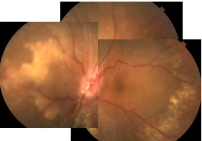
사에서 전안부에 특이소견은 없었으나 안저 검사상 시신경 유두에 인접한 비측 종괴에 의한 압박소견과 망막주름 소 견이 있었고 망막색소화, 망막혈관의 변형, 삼출성의 망막 박리 소견 등은 없었다(Fig. 2). 안구 초음파검사에서 중등 도의 내부신호를 나타내는 비균질성의 종양 소견을 나타냈 다(Fig. 3A). 조영제를 사용한 안와컴퓨터단층촬영에서 좌 안의 상비측에 접한 고강도 신호의 불규칙한 경계를 지닌 2.0×2.0×1.5 cm 크기의 안와 종양소견이 있었으나, 그 이 외의 눈물샘, 외안근, 안와뼈 등에는 특이소견은 없었다 (Fig. 3B). 이후 시행한 뇌, 흉부, 복부 등의 전신적인 영상 검사에서 이상소견은 없었다. 일주일 후 환자는 두통을 동 반한 좌안구통 및 좌안 시력 광각 인지상태의 급격한 시력 감소 발생하여 맥락막 흑색종 의심하에 치료 및 진단을 위 한 좌안구 및 종양적출술 및 안와삽입물(hydroxyapatite) 삽입술을 시행하였다. 적출된 종양은 2.5×2.5×2.5 cm 크 기의 회백질의 종괴로 경계가 불규칙하고 안구와 인접하였 으나 망막 및 시신경은 침범하지 않았다(Fig. 4). 조직검사 에서 림프구, 중성구, 대식세포 등의 염증반응세포에 둘러 싸인 중증도로 분화된 편평세포암종으로 확인되었고 고배 율 현미경에서 비전형성의 유사분열을 하는 다염색성의 핵 을 지닌 종양 세포가 관찰되었으며 편평세포암종이 공막의 부분층을 침범한 소견을 보였다(Fig. 5). 이후 보조 외부조 사 방사선 치료를 시행하였고 수술 후 6개월째 안와컴퓨터 단층촬영에서 재발소견은 없었다(Fig. 6).
www.ophthalmology.org 739 - 한대헌⋅지미정 : 안와에 발생한 고립성 편평세포암종 -
A B C
Figure 3. (A). On B-scan, a large mass with distorted echotexture can be seen. A-scan shows an initial prominent spike followed by a low-to-medium internal reflectivity with diminishing amplitude. (B, C) Enhanced axial CT demonstrated a highly-intense, irregularly- circumscribed heterogenous mass (2.0×2.0×1.5 cm sized) in the superomedial quadrant of the left eye.
Figure 1. A 75-year-old man without axial proptosis.
Figure 2. An oval shaped, juxtapapillary mass-like lesion with retinal foldings but w ithout dark pigm entations, dragging of retinal vessels, or exudative retinal detachment can be
observed. Figure 4. A 2.5×2.5×2.5 cm sized irregular shaped gray- white tumor adjacent to sclera.
고 찰
안와 내에는 상피세포로 이루어진 조직이 정상적으로 존 재하지 않기 때문에 안와에 발생한 편평세포암종은 매우 드물다. 안와에 발생한 편평세포암종의 대부분은 전이성 암 종으로 알려져 있으며 폐, 자궁경부 등의 원격 원발병소에 서 혈행성 전파에 의하거나, 부비동 편평세포암종의 직접적
인 안와내로의 전파에 의해서도 발생한다.1McNab et al4은 피부 편평세포암종이 신경초를 통해 안와 내로 침범한 21 증례를 분석하여 발표하였다. McNab et al4의 보고에 의하 면 대부분의 원발 피부 병소는 이마와 눈썹 주변부이고 신 경초를 통해 안와 내로 침범한 편평세포암종은 해면혈관종 과 얼굴신경을 동시에 침범한 경우가 대부분이기에 방사선 치료를 하는 데 제한이 있어 치료예후가 좋지 않다. 그러나 이와 같은 경우는 안와에 발생한 고립성 편평세포암종의 보고는 아니다. 안와에 발생한 전이성 고립성 편평세포암종 에 대한 이전 증례를 살펴보면 Roseman5은 다른 악성 종양 과 같이 원발 병소가 자연관해된 후 원격 전이가 된 사례를 보고하였는데 이 중 피부의 원발 병소가 자연 관해된 후 안 와에 재발한 고립성 편평세포암종 3예를 보고한 바 있다.
전이성 암종 이외에 안와에 발생한 고립성 편평세포암종 에 대한 보고는 드물지만, 대부분의 경우는 다형성 눈물샘 종 또는 유피낭종의 악성 변화로 인한 경우이다.2,3 Su et al2은 상피세포를 포함한 눈물샘종에서 기원한 원발성 편평 세포암종 1예를 보고하였다. 조직검사에서 상피세포로 이
www.ophthalmology.org 740
- 대한안과학회지 2011년 제 52 권 제 6 호 -
★
A
B
C
Figure 5. (A) Histopathology shows moderately-differentiated squamous cell carcinoma with keratinization (arrow) surrounded by inflammatory cells (asterisk) of lymphocytes, macrophages, neutrophils, eosinophils, and plasma cells (hematoxylin-eosin, ×20). (B) High power view show- ing numerous pleomorphic cell with hyperchromasia and atypical mitotic figures (hematoxylin-eosin, ×100). (C) Histopathology shows invasion of the sclera (arrow) (hematoxylin-eosin, ×4).Figure 6. Axial CT shows no evidence of local recurrence 6 months later.
루어진 낭종를 포함한 편평상피암종으로 보고하여 눈물샘종에 서 기원한 점을 지지하였다. 이외에도 Löffler and Witschel6은 양안 망막박리로 공막돌륭술을 시행 받은 63세 남자에서 술 후 13년째 좌안 공막을 침범한 편평세포암종 1예를 보 고하였다.
본 증례는 안저 검사상 맥락막흑색종의 전형적인 안저 소견인 색소 침착은 없었으나 삼출성 망막박리를 동반한
타원형의 거대 종양소견이 있었고 초음파검사에서 중등도 의 내부신호를 나타내는 비균질성의 종양소견을 보였으며 안구운동장애나 안구돌출 등의 안와조양의 흔한 임상 소견 을 보이지 않아 가장 흔한 원발성 안내종양인 맥락막흑색 종과 임상적으로 감별이 어려웠다. 맥락막흑색종은 유색인 종에는 매우 드물지만 안내종양 중 가장 흔한 원발성 종양 이다.7
이번 보고는 조직검사 결과 낭종을 포함하지 않는 중등 도로 분화된 편평세포암종으로 확인된 점과 다른 전신적인 검사에서도 특이소견이 없었다는 점에서 이전에 보고되었 던 대부분의 안와에 발생한 고립성 편평세포암종과 차이가 있다. Roseman5의 보고와 같이 본 증례 또한 피부나 그 외 원격 원발병소의 자연관해 가능성을 완전히 배제할 수는 없다고 생각한다.
결론적으로 안와에 발생한 고립성 편평세포암종은 매우 드문 질환으로 안구에 인접하여 압박할 시 맥락막흑색종과 같은 안내 종양과 감별이 어렵고 또한 원발병소를 찾기 힘 든 경우가 있어 진단 및 치료를 하는 데 주의해야 한다.
참고문헌
1) Ferry AP, Font RL. Carcinoma metastatic to the eye and orbit. I. A clinicopathologic study of 227 cases. Arch Ophthalmol 1974;92:
www.ophthalmology.org 741 - 한대헌⋅지미정 : 안와에 발생한 고립성 편평세포암종 -
276-86.
2) Su GW, Patipa M, Font RL. Primary squamous cell carcinoma aris- ing from an epithelium-lined cyst of the lacrimal gland. Ophthal Plast Reconstr Surg 2005;21:383-5.
3) Holds JB, Anderson RL, Mamalis N, et al. Invasive squamous cell carcinoma arising from asymptomatic choristomatous cysts of the orbit. Two cases and a review of the literature. Ophthalmology 1993;100:1244-52.
4) McNab AA, Francis IC, Benger R, Crompton JL. Perineural spread of cutaneous squamous cell carcinoma via the orbit. Clinical fea-
tures and outcome in 21 cases. Ophthalmology 1997;104:1457-62.
5) Roseman JM. Regression of locally recurrent squamous cell carci- noma of the skin following excision of a metastasis: with review of the literature. J Surg Oncol 1988;39:213-4.
6) Löffler KU, Witschel H. Orbital squamous cell carcinoma after retinal detachment surgery. Br J Ophthalmol 1991;75:568-71.
7) Weis E, Shah CP, Lajous M, et al. The association between host susceptibility factors and uveal melanoma: a meta-analysis. Arch Ophthalmol 2006;124:54-60.
=ABSTRACT=
A Case of Isolated Squamous Cell Carcinoma of the Orbit
Dae Heon Han, MD, Mijung Chi, MD, PhD
Department of Ophthalmology, Gachon University of Medicine and Science, Incheon, Korea
Purpose: To report a case of isolated squamous cell carcinoma of the orbit.
Case summary: A 75-year-old man with over a 50 pack-year history of smoking presented discomfort and visual dis- turbance of the left eye for several months. His best-corrected visual acuity was 0.3, intraocular pressure was 9 mm Hg, and extraocular movements were normal. Slit-lamp examinations revealed no specific findings in the anterior segment in the left eye. However, retinal exams showed an oval-shaped, juxtapapillary mass-like lesion associated with retinal folding in the left eye. A huge, distorted echoic mass with an initial prominent spike and low-to-medium internal reflectivity with diminishing amplitude was observed on ocular ultrasonography. Enhanced CT revealed a highly-intense, irregular-circumscribed heterogeneous mass (2.0 × 2.0 × 1.5) in the superomedial quadrant of the left eye. Metastatic workups, including bone scan and CT of the head, neck, chest, and abdomen, were unremarkable. One week after the initial visit, the patient experienced pain and reduced visual acuity (light perception) in the left eye. Following the diagnosis, enucleation with tumor resection and hydroxyapatite implantation was performed. Histopathologic examination revealed a moderated-differentiated squ- amous cell carcinoma invading the sclera. The patient subsequently underwent radiation treatment and no evidence of recurrence was reported 6 months after surgery.
J Korean Ophthalmol Soc 2011;52(6):738-741
Key Words: Isolated squamous cell carcinoma, Orbit, Orbital malignant tumor
Address reprint requests to Mijung Chi, MD, PhD
Department of Ophthalmology, Gachon University Gil Hospital
#1198 Guwol-dong, Namdong-gu, Incheon 405-760, Korea
Tel: 82-32-460-3751, Fax: 82-32-460-3358, E-mail: [email protected]