Fixation Strategies to Prevent Screw Cut-Out and Malreduction in Proximal
Humeral Fracture Fixation
Surena Namdari, MD, Adam J. Lipman, MD*, Eric T. Ricchetti, MD
†, Fotios P. Tjoumakaris, MD*, G. Russell Huffman, MD
‡, Samir Mehta, MD
Department of Orthopaedic Surgery, Hospital of the University of Pennsylvania, Philadelphia, PA,
*Jefferson Medical College, Rothman Institute, Philadelphia, PA,
†Cleveland Clinic Orthopaedics, Middleburg Heights, OH,
‡Penn Presbyterian Medical Center, Philadelphia, PA, USA
Technical Note
Clinics in Orthopedic Surgery 2012;4:321-324 • http://dx.doi.org/10.4055/cios.2012.4.4.321Received April 19, 2011; Accepted January 12, 2012 Correspondence to: Samir Mehta, MD
Department of Orthopaedic Surgery, Hospital of the University of Pennsyl- vania, 2 Silverstein, 3400 Spruce Street, Philadelphia, PA 19104, USA Tel: +1-215-432-7432, Fax: +1-215-349-5890
E-mail: [email protected]
Locking plate constructs have shown promising results for treating displaced and unstable proximal humerus fractures,1-3) but the use of the this technique has not been without complications.3-5) Studies that have evaluated the outcomes of patients treated with locking plates for proximal humerus fractures have shown that one of the most frequent complications of this technique is intra- articular penetration of the locking screw.3-5) No study has described the superiority of longer versus shorter locking screws placed into the humeral head for fixation of these fractures. This article describes a variation of the locking plate application that utilizes short locking screws in the humeral head in conjunction with suture fixation to the rotator cuff as a means of minimizing the potential for intra-articular screw penetration and increasing stability.
Fixation of proximal humerus fractures with precontoured, fixed angle devices has improved operative management of these dif- ficult injuries, particularly in patients with osteoporosis. However, recent data has revealed that fixation with these constructs is not without complications, particularly screw cut-out and loss of reduction. Multiple strategies have been developed to decrease the number of complications. We offer a surgical technique combining suture augmentation of the proximal humerus with locked plate fixation utilizing short screws.
Keywords: Proximal humerus, Locking plate, Screw cut-out
TECHNIQUE
The patient can be positioned supine on a radiolucent op- erating room table or placed in the beach chair position.
An image intensifier is positioned at the head of the bed.
Fluoroscopic images are obtained to confirm visualization with the image intensifier prior to prepping and draping the patient’s shoulder, neck, and arm. Full anesthetic relax- ation allows for less traumatic retraction of the deltoid and minimizes dynamic forces on the fracture fragments dur- ing reduction.
A deltopectoral exposure is used to expose the prox- imal humerus, as described previously by Badman and Mighell.6) The coracoacromial ligament may be partially or completely released. Similarly, the coracohumeral ligament is released. The long head of the biceps brachii tendon is identified at its position medial to insertion of the pectora- lis major on the humerus. The pectoralis does not typically need to be released. However, if left in situ, the long head of the biceps brachii can be a source of pain and we often tenodese it at the time of plate fixation in older patients or those with grossly poor tendon quality. The transverse hu- meral ligament is released with a knife or electrocautery as
Copyright © 2012 by The Korean Orthopaedic Association
This is an Open Access article distributed under the terms of the Creative Commons Attribution Non-Commercial License (http://creativecommons.org/licenses/by-nc/3.0) which permits unrestricted non-commercial use, distribution, and reproduction in any medium, provided the original work is properly cited.
Clinics in Orthopedic Surgery • pISSN 2005-291X eISSN 2005-4408
322
Namdari et al. Strategies in Proximal Humeral Fracture Fixation Clinics in Orthopedic Surgery • Vol. 4, No. 4, 2012 • www.ecios.org
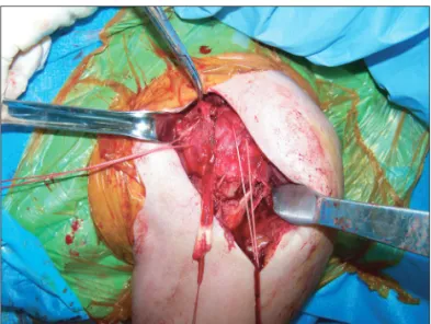
the biceps is traced superiorly, and as the tendon courses superiorly, it is used to define the rotator cuff interval. Af- ter the rotator interval is released to the base of the cora- coid process, the long head of the biceps can be released from the supraglenoid tubercle and superior glenoid la- brum if there is a plan for tenodesis. Heavy nonabsorbable sutures are placed in the subscapularis, supraspinatus, and infraspinatus tendons at the myotendinous junction. Tem- porary traction sutures are often necessary to help mobi- lize the tendons to obtain better suture purchase more me- dially. The bone quality in these patients is typically poor, and sutures should be placed in the stronger rotator cuff tendons rather than through the soft, metaphyseal bone of the tuberosities. We have found that unlocked horizontal mattress sutures are adequate. Traction sutures should be placed in the tendinous insertions to hold and reduce the fragments securely to the plate (Fig. 1).
A low-profile, precontoured, peri-articular lock- ing plate with angular stable screws and suture eyelets is selected to provide fracture fixation. Separate sutures are passed through the plate eyelets prior to applying the plate (Fig. 2). Ideally, a superior suture is placed for the supra- spinatus tendon, an anterior suture for the subscapularis, and a posterior suture for the infraspinatus tendon. The plate is applied to the proximal humerus lateral to the bi- ceps tendon. Care should be taken to avoid medial dissec- tion, which entails detaching the pectoralis major tendon, and risks injury to the posterior humeral circumflex artery.
A provisional reduction of the surgical neck can be held with Kirschner wires and confirmed with an image intensifier and by direct inspection. The tuberosities are reduced via the traction sutures with minimal manipula- tion of the metaphysis to prevent further fracture com- minution. The plate is secured to the humeral head and/
or the shaft using Kirschner wires. The initial screw should be diaphyseal, bicortical, and non-locking. This allows compression of the plate against the humeral shaft and al-
Fig. 1. Delto-pectoral exposure revealing the long head of the biceps brachii, which is released from the supraglenoid tubercle. Stay sutures are placed in the supraspinatus, infraspinatus, and subscapularis tendons to aid in fracture reduction. The fracture is exposed well lateral to the bicipital groove. The deltoid and pectoralis tendon insertions may be left completely intact, even in long fractures extending into the humeral shaft.
Fig. 2. A short, locking precontoured plate is prepared, and sutures are placed through the eyelets of the plate (3.5-mm locking compression plate Proximl Humerus Plate, Synthes Inc.) superiorly (supraspinatus tendon), anteriorly (subscapularis), and posteriorly (infraspinatus).
Fig. 3. The plate is initially secured to the humeral shaft with a non- locked, bicortical screw through the diaphyseal portion of the plate.
Sutures through the rotator cuff and through the plate are shown. The plate is positioned lateral to the bicipital groove.
323
Namdari et al. Strategies in Proximal Humeral Fracture Fixation Clinics in Orthopedic Surgery • Vol. 4, No. 4, 2012 • www.ecios.org
lows subsequent reduction of the tuberosities to the shaft via the plate (Fig. 3). The plate can be moved caudal or cephalad as needed using the oblong hole in the plate. It is critical not to reduce a fracture in internal rotation, as this will limit the patient’s ability to regain functional external rotation postoperatively. To ensure that this does not oc- cur, reduction and plating are performed with the arm in 30° of external rotation. The position of the plate is then evaluated using fluoroscopy to ensure appropriate place- ment such that the plate is not too proximal to impinge on the coracoacromial arch with shoulder abduction. Similar- ly, the plate should not be positioned too distal such that fixation into the head is limited.
Reduction techniques are employed to reduce the head to the shaft by using the sutures in the rotator cuff to gain control of the head. Alternatively, the humeral head can be reduced or stabilized by manipulation with blunt elevators or joysticks such as Kirschner wires or Schanz pins. The use of elevators or joysticks can be problematic when working with osteoporotic bone, due to poor bone quality and further fragmentation at the site of application of these reduction tools.
Once reduced, fixation into the head is limited to five or more short (32-38 mm), fixed angle screws aug- mented with suture fixation of the rotator cuff (which has control of the head) directly to the plate. Screw length is based on the overall size of the proximal humerus. Once a single screw is placed and its length is confirmed by an im- age intensifier to be > 1-2 cm from the articular surface, all other holes are drilled and screws of the same length are placed. This is done to prevent cut-out and intra-articular penetration of the screws. In addition, this can lead to decreased operative time. If the long head of the biceps tendon was released, it can be tenodesed to the pectoralis major and the rotator cuff interval prior to a layered clo- sure.
Postoperative Rehabilitation
Patients are placed in a sling for comfort only and are encouraged to perform non-load bearing activities and pendulum exercises immediately after surgery. If there are no signs of reduction loss, patients are increased to full weight-bearing with the arm over the course of 4-6 weeks.
All restrictions are lifted at 12 weeks if radiographs reveal healing.
DISCUSSION
Proximal humerus fractures account for 4-5% of all frac-
tures.7) The majority of these fractures occur in the elderly population, particularly in those with osteoporosis. Ap- proximately 85% of proximal humerus fractures are mini- mally displaced or stable and can be successfully treated with conservative management and early motion.8-10) The remaining 15% of fractures are displaced or unstable and require surgical intervention because of poor results with non-operative treatment.9) Plate and screw fixation offer the best chance for stable fixation of multi-fragmented fractures. Importantly, no study has described the biome- chanical superiority of long versus short screws placed in the humeral head for fixation of these fractures.
Locking plate constructs have shown promising re- sults for treating displaced and unstable proximal humerus fractures,1-3) but the use of this technique has not been without complications.3-5) Studies evaluating the outcomes of patients treated with locking plates for proximal hu- merus fractures have shown that one of the most frequent complications (16-23%) of this technique is intra-articular penetration of the locking screw.3-5) It was noted that this complication is more common in patients > 60 years in whom osteoporotic bone is more likely to be found. We believe that the concept of subchondral screw fixation (as in load bearing joint periarticular fractures such as femo- ral neck fractures) is a misuse of the locking design for proximal humerus fractures in which rotator cuff tissue integrity often exceeds that of the metaphyseal bone of the humeral tuberosities. For this reason, we use short, diver- gent locking screws and suture fixation to minimize the risk of varus malunion, plate failure, and intra-articular screw penetration. We have treated 53 proximal humerus fractures at our institution with this fixation technique.
None have had intra-articular screw penetration or cut- out and only two patients had an asymptomatic varus mal- union at an average follow-up of 16 months. It is our belief that such a technique reduces the incidence of screw pen- etration into the glenohumeral joint and provides stable fixation for healing. Further biomechanical and long-term clinical data are necessary to substantiate these hypoth- eses.
CONFLICT OF INTEREST
No potential conflict of interest relevant to this article was reported.
324
Namdari et al. Strategies in Proximal Humeral Fracture Fixation Clinics in Orthopedic Surgery • Vol. 4, No. 4, 2012 • www.ecios.org
1. Fankhauser F, Boldin C, Schippinger G, Haunschmid C, Szyszkowitz R. A new locking plate for unstable frac- tures of the proximal humerus. Clin Orthop Relat Res.
2005;(430):176-81.
2. Koukakis A, Apostolou CD, Taneja T, Korres DS, Amini A.
Fixation of proximal humerus fractures using the PHILOS plate: early experience. Clin Orthop Relat Res. 2006;442:115- 20.
3. Charalambous CP, Siddique I, Valluripalli K, et al. Proximal humeral internal locking system (PHILOS) for the treat- ment of proximal humeral fractures. Arch Orthop Trauma Surg. 2007;127(3):205-10.
4. Egol KA, Ong CC, Walsh M, Jazrawi LM, Tejwani NC, Zuckerman JD. Early complications in proximal humerus fractures (OTA types 11) treated with locked plates. J Or- thop Trauma. 2008;22(3):159-64.
5. Owsley KC, Gorczyca JT. Fracture displacement and screw cutout after open reduction and locked plate fixa-
tion of proximal humeral fractures. J Bone Joint Surg Am.
2008;90(2):233-40.
6. Badman BL, Mighell M. Fixed-angle locked plating of two-, three-, and four-part proximal humerus fractures. J Am Acad Orthop Surg. 2008;16(5):294-302.
7. Horak J, Nilsson BE. Epidemiology of fracture of the upper end of the humerus. Clin Orthop Relat Res. 1975;(112):250- 3.
8. Neer CS 2nd. Displaced proximal humeral fractures.
I. Classification and evaluation. J Bone Joint Surg Am.
1970;52(6):1077-89.
9. Mills HJ, Horne G. Fractures of the proximal humerus in adults. J Trauma. 1985;25(8):801-5.
10. Koval KJ, Gallagher MA, Marsicano JG, Cuomo F, McShi- nawy A, Zuckerman JD. Functional outcome after minimal- ly displaced fractures of the proximal part of the humerus. J Bone Joint Surg Am. 1997;79(2):203-7.