Maximum standardized uptake value on positron emission
tomography/computed tomography predicts clinical outcome in patients with relapsed or refractory diffuse large B-cell lymphoma
Hee Ryeong Jang
1, Moo Kon Song
2, Joo Seop Chung
2, Deok Hwan Yang
3, Jeong Ok Lee
4, Junshik Hong
5, Su Hee Cho
6, Seong Jang Kim
7, Dong Hoon Shin
8, Young Joo Park
1, Jin-Suk Kang
1, Jeong Eun Lee
1, Moon Won Lee
1, Ho-Jin Shin
21Department of Internal Medicine, Pusan National University Hospital, Department of Hematology-Oncology, 2Pusan National University Hospital, Busan, 3Chonnam National University Hwasun Hospital, Hwasun, Department of Hematology, 4Seoul National University Bundang Hospital, Seongnam, 5Gachon University Gil Hospital, Incheon, 6Department of Hematology-Oncology, Pusan National University Yangsan Hospital, Yangsan, 7Department of Nuclear Medicine, Pusan National University Hospital, Busan,
8Department of Pathology, Pusan National University Yangsan Hospital, Yangsan, Korea
p-ISSN 2287-979X / e-ISSN 2288-0011 http://dx.doi.org/10.5045/br.2015.50.2.97 Blood Res 2015;50:97-102.
Received on February 10, 2015 Revised on March 11, 2015 Accepted on April 2, 2015
Background
Few clinical studies have clarified the prognostic factors that affect clinical outcomes for patients with relapsed or refractory diffuse large B-cell lymphoma (DLBCL) after immunochemotherapy.
Methods
A total of 158 patients with relapsed or refractory DLBCL were enrolled. All patients under- went positron emission tomography/computed tomography (PET/CT) before and after salvage therapy. All enrolled patients previously received the ifosfamide, carboplatin, and etoposide regimen. Clinical outcomes were compared according to several factors (age
≥ 65 years, low age-adjusted International Prognostic Index [aa-IPI], maximum stand- ardized uptake value [SUVmax] <6.0 on PET/CT, time to relapse ≥12 months, complete response after salvage therapy). A low aa-IPI, SUVmax <6.0, and time to relapse ≥ 12 months were independent prognostic factors for survival.
Results
In univariate analysis and multivariate analysis, SUVmax below 6.0 (P<0.001 for pro- gression-free survival (PFS), P<0.001 for overall survival (OS)) and low aa-IPI (P<0.001 for PFS, P<0.001 for OS) were independent prognostic factors associated with favorable outcome.
Conclusion
The aa-IPI and initial SUVmax were powerful prognostic factors in patients with relapsed or refractory DLBCL.
Key Words Positron emission tomography, SUVmax, aa-IPI
*This study was supported by a 2-year research grant from Pusan National University.
Correspondence to Joo Seop Chung, M.D., Ph.D.
Department of Hematology-Oncology, School of Medicine, Pusan National University, 179, Gudeok-ro, Seo-gu, Busan 602-739, Korea
Tel: +82-51-240-7225 Fax: +82-51-254-3127 E-mail: [email protected]
Ⓒ 2015 Korean Society of Hematology
INTRODUCTION
The combination of rituximab with cyclophosphamide, doxorubicin, vincristine, and prednisone (R-CHOP) has pro- duced significant survival benefits, especially in elderly pa- tients with untreated diffuse large B-cell lymphoma (DLBCL), compared to the CHOP regimen [1]. Although
the immunochemotherapy improved clinical outcomes, some patients experience early refractoriness, unsatisfactory response, or relapse.
High-dose therapy (HDT) with autologous stem cell trans- plantation (ASCT) has become the standard of care for young- er patients with chemosensitive relapsed non-Hodgkin lym- phoma (NHL) and patients with primary refractory aggressive NHL [2]. In a recent study, the second-line age-adjusted
International Prognostic Index (aa-IPI) accurately reflected prognosis in a group of patients with relapsed NHL [3].
The intergroup Collaborative Trial in Relapsed Aggressive Lymphoma evaluated salvage therapy regimens in HDT/
ASCT-eligible patients, and the study identified several prog- nostic factors, including prior exposure to rituximab, early relapse (<12 months), and a secondary IPI of 2–3 [4]. In other studies, the use of 18-fluorine fluorodeoxyglucose (18F-FDG) positron emission tomography/computed tomog- raphy (PET/CT) in the relapsed setting was found to predict patient outcomes [5-9].
Although HDT/ASCT is the standard treatment for im- proving clinical outcomes in relapsed patients, the treatment is associated with an extremely poor prognosis in elderly patients with relapsed or refractory DLBCL and for patients who are ineligible for HDT/ASCT. However, little is known regarding the prognostic factors that affect the clinical out- comes of patients.
The present study investigated the relationships and clin- ical value of several prognostic parameters and clinical out- comes in patients with relapsed or refractory DLBCL who are ineligible for HDT/ASCT.
MATERIALS AND METHODS
A retrospective review of records was performed at five medical centers in Korea (Pusan National University Hospital, Chonnam National University Hwasun Hospital, Seoul National University Bundang Hospital, Gachon University Gil Medical Center, and Pusan National University Yangsan Hospital). Patients who experienced re- lapse following R-CHOP therapy for DLBCL between 2006 and 2012 were included. Patients were excluded from the study if they had received additional therapies such as ritux- imab maintenance therapy after R-CHOP or underwent ASCT. During the period, all patients received treatment with the ifosfamide, carboplatin, and etoposide (ICE) regimen. The patients who received radiotherapy, and other types of salvage chemotherapy were excluded. This study was approved by the local Institutional Review Board.
Salvage therapy schedule
The ICE regimen was also administered in an inpatient setting. Ifosfamide was administered intravenously as a single dose of 5 g/m2/day on day 2 with mesna. Carboplatin was administered at a dose of 800 mg on day 2. Etoposide was administered intravenously at a dose of 100 mg/m2/day from day 1 to day 3. These two schedules were administered every 3 weeks.
aa-IPI
For each patient, the aa-IPI was assessed using data ob- tained immediately before the initiation of salvage therapy.
The aa-IPI factors consist of elevated serum lactate de- hydrogenase (LDH) levels above the upper limit of the normal range, Ann Arbor stage III or IV, and Eastern Cooperative
Oncology Group (ECOG) performance status (PS) above grade 2. In the present study, each group was classified ac- cording to the sum of these risk factors as follows: the low-risk group had no risk factors, the intermediate-risk group had one risk factor, and the high-risk group had two or three risk factors.
18F-FDG-PET/CT scan and assessment of maximum stand- ardized uptake value
All patients underwent an initial 18F-FDG-PET/CT scan before and after salvage therapy. All of the scans were ana- lyzed using fusion software (Syngo, Siemens Medical Solutions, Malvern, PA, USA). By using the software package, the scans were evaluated in trans-axial, sagittal, and coronal views. Target lesions with a largest diameter >1 cm were identified on baseline 18F-FDG PET/CT scans. The maximum standardized uptake value (SUVmax) of each lesion was measured using the initial PET/CT scan. For each patient, the lesions with the greatest SUVmax were considered representative.
Response assessment and follow-up
After the diagnosis of relapse, the patients underwent a conventional re-staging work-up including a physical exami- nation, complete blood cell counts and blood chemistry, CT (neck, chest, and abdominal-pelvic area), and PET/CT.
The final response was assessed by using conventional CT and PET/CT. Bone marrow biopsy was performed only if it had been involved before salvage therapy. The response was assessed at 3 weeks after salvage therapy using the revised criteria of Cheson et al. [10].
Statistical analysis
Progression-free survival (PFS) was defined as the time between the beginning of salvage therapy until disease pro- gression or death. Overall survival (OS) was defined as the time from the start of salvage therapy until death. The Kaplan-Meier method was used to estimate survival, and 95% confidence intervals (CIs) were calculated. Cox pro- gression analysis was used to calculate the hazard ratio be- tween the two arms. Receiver operating characteristic (ROC) curves were prepared to estimate the accuracy of predicting the ideal cut-off of SUVmax. The statistical analysis was conducted using SPSS software version 18.0 (SPSS Inc., Chicago, IL, USA). A P-value of <0.05 was considered significant.
RESULTS
The baseline characteristics of the patients are summarized in Table 1. All 158 patients received ICE therapy. The male-to-female ratio was 102:56. The median patient age was 66 years (range, 20–85 years). One hundred patients (63.2%) were older than 60 years. In total, 102 patients (64.5%) had advanced disease (including stage III and IV), and 72 patients (45.4%) had an ECOG PS above grade 2.
Table 1. Baseline characteristics of the patients.
Characteristics N (%)
Gender
Male 102 (64.5%)
Female 56 (35.5%)
Age, median (Range) 66 (20–85)
>60 years 100 (63.2%)
Ann Arbor stage
III-IV 102 (64.5%)
ECOG, PS
≥2 72 (45.5%)
B symptoms 99 (62.6%)
Elevated LDH 94 (59.4%)
EN site involvement >1 78 (49.3%)
Age-adjusted IPI
Low (0) 23 (14.5%)
Intermediate (1) 67 (42.4%)
High (2–3) 68 (43.0%)
SUVmax on PET/CT
Median (range) 10.9 (2.9–33.0)
Time to relapse, months
<12 months 116 (73.4%)
≥12 months 42 (26.5%)
Abbreviations: ECOG, Eastern Cöoperative Oncology Group; PS, performance Status; LDH, lactate dehydrogenase; EN, extra- nodal; IPI, international prognostic index; SUVmax, maximum standardized uptake value; PET/CT, position emission tomo- graphy/computed tomography; CR, complete response.
Fig. 1. Comparison of clinical outcomes according to the age-adjusted International Prognostic Index (aa-IPI).
Ninety-nine patients (62.6%) had B symptoms, and the LDH level was elevated in 94 patients (59.4%). Forty-two patients experienced relapses that occurred 12 months after R-CHOP therapy (26.5%). Twelve patients (7.5%) achieved complete responses (CRs) after salvage therapy. The median follow-up duration was 32.6 months. During a follow-up period of 36.8 months, the PFS and OS rates were 26.9% and 33.5%, respectively, in the ICE group. The median number of che-
motherapy cycles was 4.
Risk assessment using aa-IPI
There were 23 patients in the low-risk group (0 aa-IPI risk factor). There were 67 patients in the intermediate risk group (1 risk factor), and 76 patients in the high-risk group (2–3 risk factors). Significant differences in PFS and OS were noted between the low- and high-risk groups (PFS, P<0.001 between the low- and high-risk groups; OS, P<0.001 be- tween the low- and high-risk groups, Fig. 1A-B).
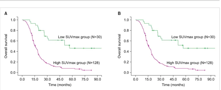
Risk assessment according to SUVmax at relapse
The median SUVmax was 10.9 (range, 2.9–33.0). The ideal cut-off for SUVmax was 6.0 according to ROC curve analysis.
The area under the ROC curve of SUVmax was 0.811. The sensitivity and specificity were 90.1 and 73.1%, respectively.
Depending on his or her SUVmax, each patient was catego- rized into either the low (<6.0) or high SUVmax group (≥6.0). According to this cut-off value, significant differ- ences in survival were observed (P<0.001 for PFS and P
<0.001 for OS, Fig. 2A-B).
Prognostic factors affecting clinical outcomes
To examine whether clinical outcomes were affected by various prognostic factors (age≥65 years, low aa-IPI, SUVmax <6.0, time-to-relapse >12 months, and CR status after salvage therapy), the factors were analyzed regarding their prognostic significance for salvage therapy.
In univariate analysis, a low aa-IPI (P<0.001 for PFS, P<0.001 for OS), SUVmax <6.0 (P<0.001 for PFS, P
<0.001 for OS), time to relapse >12 months (P=0.025 for PFS, P<0.001 for OS), and CR status after the therapy (P=0.002 for PFS, P<0.001 for OS) were associated with both PFS and OS (Table 2).
Multivariate analysis revealed that a low aa-IPI (P<0.001 for PFS, P<0.001 for OS), SUVmax <6.0 (P<0.001 for PFS, P<0.001 for OS), and time to relapse ≥12 months (P<0.057 for PFS, P<0.001 for OS) were independent prog- nostic factors associated with favorable outcomes (Table 3).
Fig. 2. Comparison of clinical outcomes according to a maximum standardized uptake value (SUVmax) cut-off of 6.0.
Table 2. Univariate analysis of the prognostic factors predicting the clinical outcomes.
Prognostic factors Progression-free survival Overall survival
HR (95% CI) P HR (95% CI) P
Low aa-IPI 0.316 (0.218–0.457) <0.001 0.483 (0.339–0.688) <0.001
SUVmax <6.0 0.249 (0.135–0.459) <0.001 0.232 (0.132–0.409) <0.001
Time to relapse ≥12 months 0.622 (0.410–0.943) 0.025 0.333 (0.216–0.512) <0.001 CR status after salvage therapy 0.305 (0.123–0.753) 0.002 0.363 (0.168–0.784) <0.001
Abbreviations: HR, hazard ratio; CI, confidence interval; aa-IPI, age adjusted-international prognostic index; SUVmax, maximum standardized uptake value; CR, complete response.
Table 3. Multivariate analysis of the prognostic factors predicting the clinical outcomes.
Prognostic factors Progression-free survival Overall survival
HR (95% CI) P HR (95% CI) P
Low aa-IPI 0.359 (0.247–0.522) <0.001 0.508 (0.353–0.730) <0.001
SUVmax <6.0 0.314 (0.167–0.589) <0.001 0.254 (0.143–0.454) <0.001
Time to relapse ≥12 months 0.664 (0.435–1.013) 0.057 0.308 (0.197–0.481) <0.001 CR status after salvage therapy 0.965 (0.356–2.614) 0.944 1.037 (0.464–2.320) 0.929
Abbreviations: HR, hazard ratio; CI, confidence interval; aa-IPI, age adjusted-international prognostic index; SUVmax, maximum standardized uptake value; CR, complete response.
DISCUSSION
Few studies have been devoted to therapeutic options for patients with relapsed DLBCL who are ineligible for HDT/ASCT. It is well known that the prognosis is extremely poor with few therapeutic possibilities. The best therapeutic intervention is palliative chemotherapy together with other interventions to preserve a high quality of life. However, some subgroups of relapsed patients with low risk achieve a second good response and experience prolonged survival.
Therefore, it is important to identify prognostic factors for predicting long-term outcomes in this population.
Several studies suggested the clinical value of certain prog- nostic factors including a tumor mass dimension larger than 10 cm, therapeutic regimens prior to ASCT, elevated LDH levels, short time to relapse, and an overall high disease burden [9-14]. However, the widely accepted single prog- nostic model in the relapsed/refractory setting could not be generalized. A recent study illustrated the prognostic significance of aa-IPI based on the clinical data of patients with relapsed NHL who underwent HDT/ASCT [15-17].
Guglielmi et al. also performed a retrospective analysis of aa-IPI factors for patients with relapsed DLBCL and identified relapse within 12 months of the initial diagnosis and aa-IPI factors as significant independent prognostic factors [18].
In addition, other clinical studies demonstrated the positive predictive value of aa-IPI [3, 19, 20]. However, these results cannot be generalized to all patients with NHL because the aa-IPI factors were mostly focused on patients who under- went HDT/ASCT. Conversely, the present study was de- signed to evaluate the prognostic value of aa-IPI for patients with relapsed or refractory DLBCL who were ineligible for HDT/ASCT, and the data suggested that a low aa-IPI is asso- ciated with favorable outcomes for HDT/ASCT-ineligible patients.
Several research groups have investigated the use of
18F-FDG PET/CT to predict the outcomes of patients with relapsed or refractory NHL as well as newly diagnosed patients.
In particular, it is well established that a CR on PET/CT or the negative conversion of FDG uptake after salvage therapy was associated with favorable outcomes [21, 22]. However, it is not known whether the degree of metabolic activity at the time of relapse influences clinical outcomes. In the present study, the clinical significance of the initial SUVmax and CR on PET/CT was evaluated in the relapsed setting.
Interestingly, an initial high SUVmax at relapse was asso- ciated with an advanced Ann Arbor stage compared to a low SUVmax, and it had significant prognostic value for predicting poor clinical outcome. Meanwhile, the CR status did not have significance in the multivariate analysis. To confirm our result, additional clinical data are needed.
Recent studies demonstrated that SUVmax on pre-treat- ment 18F-FDG PET/CT was an important prognostic factor for patients with relapsed NHL who were treated with radio- immunotherapy [10, 11]. Salvage therapeutic regimens were used in patients with relapsed or refractory DLBCL. The present study only revealed clinical outcomes after ICE sal- vage therapeutic regimens. Therefore, further well-designed studies are needed to confirm our study.
In conclusion, the present study illustrated the prognostic significance of aa-IPI and SUVmax for patients with relapsed DLBCL who did not undergo ASCT. The aa-IPI and SUVmax were available to predict the survival outcomes in patients with relapse regardless of the use of salvage therapy.
Although our study had limitations associated with its small sample size, the retrospective nature of its design, and the lack of rituximab assignment to salvage therapy, no prior study assessed the prognostic factors affecting patients with relapsed DLBCL who did not or could not undergo ASCT.
AuthorsÊ Disclosures of Potential Conflicts of Interest
No potential conflicts of interest relevant to this article were reported.
REFERENCES
1. Coiffier B, Thieblemont C, Van Den Neste E, et al. Long-term outcome of patients in the LNH-98.5 trial, the first randomized study comparing rituximab-CHOP to standard CHOP chemo-
therapy in DLBCL patients: a study by the Groupe d'Etudes des Lymphomes de l'Adulte. Blood 2010;116:2040-5.
2. Philip T, Guglielmi C, Hagenbeek A, et al. Autologous bone marrow transplantation as compared with salvage chemotherapy in relapses of chemotherapy-sensitive non-Hodgkin's lymphoma.
N Engl J Med 1995;333:1540-5.
3. Hamlin PA, Zelenetz AD, Kewalramani T, et al. Age-adjusted International Prognostic Index predicts autologous stem cell transplantation outcome for patients with relapsed or primary refractory diffuse large B-cell lymphoma. Blood 2003;102:1989-96.
4. Gisselbrecht C, Glass B, Mounier N, et al. Salvage regimens with autologous transplantation for relapsed large B-cell lymphoma in the rituximab era. J Clin Oncol 2010;28:4184-90.
5. Spaepen K, Stroobants S, Dupont P, et al. Prognostic value of pretransplantation positron emission tomography using fluorine 18-fluorodeoxyglucose in patients with aggressive lymphoma treated with high-dose chemotherapy and stem cell trans- plantation. Blood 2003;102:53-9.
6. Schot BW, Zijlstra JM, Sluiter WJ, et al. Early FDG-PET assessment in combination with clinical risk scores determines prognosis in recurring lymphoma. Blood 2007;109:486-91.
7. Dickinson M, Hoyt R, Roberts AW, et al. Improved survival for relapsed diffuse large B cell lymphoma is predicted by a negative pre-transplant FDG-PET scan following salvage chemotherapy.
Br J Haematol 2010;150:39-45.
8. Lim I, Park JY, Kang HJ, et al. Prognostic significance of pretreatment 18F-FDG PET/CT in patients with relapsed/refractory B-cell non-Hodgkin's lymphoma treated by radioimmuno- therapy using 131I-rituximab. Acta Haematol 2013;130:74-82.
9. Lopci E, Santi I, Derenzini E, et al. FDG-PET in the assessment of patients with follicular lymphoma treated by ibritumomab tiuxetan Y 90: multicentric study. Ann Oncol 2010;21:1877-83.
10. Cheson BD, Pfistner B, Juweid ME, et al. Revised response criteria for malignant lymphoma. J Clin Oncol 2007;25:579-86.
11. Vose JM, Anderson JR, Kessinger A, et al. High-dose chemotherapy and autologous hematopoietic stem-cell transplantation for aggressive non-Hodgkin's lymphoma. J Clin Oncol 1993;11:1846- 51.
12. Prince HM, Imrie K, Crump M, et al. The role of intensive therapy and autologous blood and marrow transplantation for chemo- therapy-sensitive relapsed and primary refractory non-Hodgkin's lymphoma: identification of major prognostic groups. Br J Haematol 1996;92:880-9.
13. Moskowitz CH, Bertino JR, Glassman JR, et al. Ifosfamide, carboplatin, and etoposide: a highly effective cytoreduction and peripheral-blood progenitor-cell mobilization regimen for transplant-eligible patients with non-Hodgkin's lymphoma. J Clin Oncol 1999;17:3776-85.
14. de Kreuk M, Ossenkoppele GJ, Meijer CJ, Huijgens PC. Prognostic factors for survival of non-Hodgkin's lymphoma patients treated with high-dose chemotherapy and autologous bone marrow transplantation. Bone Marrow Transplant 1996;17:963-71.
15. Rapoport AP, Rowe JM, Kouides PA, et al. One hundred autotransplants for relapsed or refractory Hodgkin's disease and lymphoma: value of pretransplant disease status for predicting outcome. J Clin Oncol 1993;11:2351-61.
16. Hoskins PJ, Le N, Gascoyne RD, et al. Advanced diffuse large-cell
lymphoma treated with 12-week combination chemotherapy:
natural history of relapse after initial complete response and prognostic variables defining outcome after relapse. Ann Oncol 1997;8:1125-32.
17. Blay J, Gomez F, Sebban C, et al. The International Prognostic Index correlates to survival in patients with aggressive lymphoma in relapse: analysis of the PARMA trial. Parma Group. Blood 1998;92:3562-8.
18. Guglielmi C, Martelli M, Federico M, et al. Risk-assessment in diffuse large cell lymphoma at first relapse. A study by the Italian Intergroup for Lymphomas. Haematologica 2001;86:941-50.
19. Jabbour E, Peslin N, Arnaud P, et al. Prognostic value of the age-adjusted International Prognostic Index in chemosensitive recurrent or refractory non-Hodgkin's lymphomas treated with
high-dose BEAM therapy and autologous stem cell transplan- tation. Leuk Lymphoma 2005;46:861-7.
20. Berglund M, Thunberg U, Amini RM, et al. Evaluation of immunophenotype in diffuse large B-cell lymphoma and its impact on prognosis. Mod Pathol 2005;18:1113-20.
21. Spaepen K, Stroobants S, Dupont P, et al. Prognostic value of positron emission tomography (PET) with fluorine-18 fluorode- oxyglucose ([18F]FDG) after first-line chemotherapy in non- Hodgkin's lymphoma: is [18F]FDG-PET a valid alternative to conventional diagnostic methods? J Clin Oncol 2001;19:414-9.
22. Mikhaeel NG, Timothy AR, Hain SF, O'Doherty MJ. 18-FDG-PET for the assessment of residual masses on CT following treatment of lymphomas. Ann Oncol 2000;11(Suppl 1):147-50.