Received on October 16, 2012. Revised on October 29, 2012. Accepted on November 2, 2012.
CC This is an open access article distributed under the terms of the Creative Commons Attribution Non-Commercial License (http://creativecommons.org/licenses/by-nc/3.0) which permits unrestricted non-commercial use, distribu- tion, and reproduction in any medium, provided the original work is properly cited.
*Corresponding Author. Sang-Bae Han, College of Pharmacy, Chungbuk National University, Cheongju, Korea. Tel:
82-43-261-2815; Fax: 82-43-268-2732; E-mail: [email protected]
Keywords: Pancreatic cancer, Adoptive immunotherapy, Cytokine-induced killer cells
Abbreviations: CIK, cytokine-induced killer; LAK, lymphokine-activated killer; ADR, adriamycin
Inhibition of Human Pancreatic Tumor Growth by
Cytokine-Induced Killer Cells in Nude Mouse Xenograft Model
Ji Sung Kim, Yun Soo Park, Ju Young Kim, Yong Guk Kim, Yeon Jin Kim, Hong Kyung Lee, Hyung Sook Kim, Jin Tae Hong, Youngsoo Kim and Sang-Bae Han*
College of Pharmacy, Chungbuk National University, Cheongju 361-763, Korea
Pancreatic cancer is the fourth commonest cause of can- cer-related deaths in the world. However, no adequate ther- apy for pancreatic cancer has yet been found. In this study, the antitumor activity of cytokine-induced killer (CIK) cells against the human pancreatic cancer was evaluated in vitro and in vivo. Human peripheral blood mononuclear cells were cultured with IL-2-containing medium in anti-CD3 for 14 days. The resulting populations of CIK cells comprised 94%
CD3+, 4% CD3−CD56+, 41% CD3+CD56+, 11% CD4+, and 73% CD8+. This heterogeneous cell population was called cytokine-induced killer (CIK) cells. At an effector-tar- get cell ratio of 100:1, CIK cells destroyed 51% of AsPC-1 human pancreatic cancer cells, as measured by the 51Cr-re- lease assay. In addition, CIK cells at doses of 3 and 10 million cells per mouse inhibited 42% and 70% of AsPC-1 tumor growth in nude mouse xenograft assays, respectively. This study suggests that CIK cells may be used as an adoptive immunotherapy for pancreatic cancer patients.
[Immune Network 2012;12(6):247-252]
INTRODUCTION
Pancreatic cancer is the fourth commonest cause of cancer-re- lated mortality across the world, with incidence equaling mor- tality (1). According to the American Cancer Society, for all stages of pancreatic cancers combined, the one-year relative survival rate is 20%, and the five-year rate is 6% (2). These low survival rates are attributable to the fact that fewer than
20% of patients' tumors are confined to the pancreas at the time of diagnosis; in most cases, the malignancy has already pro- gressed to the point where surgical removal is impossible (3).
Gemcitabine is a chemotherapeutic drug that has become the standard treatment for advanced diseases after showing supe- riority over 5-fluorouracil, while the combination of radiation and chemotherapy is still widely used (4). Although gemcita- bine-based chemotherapy is typically offered as the standard of care, most patients do not survive longer than 6 months (3). Pancreatic cancer cells that developed gemcitabine resist- ance would still be suitable targets for immunotherapy (5).
Although the combination of radio-, chemo-, and immune- therapy showed improvements in overall survival rates (6), new therapeutic approaches are still needed.
Cytokine-induced killer (CIK) cells are a population of cells derived from human peripheral blood mononuclear cells after ex vivo expansion with interferon-gamma, anti-CD3 anti- bodies, and interleukin (IL)-2 (7). A majority of CIK cells ex- press T cell receptors, and others express NK cell markers (8).
CIK cells have a higher proliferation rate, cytolytic activities and non-MHC-restricted killing of tumor cells in comparison with lymphokine-activated killer (LAK) cells, which are essen- tially activated by natural killer (NK) cells (9,10). They have been shown to target a variety of tumors and can exert their cytotoxic effects following systemic delivery (11). Although CIK cells might be similar to LAK cells, there were several differences. First, LAK cells are originated from NK cells, but
CIK cells are generated from CD8+ T cells (8). Second, LAK cell population mainly include CD3−CD56+ cells, but CIK cells include more than 30% CD3+CD8+CD56+ cells.
Many preclinical and clinical studies showed that CIK cells showed promising anti-tumor effects against various cancers, including hepatoma (12,13), leukemia (14), lung (11,15), ovarian (16), renal (17), and gastric cancer (18). In this study, we examined the antitumor activity of CIK cells against hu- man pancreatic cancer to broaden the clinical applicability of CIK cells. We generated CIK cells from human peripheral blood mononuclear cells, characterized their phenotypes, and evaluated the anti-tumor activity of CIK cells in vitro and in vivo.
MATERIALS AND METHODS Cells
Human pancreatic cancer AsPC-1 cells and human leukemia K562 cells were grown in RPMI1640 medium supplemented with 10% fetal bovine serum, 100 U/ml penicillin and 100μg/
ml streptomycin. CIK cells were generated from the PBMCs of volunteers. After the volunteers gave informed consent, 10
∼20 ml of blood was drawn in evacuated tubes with heparin (Vacutainer, BD Biosciences). The PBMCs were obtained from buffy coats by Ficoll-Hypaque density centrifugation and washed three times with PBS (13). Next, the PBMCs were re-suspended at 1×10⁶ cells/ml in Lymphomedia (Lympho- tech) containing 10% fetal bovine serum (Invitrogen) and cul- tured in the presence of immobilized anti-CD3 antibody (5μg/
ml, BD Pharmingen) and recombinant human IL-2 (700 U/ml, R&D Systems) for 5 days. Following this, the cell suspension was further incubated in Lymphomedia containing recombi- nant human IL-2 only (170 U/ml) for 9 days. Fresh IL-2 and medium were replenished every 2∼3 days (16). During the generation period, the cell number was maintained at approx- imately one million cells per milliliter. The viability of ex- panded cell populations on day 14 was 85∼90%.
Phenotype analysis
Cells were obtained from CIK cultures for phenotype analysis with appropriate monoclonal antibodies, including CD3-FITC as a T cell marker, CD4-FITC as a helper T cell marker, CD8-PE as a cytotoxic T cell marker, and CD56-PE as a natu- ral killer cell marker. One million CIK cells were washed once with PBS containing 1% bovine serum albumin (BSA) and resuspended in 100μl of PBS/BSA buffer. The cells were
incubated with various conjugated monoclonal antibodies for 20 min at 4oC, washed twice with PBS, and resuspended in 400μl of PBS. Flow cytometric analysis was performed on a FACSCalibur flow cytometer (BD Biosciences), and the data were analyzed using the WinMDI statistical software (Scripps).
Cell viability was examined using the propidium iodide (PI) nuclear staining method. Cells were stained with 1μg/ml of PI and analyzed with FACSCalibur flow cytometer. Cells stained with PI were considered dead (11).
51Cr release assay
The lysis of human cancer cells by CIK cells was measured in a 4-h 51Cr release assay (11,19). Briefly, two million target cells were labeled with 100μCi of sodium chromate (Dupont-NEN) for 4 h at 37oC. The labeled cells were washed three times in PBS and resuspended in 10 ml of RPMI 1640 medium supplemented with 10% fetal bovine serum. The la- beled cells were plated in triplicate in round bottom 96-well plates at 1×10⁴ cells/100μl per well. CIK cells were added at specified effector:target (E:T) ratios (1:1, 3:1, 10:1, 30:1 and 100:1) and incubated for 4 h. The supernatant was removed and the radioactivity was measured in a gamma counter. The percentage of specific lysis was calculated accord- ing to the following equation: cytotoxicity=[(sample-sponta- neous)/(maximum-spontaneous)]×100. Spontaneous release was obtained by incubating target cells in medium alone, whereas maximal release was obtained by treatment with 2%
Nonidet P-40 (Sigma).
Nude mouse xenograft assay
Nude mice (6∼8 weeks old) were obtained from Charles River and were housed under specific pathogen free con- ditions according to the guidelines of the Animal Care Committee at the Chungbuk National University. On day 0, nine million AsPC-1 cells in 300μl of PBS were injected sub- cutaneously into nude mice. Following this, CIK cells were injected intravenously once a week at doses of 1, 3, and 10 million cells per mouse. Adriamycin was injected intra- venously once a week at a dose of 2 mg/kg. Tumor volumes were estimated by the formula: length (mm)×width (mm)×height (mm)/2. On day 25, the mice were sacrificed and the tumor mass was weighed. To determine animal tox- icity, the body weights of the animals were measured (20).
Statistics
The results represent the mean values of more than three
Figure 2. The in vitro cytotoxicity of CIK cells. Human PBMCs were cultured in the presence of IL-2 and anti-CD3 antibody for 14 days and the resulting CIK cell populations were used to examine cytotoxicity to K562 (A) and AsPC-1 (B). These target cells were labeled with 51Cr and incubated for 4 h with CIK cells at effector- to-target ratios of 1-100:1.
Figure 1. The phenotypes of CIK cells. Human PBMCs were cultured in the presence of IL-2 and anti-CD3 antibody for 14 days and the resulting CIK cell populations were stained with human monoclonal antibodies, such as anti-CD3-FITC plus anti-CD56-PE (A), anti-CD4- FITC plus anti-CD8-PE (B).
samples and seven mice. Standard deviations (SD) were cal- culated using the Student’s t test and p-values were calculated by ANOVA (GraphPad Software) (21).
RESULTS AND DISCUSSION
After 14 days, the absolute number of human PBMC cultured in the presence of IL-2 and anti-CD3 antibodies increased by more than 230-fold from 30 million PBMCs to 6,900 million CIK cells. When we examined the phenotypes of the cultured cell population with fluorescence-activated cell sorting analy- ses, the cell population was composed of 94% CD3+, 43%
CD3+CD56+, 41% CD3+CD56+, 11% CD4+, and 73% CD8+ (Fig. 1A and B). Interestingly, most CIK cells were CD8+, but not CD4+. Fresh PBMC usually include less than 5% CD3
+CD56+ cells. These results suggested that CIK cells CD3+ T cell population containing CD3+CD56+ cells, as previously reported that CIK cells were generated from CD3+CD8+ pre- cursors (8).
Next, we examined the cytotoxicity of CIK cells under in vitro conditions in a 4-h Cr51 release assay. CIK cells have been demonstrated to have strong cytotoxicity in a non- MHC-restricted manner. At E:T cell ratios of 10:1, 30:1, and 100:1, CIK cells destroyed 22%, 37%, and 51% of AsPC-1 cells, respectively (Fig. 2B). These cells also de- stroyed 46%, 70%, and 77% of K-562 target cells, respectively (Fig. 2A). We also examined the cytotoxicity of fresh PBMC against these target cells and observed that fresh PBMC de- stroyed less than 4% of AsPC-1 and K562 cells.
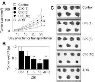
Finally, we evaluated the anti-tumor activity of CIK cells in nude mouse xenograft assays. Preliminary experiments re- vealed that one hundred million CIK cells did not produce
any observable toxicity in nude or SCID mice. Both mice did not exhibit hair ruffling, lowered morbidity, or weight loss (data not shown). Thus, we injected CIK cells intravenously at doses of less than one hundred million cells. Nine million AsPC-1 cells were injected subcutaneously into nude mice and grew to a tumor volume of 251±50 mm3 (n=7) 25 days after implantation (Fig. 3A). CIK cells were injected intra- venously at doses of 1, 3, and 10 million cells per mouse and inhibited in vivo tumor growth by 23%, 42%, and 70%, respectively. Adriamycin (ADR), used as a reference drug, strongly inhibited the growth of AsPC-1 tumors.
On day 25, all tumors were isolated from nude mice and weighed, which demonstrated the strong anti-tumor effect of CIK cells against AsPC-1 tumors (Fig. 3B and C). The weight of AsPC-1 tumors increased to 790+/−193 mg 25 days after implantation. CIK cells that were injected intravenously at doses of 3 and 10 million cells per mouse inhibited tumor weight by 42% and 66%, respectively. Adriamycin (ADR), used as a reference drug, inhibited tumor growth by 44%. The body weights of tumor-bearing nude mice were examined to assess the toxicity. Overall, the nude mice used in this study exhibited body weight gains of 120∼130%, suggesting that CIK cell therapy does not produce animal toxicity (Fig. 4).
The goal of immune cell-based cancer therapy is to elimi- nate cancer cells through the transfer of ex vivo expanded and activated immune cells. Immune cells such as dendritic cells (DC) (22), LAK cells (23), natural killer (NK) cells (24), cytotoxic T lymphocytes (CTL) (17), and cytokine-induced killer (CIK) cells (25) have been explored for adoptive im- munotherapy of cancer. NK and LAK cell therapy has been hindered by the inherently low anti-tumor activity (9). CTL therapy, in turn, was hindered by the MHC-restricted mecha- nism, a limited number of tumor-associated antigens, and a
Figure 3. Inhibition of AsPC-1 tumor growth by CIK cells in nude mouse xenograft models. Nude mice (n=7) were implanted sub- cutaneously with nine million AsPC-1 cancer cells. CIK cells at doses of 1 (CIK 1), 3 (CIK 3), and 10 (CIK 10)×106 cells/mouse were injected intravenously once a week. Adriamycin (ADR) was injected intra- venously at 2 mg/kg. Tumor volumes were estimated by the formula:
length (mm)×width (mm)×height (mm)/2 (A). On day 20, the mice were sacrificed and the tumor weights were measured (B). Repre- sentative photographs are shown (C). Statistical significance was determined using the ANOVA test versus PBS-treated control group (*p<0.05, **p<0.01).
Figure 4. Body weight changes of tumor-bearing nude mice. Nude mice (n=7) were implanted subcutaneously with nine million AsPC-1 cancer cells. CIK cells at doses of 1 (CIK 1), 3 (CIK 3), and 10 (CIK 10)×106 cells/mouse were injected intravenously once a week.
Adriamycin (ADR) was injected intravenously at 2 mg/kg. The body weights of the tumor-bearing nude mice were measured to estimate toxicity.
low number of tumor-specific CTL (26). In the case of DC therapy, it may be difficult for transplanted DC’s to activate effector T cells, which were usually constrained by the severe chemotherapy (11). In contrast, CIK cells had several attrac- tive advantages. First, it is very easy to generate a large num- ber of CIK cells ex vivo and they are readily expandable from PBMC's of cancer patients (27). Second, compared to LAK cells, CIK cells exhibit enhanced cytotoxic activity (28). Third, cytotoxicity is MHC-unrestricted (9,10,29). Fourth, CIK cells are the final effector cells, which are able to directly kill can- cer cells (7).
Here, we showed that after 14 days of culturing human PBMC in the presence of IL-2 and anti-CD3 antibodies, the absolute number of cells increased by more than 200-fold.
Anti-CD3 antibody has been shown to trigger T cell pro- liferation (28). As a key cytokine in CIK cell generation, IL-2 increased the cell number, maintained cell viability, and aug- mented cytotoxicity. From day 6, we incubated cells with on- ly IL-2, resulting in generation of CD3+CD8+CD4 CD56 or CD3+CD8+CD4 CD56+ cells. Although IFN-γ was used in general protocols, we did not use it because our previous ob- servations showed that CIK cells themselves produce high
amounts of IFN-γ, which might stimulate the autocrine path- way (11). We simply added fresh medium to the culture bags every 2 or 3 days and split them without removing the old medium (30). Thus it is possible that CIK cell-derived cyto- kines, especially IFN-γ, accumulated in culture medium.
CIK cells had an effect on various cancers, including hep- atoma, leukemia, lung, ovarian, renal, and gastric cancer, in pre-clinical and clinical studies (11-18). Here, we provide ad- ditional evidences that CIK cell immunotherapy can be effec- tive in eradicating pancreatic cancers. CIK cells broke down AsPC-1 pancreatic cancer cells in vitro and in vivo. This study suggests that CIK cells are good candidates for cell-based im- munotherapy in pancreatic cancer patients.
ACKNOWLEDGEMENTS
This work was supported by the research grant of the Chungbuk National University in 2011.
CONFLICTS OF INTEREST
The authors have no financial conflict of interest.
REFERENCES
1. Hariharan, D., A. Saied, and H. M. Kocher. 2008. Analysis of mortality rates for pancreatic cancer across the world. HPB (Oxford). 10: 58-62.
2. Jemal, A., R. Siegel, J. Xu, and E. Ward. 2010. Cancer sta- tistics, 2010. CA Cancer J. Clin. 60: 277-300.
3. Koido, S., S. Homma, A. Takahara, Y. Namiki, S. Tsukinaga, J. Mitobe, S. Odahara, T. Yukawa, H. Matsudaira, K.
Nagatsuma, K. Uchiyama, K. Satoh, M. Ito, H. Komita, H.
Arakawa, T. Ohkusa, J. Gong, and H. Tajiri. 2011. Current immunotherapeutic approaches in pancreatic cancer. Clin. Dev. Immunol. 2011: 267539.
4. Burris, H. A. 3rd., M. J. Moore, J. Andersen, M. R. Green, M. L. Rothenberg, M. R. Modiano, M. C. Cripps, R. K.
Portenoy, A. M. Storniolo, P. Tarassoff, R. Nelson, F. A. Dorr, C. D. Stephens, and D. D. Von Hoff. 1997. Improvements in survival and clinical benefit with gemcitabine as first-line therapy for patients with advanced pancreas cancer: a randomized trial. J. Clin. Oncol. 15: 2403-2413.
5. Weng, D., B. Song, J. Durfee, V. Sugiyama, Z. Wu, S. Koido, S. K. Calderwood, and J. Gong. 2011. Induction of cytotoxic T lymphocytes against ovarian cancer-initiating cells. Int. J.
Cancer 129: 1990-2001.
6. Neoptolemos, J. P., D. D. Stocken, H. Friess, C. Bassi, J.
A. Dunn, H. Hickey, H. Beger, L. Fernandez-Cruz, C.
Dervenis, F. Lacaine, M. Falconi, P. Pederzoli, A. Pap, D.
Spooner, D. J. Kerr, and M. W. Büchler; European Study Group for Pancreatic Cancer. 2004. A randomized trial of chemoradiotherapy and chemotherapy after resection of pan- creatic cancer. N. Engl. J. Med. 350: 1200-1210.
7. Kim, H. M., J. S. Kang, J. Lim, J. Y. Kim, Y. J. Kim, S. J.
Lee, S. Song, J. T. Hong, Y. Kim, and S. B. Han. 2009.
Antitumor activity of cytokine-induced killer cells in nude mouse xenograft model. Arch. Pharm. Res. 32: 781-787.
8. Franceschetti, M., A. Pievani, G. Borleri, L. Vago, K. Fleischha- uer, J. Golay, and M. Introna. 2009. Cytokine-induced killer cells are terminally differentiated activated CD8 cytotoxic T-EMRA lymphocytes. Exp. Hematol. 37: 616-628.e2.
9. Schmidt-Wolf, I. G., R. S. Negrin, H. P. Kiem, K. G. Blume, and I. L. Weissman. 1991. Use of a SCID mouse/human lym- phoma model to evaluate cytokine-induced killer cells with potent antitumor cell activity. J. Exp. Med. 174: 139-149.
10. Schmidt-Wolf, I. G., P. Lefterova, B. A. Mehta, L. P.
Fernandez, D. Huhn, K. G. Blume, I. L. Weissman, and R.
S. Negrin. 1993. Phenotypic characterization and identi- fication of effector cells involved in tumor cell recognition of cytokine-induced killer cells. Exp. Hematol. 21: 1673-1679.
11. Kim, H. M., J. Lim, S. K. Park, J. S. Kang, K. Lee, C. W.
Lee, K. H. Lee, M. J. Yun, K. H. Yang, G. Han, S. W. Kwon, Y. Kim, and S. B. Han. 2007. Antitumor activity of cyto- kine-induced killer cells against human lung cancer. Int.
Immunopharmacol. 7: 1802-1807.
12. Kim, H. M., J. Lim, Y. D. Yoon, J. M. Ahn, J. S. Kang, K.
Lee, S. K. Park, Y. J. Jeong, J. M. Kim, G. Han, K. H. Yang, Y. J. Kim, Y. Kim, and S. B. Han. 2007. Anti-tumor activity of ex vivo expanded cytokine-induced killer cells against hu- man hepatocellular carcinoma. Int. Immunopharmacol. 7:
1793-1801.
13. Takayama, T., T. Sekine, M. Makuuchi, S. Yamasaki, T.
Kosuge, J. Yamamoto, K. Shimada, M. Sakamoto, S. Hiroha- shi, Y. Ohashi, and T. Kakizoe. 2000. Adoptive immuno- therapy to lower postsurgical recurrence rates of hep- atocellular carcinoma: a randomised trial. Lancet 356: 802- 807.
14. Kornacker, M., G. Moldenhauer, M. Herbst, E. Weilguni, F.
Tita-Nwa, C. Harter, M. Hensel, and A. D. Ho. 2006.
Cytokine-induced killer cells against autologous CLL: direct cytotoxic effects and induction of immune accessory mole- cules by interferon-gamma. Int. J. Cancer 119: 1377-1382.
15. Yang, X. J., J. A. Huang, W. Lei, Y. B. Zhu, and X. G.
Zhang. 2006. Antitumor effects of cocultured dendritic cells and cytokine-induced killer cells on lung cancer in vitro and in vivo. Ai Zheng 25: 1329-1333.
16. Kim, H. M., J. S. Kang, J. Lim, S. K. Park, K. Lee, Y. D.
Yoon, C. W. Lee, K. H. Lee, G. Han, K. H. Yang, Y. J. Kim, Y. Kim, and S. B. Han. 2007. Inhibition of human ovarian tumor growth by cytokine-induced killer cells. Arch. Pharm.
Res. 30: 1464-1470.
17. Wang, W., J. Epler, L. G. Salazar, and S. R. Riddell. 2006.
Recognition of breast cancer cells by CD8+ cytotoxic T-cell clones specific for NY-BR-1. Cancer Res. 66: 6826-6833.
18. Sun, S., X. M. Li, X. D. Li, and W. S. Yang. 2005. Studies on inducing apoptosis effects and mechanism of CIK cells for MGC-803 gastric cancer cell lines. Cancer Biother. Radio- pharm. 20: 173-180.
19. Verneris, M. R., M. Karami, J. Baker, A. Jayaswal, and R. S.
Negrin. 2004. Role of NKG2D signaling in the cytotoxicity of activated and expanded CD8+ T cells. Blood 103: 3065-3072.
20. Han, S. B., C. W. Lee, Y. J. Jeon, N. D. Hong, I. D. Yoo, K. H. Yang, and H. M. Kim. 1999. The inhibitory effect of polysaccharides isolated from Phellinus linteus on tumor growth and metastasis. Immunopharmacology 41: 157-164.
21. Kim, J. Y., Y. D. Yoon, J. M. Ahn, J. S. Kang, S. K. Park, K. Lee, K. B. Song, H. M. Kim, and S. B. Han. 2007. Angelan isolated from Angelica gigas Nakai induces dendritic cell ma- turation through toll-like receptor 4. Int. Immunopharmacol. 7: 78-87.
22. Kalinski, P., Y. Nakamura, P. Watchmaker, A. Giermasz, R.
Muthuswamy, and R. B. Mailliard. 2006. Helper roles of NK and CD8+ T cells in the induction of tumor immunity.
Polarized dendritic cells as cancer vaccines. Immunol. Res. 36: 137-146.
23. Takashima, K., H. Fujiwara, S. Inada, K. Atsuji, Y. Araki, T.
Kubota, and H. Yamagishi. 2006. Tracking of green fluo- rescent protein (GFP)-labeled LAK cells in mice carrying B16 melanoma metastases. Anticancer Res. 26: 3327-3332.
24. Raja Gabaglia, C., Y. Diaz de Durana, F. L. Graham, J.
Gauldie, E. E. Sercarz, and T. A. Braciak. 2007. Attenuation of the glucocorticoid response during Ad5IL-12 adenovirus vector treatment enhances natural killer cell-mediated killing of MHC class I-negative LNCaP prostate tumors. Cancer Res. 67: 2290-2297.
25. Thorne, S. H., R. S. Negrin, and C. H. Contag. 2006. Syner- gistic antitumor effects of immune cell-viral biotherapy. Sci- ence 311: 1780-1784.
26. Grabert, R. C., L. P. Cousens, J. A. Smith, S. Olson, J. Gall, W. B. Young, P. A. Davol, and L. G. Lum. 2006. Human T cells armed with Her2/neu bispecific antibodies divide, are cytotoxic, and secrete cytokines with repeated stimulation.
Clin. Cancer Res. 12: 569-576.
27. Alvarnas, J. C., Y. C. Linn, E. G. Hope, and R. S. Negrin.
2001. Expansion of cytotoxic CD3+ CD56+ cells from periph-
eral blood progenitor cells of patients undergoing autologous hematopoietic cell transplantation. Biol. Blood Marrow Trans- plant. 7: 216-222.
28. Lu, P. H. and R. S. Negrin. 1994. A novel population of ex- panded human CD3+CD56+ cells derived from T cells with potent in vivo antitumor activity in mice with severe com- bined immunodeficiency. J. Immunol. 153: 1687-1696.
29. Pievani, A., G. Borleri, D. Pende, L. Moretta, A. Rambaldi, J. Golay, and M. Introna. 2011. Dual-functional capability of
CD3+CD56+ CIK cells, a T-cell subset that acquires NK func- tion and retains TCR-mediated specific cytotoxicity. Blood 118: 3301-3310.
30. Kim, H. M., J. Lim, J. S. Kang, S. K. Park, K. Lee, J. Y. Kim, Y. J. Kim, J. T. Hong, Y. Kim, and S. B. Han. 2009.
Inhibition of human cervical carcinoma growth by cyto- kine-induced killer cells in nude mouse xenograft model. Int.
Immunopharmacol. 9: 375-380.