Urological Oncology
Factors Affecting the Time to Recurrence After Radical Nephrectomy for Localized Renal Cell Carcinoma
Hee-Seo Son, Seung Hyun Jeon, Sung-Goo Chang
Department of Urology, Kyung Hee University School of Medicine, Seoul, Korea
Purpose: The objective of this study was to determine the factors affecting the time to recurrence after radical nephrectomy for localized renal cell carcinoma.
Materials and Methods: We retrospectively evaluated 321 patients who received radi- cal nephrectomies for localized renal cell carcinoma (pT1a–pT2b N0M0). Of 29 patients with disease recurrence, 9 had recurrence more than 5 years after radical nephrectomy.
We evaluated the clinicopathological factors, with the use of a retrospective study design.
Results: Tumor necrosis was statistically different between the late recurrence group and the recurrence free group (Fisher exact test, p=0.046). Hematuria at diagnosis (chi-square test, p=0.045) was statistically significant in early recurrence. In the uni- variate logistic regression analysis, tumor necrosis (odds ratio [OR], 4.629; 95% con- fidence interval [CI], 1.106 to 19.379; p=0.036) and pT stage>1 (OR, 7.232; 95% CI, 1.727 to 30.280; p=0.007) were risk factors of late recurrence. In the multivariable logis- tic regression analysis, pT stage>1 (OR, 7.143; 5% CI 1.706 to 29.912, p=0.007) was associated with late recurrence. Regarding early recurrence, initial symptoms at diag- nosis and pathologic T stage>1 were statistically significant in both univariate and multivariable logistic regression analysis. In terms of recurrence site, patients with late recurrence tended to have unusual metastasis sites other than lung, liver or bone (chi-square test, p=0.012).
Conclusions: These data suggest that tumor necrosis may affect late disease recurrence.
Patients with initial symptoms and hematuria at diagnosis are vulnerable to re- currence in a shorter period after nephrectomy. Patients with late recurrence showed a tendency to have unusual metastasis site other than lung, liver or bone.
Keywords: Disease-free survival; Recurrence; Renal cell carcinoma
This is an Open Access article distributed under the terms of the Creative Commons Attribution Non-Commercial License (http://creativecommons.org/licenses/by-nc/3.0) which permits unrestricted non-commercial use, distribution, and reproduction in any medium, provided the original work is properly cited.
Article History:
received 21 June, 2013 accepted 9 August, 2013
Corresponding Author:
Sung-Goo Chang
Department of Urology, Kyung Hee University School of Medicine, 26 Kyungheedae-ro, Dongdaemun-gu, Seoul 130-701, Korea
TEL: +82-2-958-8533 FAX: +82-2-959-6048 E-mail: [email protected]
INTRODUCTION
Worldwide, over 200,000 new cases of kidney cancer are di- agnosed and approximately 100,000 deaths occur from this disease each year. Renal cell carcinoma (RCC) constitutes up to 85% of renal malignancies in adults. Despite the es- tablished role of radical or partial nephrectomy as a stand- ard of treatment, a fair number of patients with localized tumors, ranging from 20% to 40%, will experience disease relapse [1]. In patients with recurrent RCC, the clinical course can vary, and survival can be stratified by an ob-
jective parameter called the memorial sloan-kettering can- cer center risk score, which includes time to recurrence, lac- tate dehydrogenase, hemoglobin, corrected calcium, and performance status. However, limited information is avail- able on clinical characteristics, prognostic factors, and out- comes in patients with late-recurring RCC [2,3]. In this study, we evaluated patients with disease recurrence after radical nephrectomy with respect to clinicopathological characteristics and focused on determining the predictive factors affecting different cancer-free intervals.
MATERIALS AND METHODS 1. Patients
From January 1990 to May 2012, a total of 363 patients un- derwent radical or partial nephrectomy for RCC with cura- tive intent at Kyung Hee University Medical Center. We retrospectively evaluated 321 patients who underwent radical nephrectomy for clinically localized RCC. We de- fined clinically localized RCC as pathologically proven RCC of T stage 1a–2b without lymph node enlargement or metastasis at diagnosis. The pathologic stage was reas- signed according to the 2009 Union Internatinale Contre le Cancer and the American Joint Committee on Cancer TNM staging system. Histological subtypes were de- termined according to the Heidelberg classification of renal tumors. Tumor cell differentiation was assessed according to Fuhrman grading system. Patients were generally fol- lowed every 3 to 6 months for the first 2 years following sur- gery, every 6 months from the 3rd through the 5th year, and annually thereafter. Follow-up evaluation consisted of his- tory taking, physical examination, routine blood tests with serum metabolic panels, and imaging evaluation. Abdo- men and chest computerized tomography scans, bone scin- tigraphy, and brain imaging were conducted in clinically indicated cases. Unscheduled evaluations were done when the patient presented with symptoms suspicious of cancer recurrence. Disease recurrence was defined as tumor re- lapse according to the radiographic evidence. Cause of death (cancer-specific death) was determined by chart re- view or death certificate. Of 321 patients who underwent radical nephrectomy for localized RCC, 29 patients experi- enced recurrence. These patients were divided into two groups according to the recurrence-free period after nephrectomy. Patients who were diagnosed with re- currence within 5 years after radical nephrectomy (n=20) were grouped into the ‘early recurrence’ group. Patients with recurrence more than 5 years after radical neph- rectomy (n=9) were included into the ‘late recurrence’
group. Mean recurrence-free survival was 22.1 months (range, 1 to 56 months) in the early recurrence group and 113.3 months (range, 64 to 166 months) in the late re- currence group. Among 292 patients without disease re- currence, 95 patients with more than 5 years of follow-up were enrolled as a control group. The mean follow-up period for the control group patients was 114.1 months (range, 61 to 237 months).
2. Statistical analysis
In comparing demographics and clinicopathological data among the three groups, analysis of variance was used for continuous variables and post hoc analysis was conducted with Bonferroni method. Chi-square test or Fischer exact test was used for categorical variables. Logistic regression analysis was applied to define clinicopathological factors affecting time to recurrence after radical nephrectomy.
Data were analyzed by using IBM SPSS ver. 20.0 (IBM Co., Armonk, NY, USA). Reported p-values are two sided and
p<0.05 was considered statistically significant.
RESULTS
1. Comparison among recurrence-free vs. early recurrence vs. late recurrence
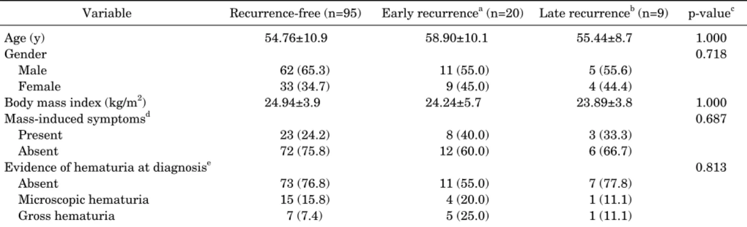
Of a total of 321 patients who underwent radical neph- rectomy for localized RCC, 29 patients were diagnosed with cancer recurrence. Of these, 20 patients (6.1%) were diag- nosed with recurrence within 5 years after radical neph- rectomy (early recurrence) and 9 patients (2.8%) were diag- nosed with recurrence more than 5 years after radical nephrectomy (late recurrence). Tables 1, 2 list the demo- graphic and histopathological characteristics of the three patient groups. According to our data, there were no sig- nificant differences in age, gender, body mass index, or his- tological subtypes among the three groups. Tumor necrosis was statistically significantly different between the late re- currence group and the recurrence-free group (Fisher exact test, p=0.046). On the other hand, tumor necrosis was not significant in the early recurrence group (Fisher exact test, p=0.113). Hematuria at presentation (chi-square test, p=0.045) was statistically significantly different between the early recurrence group and the recurrence-free group but was not significant in the late recurrence group.
Fuhrman grade (Fisher exact test, p=0.012), tumor size, and pT stage were statistically significant in both the early (Fuhrman grade: Fisher exact test, p=0.021; tumor size:
Bonferroni test, p=0.001; T stage: Fisher exact test, p
<0.001) and late (Fuhrman grade: Fisher exact test, p=0.046; tumor size: Bonferroni test, p=0.006; T stage:
Fisher exact test, p=0.002) recurrence groups compared with the recurrence-free group. However, there was no statistically significant difference in any clinicopatho- logical variables between the early and late recurrence groups.
2. Risk factors affecting recurrence after radical neph- rectomy
In the univariate logistic regression analysis, tumor ne- crosis (odds ratio [OR], 4.629; 95% confidence interval [CI], 1.106 to 19.379, p=0.036) and pT stage >1 (OR, 7.232; 95%
CI, 1.727 to 30.280; p=0.007) were risk factors of late recurrence. In multivariable logistic regression analysis, pT stage >1 (OR, 7.143; 95% CI, 1.706 to 29.912; p=0.007) was associated with late recurrence. Regarding early re- currence, initial symptoms at diagnosis (univariate analy- sis [OR, 3.414; 95% CI, 1.262 to 9.238; p=0.016], multi- variable analysis [OR, 3.609; 95% CI, 1.298 to 10.032;
p=0.014]) and pT stage >1 (univariate analysis [OR, 3.115;
95% CI, 1.058 to 9.172; p=0.039], multivariable analysis [OR, 2.920; 95% CI, 1.028 to 8.298; p=0.044]) were statisti- cally significant in both the univariate and multivariable logistic regression analyses (Table 3).
3. Analysis of sites of recurrence
The sites of recurrence were diverse, and we found a pre-
TABLE 1. Demographic and clinical characteristics of the patients
Variable Recurrence-free (n=95) Early recurrencea (n=20) Late recurrenceb (n=9) p-valuec Age (y)
Gender Male Female
Body mass index (kg/m2) Mass-induced symptomsd Present
Absent
Evidence of hematuria at diagnosise Absent
Microscopic hematuria Gross hematuria
54.76±10.9 62 (65.3) 33 (34.7) 24.94±3.9
23 (24.2) 72 (75.8) 73 (76.8) 15 (15.8) 7 (7.4)
58.90±10.1 11 (55.0) 9 (45.0) 24.24±5.7
8 (40.0) 12 (60.0) 11 (55.0) 4 (20.0) 5 (25.0)
55.44±8.7 5 (55.6) 4 (44.4) 23.89±3.8 3 (33.3) 6 (66.7) 7 (77.8) 1 (11.1) 1 (11.1)
1.000 0.718
1.000 0.687 0.813
Values are presented as mean±standard deviation or number (%).
a:Recurrence with metastasis less than 5 years after radical nephrectomy. b:Recurrence with metastasis more than 5 years after radical nephrectomy. c:For late recurrence vs. recurrence-free group. d:Flank pain, abdominal discomfort, palpable mass. e:p<0.05, for early recurrence vs. recurrence-free group.
TABLE 2. Histopathological characteristics of the patients
Variable Recurrence-free (n=95) Early recurrence (n=20) Late recurrence (n=9) p-valuea Tumor size (cm)b
Pathological stageb pT1a
pT1b pT2a pT2b
Histological subtype Clear cell Papillary Chromophobe Others Fuhrman gradeb G1
G2 G3 G4 Unknown
Sarcomatoid differentiation Present
Absent Tumor necrosisc Present Absent
4.39±2.6 59 (62.1) 22 (23.2) 12 (12.6) 2 (2.1) 78 (82.1) 4 (4.2) 10 (10.5) 3 (3.2) 9 (9.5) 40 (42.1) 20 (21.1) 2 (2.1) 24 (25.3) 0 (0) 95 (100) 14 (14.7) 81 (85.3)
6.85±3.3 3 (15.0) 10 (50.0) 4 (20.0) 3 (15.0) 20 (100.0) 0 (0) 0 (0) 0 (0) 0 (0) 3 (15.0) 7 (35.0) 2 (10.0) 8 (40.0) 1 (5.0) 19 (95.0) 6 (30.0) 14 (70.0)
7.44±2.7 1 (11.1) 3 (33.3) 3 (33.3) 2 (22.2) 8 (88.9) 1 (11.1) 0 (0) 0 (0) 0 (0) 2 (22.2) 1 (11.1) 2 (22.2) 4 (44.4) 0 (0) 9 (100) 4 (44.4) 5 (55.6)
0.006 0.002
0.542
0.046
1.000 0.046
Values are presented as mean±standard deviation or number (%).
a:For late recurrence vs. recurrence-free group. b:p<0.05, for early recurrence vs. recurrence-free group. c:Tumor necrosis: coagulative necrosis under microscopic 400 times magnification.
dominance of unusual sites, other than lung, liver, or bone, in the late recurrence group (chi-square test, p=0.012;
Table 4). Of a total of 29 patients with recurrence, 6 patients (30.0%) and 3 patients (33.3%) had multiple sites of meta- stasis in the early and late recurrence groups, respectively.
In the early recurrence group, lung (n=11, 36.7%) was the most frequent site of recurrence, followed by bone (n=7,
23.3%), liver (n=4, 13.3%), lymph node (n=3, 10.0 %), mus- cle (n=2, 6.67%), contralateral kidney (n=1, 3.3%), spleen (n=1, 3.3%), and peritoneum (n=1, 3.3%). In the late re- currence group, lung was also the most frequent organ of recurrence (n=6, 28.6%). On the other hand, more diverse distribution of metastasis was observed in the late re- currence group: brain (n=3, 14.3%), contralateral kidney
TABLE 3. Logistic regression analysis of factors associated with early recurrence and late recurrence Factor
Early recurrencea Late recurrenceb
Univariate analysis Multivariate analysis Univariate analysis Multivariate analysis OR (95% CI) p-value OR (95% CI) p-value OR (95% CI) p-value OR (95% CI) p-value Initial symptom
pT>1 Clear cell type
Fuhrman nuclear grade 3–4 Tumor necrosis
3.414 (1.262–9.238)
3.115 (1.058–9.172)
2.715 (0.997–7.392)
2.480 (0.816–7.539)
0.016 0.039
0.051 0.109
3.609 (1.298–10.032)
2.920 (1.028–8.298)
0.014 0.044
2.845 (0.712 –11.369)
7.232 (1.727–30.280)
1.641 (0.192–14.050)
1.659 (0.383–7.184)
4.629 (1.106–19.379)
0.139 0.007 0.651 0.498 0.036
7.143
(1.706–29.912) 0.007
Multivariate logistic regression analysis after backward stepwise elimination with variables eliminated at p<0.1.
OR, odds ratio; CI, confidence interval.
a:Recurrence with metastasis less than 5 years after radical nephrectomy. b:Recurrence with metastasis more than 5 years after radical nephrectomy.
TABLE 4. Site of recurrence and clinical information associated with recurrence by patient group
Variable Early
recurrence
Late
recurrence p-value No. of sites
Single site Multiple Site of recurrencea Lung
Liver Bone Other siteb Symptoms related to
metastasis Present Absent
14 (70.0) 6 (30.0) 11 (36.7) 4 (13.3) 7 (23.3) 8 (26.7)
7 (35.0) 13 (65.0)
6 (66.7) 3 (33.3) 6 (28.6) 1 (4.8) 1 (4.8) 13 (61.9)
4 (44.4) 5 (55.6)
1.000 0.012c
0.694
Values are presented as number (%).
a:Some patients had more than one recurrent site. b:Other site:
lymph node, brain, pancreas, gall bladder, stomach, duodenum, colon, muscle, contra-lateral kidney, spleen, peritoneum, pleura.
c:Chi-square test in comparison of ‘other sites’ with ‘lung, liver or bone.’
(n=2, 9.5%), pancreas (n=1, 4.8%), gall bladder (n=1, 4.8%), stomach (n=1, 4.8%), colon (n=1, 4.8%), duodenum (n=1, 4.8%), pleura (n=1, 4.8%), muscle (n=1, 4.8%), lymph node (n=1, 4.8%), liver (n=1, 4.8%), and bone (n=1, 4.8%).
DISCUSSION
Disease recurrence in patients with localized RCC after cu- ratively intended radical nephrectomy can occur at any time. However, late recurrence after radical nephrectomy is not common. The definition of ‘late recurrence’ in RCC is not clearly established. The reason we determined 5
years as a cutoff value was because surveillance patterns change at 5 years after curative treatment for RCC [4].
Also, some have suggested that surveillance after 5 years is no longer necessary for cost-effectiveness in low-risk pa- tients [3,4]. Nevertheless, about 10% to 20% of patients with disease recurrence develop late recurrence more than 5 years after nephrectomy [2]. The final objective of our study was to determine the risk factors predictive of late recurrence, at the point of radical nephrectomy, which could thus be incorporated into the postoperative surveil- lance guideline. In our studies, of a total of 104 patients with follow-up for more than 5 years after radical neph- rectomy for localized RCC, 9 patients (9.1%) developed late recurrence. To date, several studies have been conducted to determine the differential characteristics of late re- currence of localized RCC after radical nephrectomy.
However, on the basis of the current medical literature, no consensus has been reached about the parameters predict- ing late recurrence of localized RCC because of the small patient numbers [2-16]. According to the study by Adamy et al. [3], which was conducted with 44 patients with late recurrence (beyond 5 years after nephrectomy), patients with late recurrence tended to have fewer initial symp- toms, smaller tumor size, and less aggressive disease (pT1) compared with patients with early recurrence. Adamy et al. [3] also suggested that patients with late recurrence tend to be in an MSKCC favorable risk group. Park et al.
[5] evaluated 41 patients with late recurrence (beyond 5 years after nephrectomy) and suggested that old age and high high-sensitivity C-reactive protein levels at the time of operation were independent predictive factors for late recurrence. Brookman-May et al. [2] studied a total of 310 patients with cancer recurrence more than 5 years after radical nephrectomy and compared the characteristics of these patients with recurrence-free patients. They proved that lymphovascular invasion, Fuhrman grade 3–4, and pT
stage>pT1 were significantly associated with late recurrence. Ha et al. [6] evaluated 14 patients with disease recurrence more than 5 years after radical or partial neph- rectomy among 423 patients with pathologically confirmed stage T1 clear cell RCC and showed that symptoms at diag- nosis and pT stage were independent predictive factors for late recurrence. In the present study, unlike in previous studies, we tried to determine the factors that affected time to recurrence after radical nephrectomy and to discover the differential characteristics of patients in the early and late recurrence groups, respectively. We found that tumor ne- crosis was associated with late disease recurrence and ini- tial clinical symptoms were associated with early recurren- ce. Large tumor size, advanced pathologic stage, and ad- vanced Fuhrman nuclear grade were associated with both early and late recurrence. We also evaluated the correla- tion of tumor size and tumor necrosis in all the patients in- volved in this study. According to the logistic regression analysis, tumor size was shown to be a risk factor of tumor necrosis (OR, 1.261; 95% CI, 1.086 to 1.463; p=0.002).
Tumor necrosis was initially recognized in the 1970s as a predictor of aggressive RCC behavior [17]. Minervini et al. [18] confirmed that histological tumor necrosis is a stat- istically significant prognostic factor in patients with non- metastatic clear cell RCC. Kim et al. [7] suggested that the survival rate of patients with tumor necrosis was sig- nificantly lower than that of patients without tumor necrosis. According to the previously published medical lit- erature, the definitions for tumor necrosis are diverse.
However, most investigators have defined histological ne- crosis, as a prognostic factor, as the presence of any micro- scopic coagulative tumor necrosis, without consideration of degenerative changes such as hyalinization, hemor- rhage, and fibrosis [17,18]. We also determined tumor ne- crosis as the presence of microscopic coagulative tumor necrosis. As we showed in Tables 2, 3, tumor necrosis was statistically significant only in the late recurrence group.
In consideration of the hypothesis that rapid tumor cell growth outgrows its own blood supply and subsequently creates a hypoxic condition and resultant tumor necrosis, this result might be contradictory. However, in several re- cent reports, a high proliferation index or insufficient oxy- gen supply (hypoxia inducible factor-1, or HIF-1a) and high Ki-67 (a proliferation marker) expression were not inter- changeable with tumor necrosis. Some medical literature suggests that host immunologic factors, such as differ- ential expression of chemokines, may be involved in tumor necrosis [19]. Although the exact nature of immunologic triggers and tumor necrosis remain to be clarified, there might be a kind of ‘time-related function’ between ‘tumor necrosis’ and a ‘tumor dormancy state’.
This study also showed the diversity of the recurrence site in the late recurrence group. The lung, liver, and bone are known as the usual recurrence organs of malignant ne- oplasms, including RCC [20], and we found such a tendency in the early recurrence group. However, there was a pre- dominance of cancer recurrence at other sites in the late
recurrence group, such as the lymph node, brain, pancreas, gall bladder, stomach, duodenum, colon, muscle, con- tralateral kidney, and pleura. The question of differences in metastasis site according to recurrence-free interval re- mains to be answered. Bruin et al. [21] suggested that or- gan-specific metastasis localization can be predicted by specific genomic aberrations in primary colorectal cancer [5]. Yerushalmi et al. [22] showed that cancer antigen 125 levels varied among the different sites of metastasis in breast cancer [5]. Koo et al. [23] reported that metastatic breast cancer showed different phenotypes of estrogen re- ceptor, progesterone receptor, and human epidermal growth factor receptor 2 according to the different re- currence sites [5]. Therefore, understanding the molecular biological mechanisms underlying RCC might solve these questions.
The potential limitations of our study lie in its retro- spective design. There was no standardized postoperative follow-up protocol and the quality of imaging modality has improved substantially during the past 30 years. No in- corporation of molecular markers is another limitation of this study. Furthermore, the small number of patients who were evaluated in a single institution might have affected our study data. For this reason, we are planning to conduct a multicenter study with a large patient pool to minimize these biases.
CONCLUSIONS
This study suggested that the presence of microscopic coag- ulative necrosis in the resected specimen may be a pre- dictive factor of late recurrence after radical nephrectomy for localized RCC. Also, clinical symptoms such as hema- turia, flank pain, and a palpable mass at diagnosis may pre- dict disease recurrence in a short period after radical nephrectomy. Large tumor size, advanced pathologic stage, and advanced Fuhrman nuclear grade may be risk factors for both early and late recurrence. Therefore, we suggest that patients with tumor necrosis may need to un- dergo long-term, thorough surveillance after radical neph- rectomy for localized RCC.
CONFLICTS OF INTEREST The authors have nothing to disclose.
REFERENCES
1. Rodriguez-Covarrubias F, Gomez-Alvarado MO, Sotomayor M, Castillejos-Molina R, Mendez-Probst CE, Gabilondo F, et al. Time to recurrence after nephrectomy as a predictor of cancer-specific survival in localized clear-cell renal cell carcinoma. Urol Int 2011;86:47-52.
2. Brookman-May S, May M, Shariat SF, Xylinas E, Stief C, Zigeuner R, et al. Features associated with recurrence beyond 5 years after nephrectomy and nephron-sparing surgery for renal cell carcinoma: development and internal validation of a risk mod- el (PRELANE score) to predict late recurrence based on a large multicenter database (CORONA/SATURN Project). Eur Urol
2013;64:472-7.
3. Adamy A, Chong KT, Chade D, Costaras J, Russo G, Kaag MG, et al. Clinical characteristics and outcomes of patients with re- currence 5 years after nephrectomy for localized renal cell carcinoma. J Urol 2011;185:433-8.
4. Babjuk M, Burger M, Zigeuner R, Shariat S, Van Rhijn B, Comperat E, et al. Guidelines on non-muscle-invasive bladder cancer (TaT1 and CIS). Updated 2013 [Internet]. Arnhem;
European Association of Urology; c2013 [cited 2013 Jul 22].
Available from: http://www.uroweb.org/fileadmin/guidelines/
Total_file_2013_large_guidelines_prints.pdf.
5. Park YH, Baik KD, Lee YJ, Ku JH, Kim HH, Kwak C. Late re- currence of renal cell carcinoma >5 years after surgery: clin- icopathological characteristics and prognosis. BJU Int 2012;110 (11 Pt B):E553-8.
6. Ha YS, Park YH, Kang SH, Hong SH, Hwang TK, Byun SS, et al.
Predictive factors for late recurrence in patients with stage T1 clear cell renal cell carcinoma: a multiinstitutional study. Clin Genitourin Cancer 2013;11:51-5.
7. Kim JM, Song PH, Kim HT, Park TC. The prognostic factors for patients with pT1a renal cell carcinoma. Korean J Urol 2010;51:233-8.
8. Abel EJ, Culp SH, Meissner M, Matin SF, Tamboli P, Wood CG.
Identifying the risk of disease progression after surgery for lo- calized renal cell carcinoma. BJU Int 2010;106:1277-83.
9. Miyao N, Naito S, Ozono S, Shinohara N, Masumori N, Igarashi T, et al. Late recurrence of renal cell carcinoma: retrospective and collaborative study of the Japanese Society of Renal Cancer.
Urology 2011;77:379-84.
10. Kim SP, Weight CJ, Leibovich BC, Thompson RH, Costello BA, Cheville JC, et al. Outcomes and clinicopathologic variables asso- ciated with late recurrence after nephrectomy for localized renal cell carcinoma. Urology 2011;78:1101-6.
11. Margulis V, McDonald M, Tamboli P, Swanson DA, Wood CG.
Predictors of oncological outcome after resection of locally re- current renal cell carcinoma. J Urol 2009;181:2044-51.
12. Eggener SE, Yossepowitch O, Pettus JA, Snyder ME, Motzer RJ, Russo P. Renal cell carcinoma recurrence after nephrectomy for localized disease: predicting survival from time of recurrence. J Clin Oncol 2006;24:3101-6.
13. May M, Brookman-Amissah S, Kendel F, Knoll N, Roigas J, Hoschke B, et al. Validation of a postoperative prognostic model consisting of tumor microvascular invasion, size, and grade to pre- dict disease-free and cancer-specific survival of patients with sur- gically resected renal cell carcinoma. Int J Urol 2009;16:616-21.
14. Thompson RH, Leibovich BC, Lohse CM, Cheville JC, Zincke H, Blute ML, et al. Dynamic outcome prediction in patients with clear cell renal cell carcinoma treated with radical nephrectomy:
the D-SSIGN score. J Urol 2007;177:477-80.
15. Kume H, Teramoto S, Kitamura T. Metachronous bilateral renal cell carcinoma with an interval of more than 10 years. Int Urol Nephrol 2009;41:843-6.
16. Lane BR, Kattan MW. Prognostic models and algorithms in renal cell carcinoma. Urol Clin North Am 2008;35:613-25.
17. Lee SE, Byun SS, Oh JK, Lee SC, Chang IH, Choe G, et al.
Significance of macroscopic tumor necrosis as a prognostic in- dicator for renal cell carcinoma. J Urol 2006;176(4 Pt 1):1332-7.
18. Minervini A, Di Cristofano C, Gacci M, Serni S, Menicagli M, Lanciotti M, et al. Prognostic role of histological necrosis for non- metastatic clear cell renal cell carcinoma: correlation with patho- logical features and molecular markers. J Urol 2008;180:1284-9.
19. Pichler M, Hutterer GC, Chromecki TF, Jesche J, Kampel- Kettner K, Rehak P, et al. Histologic tumor necrosis is an in- dependent prognostic indicator for clear cell and papillary renal cell carcinoma. Am J Clin Pathol 2012;137:283-9.
20. Bianchi M, Sun M, Jeldres C, Shariat SF, Trinh QD, Briganti A, et al. Distribution of metastatic sites in renal cell carcinoma: a population-based analysis. Ann Oncol 2012;23:973-80.
21. Bruin SC, Klijn C, Liefers GJ, Braaf LM, Joosse SA, van Beers EH, et al. Specific genomic aberrations in primary colorectal can- cer are associated with liver metastases. BMC Cancer 2010;
10:662.
22. Yerushalmi R, Tyldesley S, Kennecke H, Speers C, Woods R, Knight B, et al. Tumor markers in metastatic breast cancer sub- types: frequency of elevation and correlation with outcome. Ann Oncol 2012;23:338-45.
23. Koo JS, Jung W, Jeong J. Metastatic breast cancer shows different immunohistochemical phenotype according to metastatic site.
Tumori 2010;96:424-32.