247
Metachronous Mantle Cell Lymphoma with a Leukemic Presentation in a Patient with Early Gastric Cancer:
A Case Report
Wan Kyu Eo, M.D.1, Kyu Jeung Ahn, M.D.1, Woo In Lee, M.D.2, Sung Jig Lim, M.D.3 and Jong Soo Jeong, O.M.D.4
Departments of 1Internal Medicine, 2Laboratory Medicine, and 3Pathology, Kyung Hee University School of Medicine, and Department of 4Internal Medicine, College of Oriental Medicine, Kyung Hee University, Seoul, Korea
We report here on a case of metachronous second primary non-Hodgkin’s lymphoma (NHL) that was diagnosed 6 years after performing subtotal gastrectomy for treating early gastric cancer (EGC). The sub- type analysis revealed mantle cell lymphoma (MCL) of the blastic variant with a leukemic presentation, which was composed of mixed small and medium-sized cells. The immunohistochemical staining for cy- clin-D1 was positive. The cytogenetic study revealed t(4;6). In Korea, the risk of developing a second primary cancer following gastric cancer was reported to be less than 3.4%, and NHL comprised less than 6.3% of this second primary cancer. Furthermore, MCL represents about 2% of all lymphomas in Korea. To the best of our knowledge, this is the first report of metachronous primary MCL with a leuke- mic presentation following curative resection of EGC. (Korean J Hematol 2008;43:247-252.)
Key Words: Gastric cancer, Metachronous second primary neoplasms, Mantle cell lymphoma, Leukemia
접수:2008년 7월 12일, 수정:2008년 10월 6일 승인:2008년 10월 15일
교신저자:어완규, 서울시 강동구 상일동 149
134-090, 경희대학교 동서신의학병원 혈액종 양내과
Tel: 02-440-6122, Fax: 02-440-7287 E-mail: [email protected]
Correspondence to:Wan Kyu Eo, M.D.
Department of Internal Medicine, East-West Neo Medical Center, Kyung Hee University
149, Sangil-dong, Gangdong-gu, Seoul 134-090, Korea Tel: +82-2-440-6122, Fax: +82-2-440-7287
E-mail: [email protected] INTRODUCTION
During the past two decades, the survival rate of patients with gastric cancer has improved sig- nificantly, and this led to an increased incidence of second primary cancers. The risk of developing a second primary cancer following gastric cancer was reported less than 3.4% in Korea, and sig- nificantly elevated risks were observed for cancers of the colorectum, lung and liver. The non- Hodgkin’s lymphoma (NHL) comprised less than 6.3% of second primary cancers following gastric cancer, and about 25∼40% of the NHL cases
were considered to be metachronous.1,2)
In this article, we report a case of metachro- nous second primary mantle cell lymphoma (MCL) of the blastic variant with a leukemic presen- tation that occurred 6 years after subtotal gas- trectomy for early gastric cancer (EGC). In Korea, MCL, a B-cell lymphoma, represents about 2% of lymphomas. Furthermore, MCL with leukemic presentation is extremely rare. To the best of our knowledge, this is the first report of MCL with leukemic presentation as a second primary malig- nancy following curative resection of EGC.
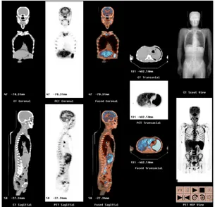
Fig. 1. The increased FDG uptake in the axial skeleton of whole body is noted. The multifocal increase of FDG uptake in multiple lymph node areas including jugular, para- tracheal, internal mammary, subcarinal, hilar, anterior and middle diaphragmatic, portocaval, paraaortic, iliac, inguinal, and mesenteric lymph node areas is noted. The increase of FDG uptake in the liver and spleen is also seen.
CASE REPORT
A 64-year-old male patient was referred to a university hospital in January 2000 for further evaluation of suspicious EGC. Gastric fibroscopy revealed slightly elevated lesion located in poste- rior wall of antrum of the stomach, and tissue bi- opsy revealed adenocarcinoma with moderate dif- ferentiation. Subtotal gastrectomy with Billoth-1 anastomosis was carried out. The tumor was 4×4 cm in size, and it extended to muscularis mucosa without lymph node metastasis (T1N0M0). EGC with IIb+IIc type was pathologically confirmed.
He had been in symptom-free state until Fe- bruary 2006 when he complained of postprandial epigastric pain. Gastric fibroscopy showed nega- tive. In May 2006, the abdominopelvic CT scan for evaluation of the periumbilical pain showed multiple enlarged lymph nodes in porta hepatis, portocaval and retroperitoneal space, and the PET-CT scan showed increased FDG uptake in the axial skeleton of whole body, liver, spleen and
multifocal lymph nodes (Fig. 1). Clinical diag- nosis of relapsed gastric cancer was made. He re- fused further evaluation and visited Integrative Cancer Center, Kyung-Hee University for herbal medication in July 2006.
At initial evaluation, there was a significant weight loss of 4∼5kg over one month. The pe- ripheral blood showed WBC 2,900/μL, hemoglo- bin 11.3g/dL, hematocrit 33.4%, MCV 94.1fL, platelet 92,000/μL, neutrophil 24.0%, band 3.0%, monocyte 5.0%, eosinophil 1.0%, immature cell 67% and reticulocyte 2.5%. The serum levels of uric acid (12.3mg/dL) and β2-microglobulin (5.7 mg/L) were increased. The serum levels of CEA, CA 19-9, and CA72-4 2.2 were within normal limits.
The gastric fibroscopy showed no evidence of tumor. The neck and chest CT scan showed some enlarged lymph nodes in supraclavicular fossa, anterior mediastinal and bilateral paratracheal area. The abdominopelvic CT scan showed sple- nomegaly and multiple lymph nodes in the hep- atic artery, retropancreatic, paraaortic, and in- guinal areas.
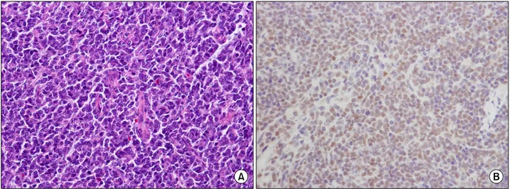
The microscopic examination of the lymph node from the inguinal area showed diffuse pro- liferation of small to medium-sized lymphoid cells with irregular nuclar contour, and admixed lymphoid cells with dispersed chromatin resem- bling lymphoblasts. The immunohistochemical staining revealed positive expression of cy- clin-D1, bcl-2, CD5 (weak), CD20, CD79a and KI-67, and negative expression of CD10, CD23 and CD3 (Fig. 2).
The cellularity of the bone marrow was from 50 to 60%. Bone marrow aspiration revealed blast cells comprising about 77.4% of all hematopoietic nucleated cells. Those cells were mixed small to medium in size with dispersed chromatin, bi- lobed nucleus, high nucleus/cytoplasm ratio and prominent nucleoli. The staining for myeloperox- idase, PAS, and non-specific esterase was nega- tive. The staining for iron revealed decreased to normal storage iron in marrow particles (Grade
Fig. 2. The microscopic examination of the lymph node from the inguinal area shows diffuse proliferation of small to me- dium-sized lymphoid cells with irregular nuclear contour, and admixed lymphoid cells with dispersed chromatin resembling lymphoblasts (H-E stain, ×200) (A). The immunohistochemical staining for cyclin-D1 reveals positive result (×400) (B).
Fig. 3. (A) The Bone marrow aspiration shows many blast cells of small to medium size with dispersed chromatin, high nucleus/cytoplasmic ratio and prominent nucleoli (Wright stain, ×1,000). (B) Cytogenetic study shows chromosomal abnor- mality of 46,XY, t(4;6)(q21.2;q23.2)[3]/46,XY[37].
I/III). The immunophenotyping study by flow cy- tometry revealed positive expression of CD20, CD19, FMC7, CD5, CD45, and Sm kappa, and negative expression of CD13, CD33, and myelo- peroxidase. The cytogenetic study revealed 46, XY,t(4;6)(q21.2;q23.2)[3]/46,XY[37] (Fig. 3).
Finally, the diagnosis of MCL of the blastic variant with a leukemic presentation which is composed of mixed small and medium-sized cells was made. The lymphoma was considered as a metachronous second primary malignancy, and the onset was 6 years after subtotal gastrectomy for EGC. Considering poor prognostic factors for
MCL including poor performance status, old age, splenomegaly, advanced stage, high LDH, pos- itive cyclin-D1, high serum β2-microglobulin and poor natural course of the MCL with a leuke- mic presentation, palliative chemotherapy of cy- clophosphamide, vicristine and prednisone (CVP) was started in August 2006. Unfortunately the disease was refractory to the first course of CVP, and further chemotherapy was stopped, and after that, only supportive care including hydroxyurea, blood transfusion, nutritional care and antibiotics was given. He died in September 2006.
DISCUSSION
Development of second primary cancers has become an important problem in cancer treat- ment. The incidence of second primary cancer following gastric cancer had been reported less than 3.4% in Korea1,2), which is comparable to the report from Japan.3) Significantly elevated risks were observed for cancers of the colorectum, lung and liver.1,2) Meanwhile, the NHL comprised less than 6.3%, and this suggests that the in- cidence of second primary NHL is very rare.1-3) About 25~40% of the second primary NHL cases were considered to be metachronous, de- fined as those occurring more than 6 months af- ter the diagnosis of first primary malignancy, and they comprised about 3.6~5.9% of the total of metachoronous cancers following gastric can- cers.1,2) While Ryu et al. reported 28 month inter- val between development of gastric cancer and NHL,2) in a report of two cases of lymphoma af- ter Tx of gastric cancer by Nakamura et al, the interval was 7 years in immunoblastic lymphoma subtype and 13 months in MALToma subtype.4) Metachronous occurrence of lymphoma and ad- enocarcinoma, both in the stomach, has been reported. Hamaloglu et al. reported that initial malignancy was NHL in 28 of 30 cases (93.3%), and only 2 cases were diagnosed as NHL after the treatment of adenocarcinoma.4) This trend was supported by many authors.5,6) Chemotherapy, ra- diotherapy, or combined administration of che- motherapy and radiotherapy can increases the oc- currence of second malignancies. Gastroesophag- eal reflux, formation of nitrosamine, and un- treated H. pylori infection may play a role in de- velopment of secondary cancer after gastric surgery.4)
MCL is a B-cell malignancy with distinct mo- lecular genetics and pathologic features. MCL represents 5∼10% of all lymphoma cases in west- ern series, but it comprises about 2% of lympho- mas in Korea and Japan.7) MCL is a neoplasm
composed of cells resembling those residing in follicular mantle zones. The mantle cells have a distinctive immunophenotype, with expression of pan B-cell markers (CD19, CD20, and CD22) and the T-cell marker CD5; these cells do not ex- press CD23 and CD10 antigens.7) Rendering a di- agnosis of MCL has been greatly facilitated by the immunohistochemical demonstration of cy- clin-D1, because this marker is expressed rarely by other lymphoma types.8)
MCL of the blastic variant is characterized by monomorphic sheets of small to medium-sized blasts with fine dispersed chromatin mimicking lymphoblastic lymphoma/acute lymphoblastic leu- kemia. It has narrow rim of cytoplasm and fre- quent apoptotic bodies. The blastic variant was encountered in 7.2∼17% of cases.9) In a report, patients with blastic variants presented with more mitoses/HPF, higher LDH levels, and less fre- quent bone marrow involvement than those with other histologic variants.10) In another report by Bernard, 66% had extranodal site involvement, 85% had an Ann Arbor stage IV, and 82% had peripheral lymphadenopathy.9) In terms of prog- nosis, the blastic variant was characterized by a shorter overall duration of response after first- line therapy and shorter overall survival.11) In our case, blast cells were found in the bone marrow and circulation constituting a leukemic presentation. The MCL presenting with leukemia appears to be more aggressive and, in several re- ports, the prognosis was poorer than that of local- ization to lymph node or extranodal sites.8-10,12,13)
The clinical and laboratory variables shown to be of prognostic significance in unselected MCL cas- es may not appear to have such an impact in those cases presenting with overt leukemia.13) In this case, we made a decision to start palliative chemotherapy of CVP considering poor perform- ance status, old age and advanced stage. If the pa- tient is elderly and is not a stem cell transplant candidate, the use of rituximab-containing che- motherapy can be appropriate. Purine nucleoside analogs with rituximab may be especially useful
in the elderly patient population or those who can not tolerate anthracyclines.14) In patients with relapsed or refractory MCL, the proteasome in- hibitor bortezomib may be useful. Even with the most aggressive approaches, a minority of pa- tients with relapsed MCL are cured.
According to the literature, only 40∼50% of the patients of MCL exhibited the hallmark t(11;14)(q13;q32), and various chromosomal ab- normalities have been reported as well. These in- clude abnormalities involving chromosome 1, 7q, 12, and 10p.13) In our case, cytogenetic study re- vealed 46,XY,t(4;6)(q21.2;q23.2)[3]/46,XY[37] even though the cyclin D1 was overexpressed. In a re- port by Nam et al, no one showed t(11;14) in G-banding analysis in 12 cases of MCL with mar- row infiltration that were diagnosed for over 5 years in a center.15) The possible reason for the absence of t(11;14) in spite of the expression of cyclin-D1 includes submicroscopic changes in- volving t(11;14).8) Alternatively, t(11;14)-negative MCL can be a new disease entity.15) The chromo- somal abnormality of t(4;6) in this case is an un- usual finding and, to the best of our knowledge, there has been no report of such kind of chromo- somal abnormality in MCL.
This case is of interest for three reasons. First, metachronous second primary MCL with leuke- mic presentation following curative resection of EGC is very rare and, to the best of our knowl- edge, this is the first report of that occasion.
Second, MCL was accompanied by unusual cyto- genetic abnormality of t(4;6), to the best of our knowledge, that has not been reported yet. Third, the clinician must be astute and aggressive when evaluating a patient with is a history of gastric cancer. Clinical diagnosis of relapsed gastric can- cer without pathologic confirmation should be avoided when evaluating patients with lympha- denopathy and abnormal bone uptake.
요 약
저자들은 조기위암에 대한 대부분위절제술 후 6년이
경과된 시점에서 발생한 속발성 비호지킨림프종 한 예 를 보고한다. 림프종 아형 분석 결과 외투세포림프종의 모세포 변이로 밝혀졌고, 백혈병 발현을 보였다. 모세포 들은 작거나 중간 크기의 세포로 이루어져 있었다.
Cyclin-D1에 대한 면역조직화학염색검사는 양성이었지 만, 세포유전학검사에서는 t(4;6) 염색체 이상이 보였다.
국내 보고에서 위암환자에서 속발성 종양의 발생률은 3.4% 미만인데, 림프종은 속발성 종양 중 6.3% 미만을 차지한다. 또한 국내 보고에서 외투세포림프종은 전체 림프종의 2% 미만이고, 모세포 변이의 발현은 흔하지 않 다. 저자들이 알기로는 조기위암의 절제술 후 이시성 (metachronous) 외투막세포림프종의 백혈병 발현을 보 고한 예는 없다. 따라서 본 보고는 이에 대한 첫 증례 보고로 판단된다.
REFERENCES
1) Eom BW, Lee HJ, Yoo MW, et al. Synchronous and metachronous cancers in patients with gastric cancer.
J Surg Oncol 2008;98:106-10.
2) Ryu DD, Um JW, Son GS, et al. Multiple primary malignant tumors in patients with gastric cancer. J Korean Gastric Cancer Assoc 2003;3:139-44.
3) Hiyama T, Hanai A, Fujimoto I. Second primary cancer after diagnosis of stomach cancer in Osaka, Japan. Jpn J Cancer Res 1991;82:762-70.
4) Hamaloglu E, Topaloglu S, Ozdemir A, Ozenc A.
Synchronous and metachronous occurrence of gas- tric adenocarcinoma and gastric lymphoma: a review of the literature. World J Gastroenterol 2006;12:
3564-74.
5) Prabhash K, Biswas G, Nair R, et al. Metachronous gastric diffuse large B-cell lymphoma and adeno- carcinoma. Indian J Gastroenterol 2006;25:261-2.
6) Raderer M, Streubel B, Wöhrer S, Chott A. Meta- chronous gastric MALT lymphoma and early gastric cancer. Ann Oncol 2006;17:724.
7) Banks PM, Chan J, Cleary ML, et al. Mantle cell lymphoma. A proposal for unification of morpho- logic, immunologic, and molecular data. Am J Surg Pathol 1992;16:637-40.
8) Wong KF, Chan JK, So JC, Yu PH. Mantle cell lym- phoma in leukemic phase: characterization of its broad cytologic spectrum with emphasis on the im- portance of distinction from other chronic lympho- proliferative disorders. Cancer 1999;86:850-7.
9) Bernard M, Gressin R, Lefrère F, et al. Blastic var- iant of mantle cell lymphoma: a rare but highly ag-
gressive subtype. Leukemia 2001;15:1785-91.
10) Bosch F, López-Guillermo A, Campo E, et al. Man- tle cell lymphoma: presenting features, response to therapy, and prognostic factors. Cancer 1998;82:
567-75.
11) Parrens M, Belaud-Rotureau MA, Fitoussi O, et al.
Blastoid and common variants of mantle cell lym- phoma exhibit distinct immunophenotypic and in- terphase FISH features. Histopathology 2006;48:
353-62.
12) Oinonen R, Franssila K, Teerenhovi L, Lappalainen K, Elonen E. Mantle cell lymphoma: clinical features,
treatment and prognosis of 94 patients. Eur J Cancer 1998;34:329-36.
13) Matutes E, Parry-Jones N, Brito-Babapulle V, et al.
The leukemic presentation of mantle-cell lymphoma:
disease features and prognostic factors in 58 pa- tients. Leuk Lymphoma 2004;45:2007-15.
14) Witzig TE. Current treatment approaches for man- tle-cell lymphoma. J Clin Oncol 2005;23:6409-14.
15) Nam MH, Woo HY, Park Q, et al. Mantle cell lym- phoma/leukemia in bone marrow: lacking evidence of t(11;14). Korean J Clin Pathol 2001;21:437-44.