pISSN 2093-596X · eISSN 2093-5978
Article
Lobeglitazone, a Novel Peroxisome Proliferator-Activated Receptor γ Agonist, Attenuates Renal Fibrosis Caused by Unilateral Ureteral Obstruction in Mice
Kwi-Hyun Bae1,2,*, Jung Beom Seo1,*, Yun-A Jung1, Hye-Young Seo3, Sun Hee Kang4, Hui-Jeon Jeon2, Jae Man Lee5, Sungwoo Lee6, Jung-Guk Kim1, In-Kyu Lee1,2, Gwon-Soo Jung1, Keun-Gyu Park1,2
1Department of Internal Medicine, Kyungpook National University School of Medicine; 2Leading-Edge Research Center for Drug Discovery and Development for Diabetes and Metabolic Disease, Kyungpook National University Hospital, Kyungpook National University School of Medicine; Departments of 3Internal Medicine, 4Surgery, Keimyung University School of Medicine; 5Department of Biochemistry and Cell Biology, Cell and Matrix Research Institute, Kyungpook National University School of Medicine; 6New Drug Development Center, Daegu-Gyeongbuk Medical Innovation Foundation, Daegu, Korea Background: Renal tubulointerstitial fibrosis is a common feature of the final stage of nearly all cause types of chronic kidney dis- ease. Although classic peroxisome proliferator-activated receptor γ (PPARγ) agonists have a protective effect on diabetic nephropa- thy, much less is known about their direct effects in renal fibrosis. This study aimed to investigate possible beneficial effects of lobe- glitazone, a novel PPARγ agonist, on renal fibrosis in mice.
Methods: We examined the effects of lobeglitazone on renal tubulointerstitial fibrosis in unilateral ureteral obstruction (UUO) in- duced renal fibrosis mice. We further defined the role of lobeglitazone on transforming growth factor (TGF)-signaling pathways in renal tubulointerstitial fibrosis through in vivo and in vitro study.
Results: Through hematoxylin/eosin and sirius red staining, we observed that lobeglitazone effectively attenuates UUO-induced re- nal atrophy and fibrosis. Immunohistochemical analysis in conjunction with quantitative reverse transcription polymerase chain re- action and Western blot analysis revealed that lobeglitazone treatment inhibited UUO-induced upregulation of renal Smad-3 phos- phorylation, α-smooth muscle actin, plasminogen activator inhibitor 1, and type 1 collagen. In vitro experiments with rat mesangial cells and NRK-49F renal fibroblast cells suggested that the effects of lobeglitazone on UUO-induced renal fibrosis are mediated by inhibition of the TGF-β/Smad signaling pathway.
Conclusion: The present study demonstrates that lobeglitazone has a protective effect on UUO-induced renal fibrosis, suggesting that its clinical applications could extend to the treatment of non-diabetic origin renal disease.
Keywords: Renal tubulointerstitial fibrosis; Lobeglitazone; Transforming growth factor beta; Unilateral ureteral obstruction
Received: 23 November 2016, Revised: 12 January 2017, Accepted: 16 January 2017
Corresponding authors: Keun-Gyu Park
Department of Internal Medicine, Kyungpook National University School of Medicine, 680 Gukchaebosang-ro, Jung-gu, Daegu 41944, Korea
Tel: +82-53-200-6953, Fax: +82-53-426-6722, E-mail: [email protected] Gwon-Soo Jung
Department of Internal Medicine, Kyungpook National University School of Medicine, 680 Gukchaebosang-ro, Jung-gu, Daegu 41944, Korea
Tel: +82-53-200-6722, Fax: +82-53-426-6722, E-mail: [email protected]
*These authors contributed equally to this work.
Copyright © 2017 Korean Endocrine Society
This is an Open Access article distributed under the terms of the Creative Com- mons Attribution Non-Commercial License (http://creativecommons.org/
licenses/by-nc/4.0/) which permits unrestricted non-commercial use, distribu- tion, and reproduction in any medium, provided the original work is properly cited.
INTRODUCTION
The incidence of chronic kidney disease (CKD) has increased in recent years, with more people suffering from end stage renal failure [1]. A common pathologic feature of end stage kidney disease is renal tubulointerstitial fibrosis characterized by trans- forming growth factor β (TGF-β)/Smad signaling-mediated ex- tracellular matrix (ECM) accumulation [2,3]. TGF-β/Smad sig- naling is a master regulatory pathway of profibrotic genes such as α-smooth muscle actin (α-SMA), plasminogen activator in- hibitor 1 (PAI-1), and type 1 collagen [4,5]. For this reason, much effort has been placed into finding an effective strategy for inhibiting TGF-β/Smad signaling to treat renal tubulointer- stitial fibrosis [6,7].
Thiazolidinediones (TZDs), synthetic peroxisome prolifera- tor-activated receptor γ (PPARγ) agonists, are often used to manage type 2 diabetes mellitus (T2DM) via regulation of glu- cose and lipid metabolism [8]. TZDs also affect a diverse range of activities including cell proliferation, apoptosis, inflamma- tion, and oxidative stress responses [9,10]. Recent studies have demonstrated beneficial effects of TZD on various renal inju- ries. Treatment with PPARγ agonists has protective effects against both diabetic and non-diabetic origin CKD [11,12]. Al- though glycemic and lipid control can contribute to their protec- tive renal effect, recent evidence suggests that upregulation of PPARγ expression in the kidney itself provides additional renal benefits by reducing TGF-β-induced ECM production, main- taining podocyte numbers and function, and regulating inflam- matory cell infiltration [13,14].
Lobeglitazone is a new PPARγ agonist with a TZD moiety and substituted pyrimidines, currently used to treat T2DM after completing clinical trials [15]. Phase III clinical trial data show that lobeglitazone treatment resulted in an approximately 0.6%
to 0.74% decrease in glycated hemoglobin compared with that of the placebo [16,17]. It is administered as a once-daily dose and mainly excreted in feces, reducing concerns of bladder can- cer unlike the classic TZD pioglitazone. As another antidiabetic agent, it is necessary to evaluate additional effects of lobegli- tazone on diabetic micro/macrovascular complications. A recent study has provided evidence on the cardiovascular protective role of lobeglitazone in the proliferation and migration of vas- cular cells. In the balloon injury rat model, lobeglitazone-treated rats showed less neointimal formation in the carotid artery than placebo-treated rats. Lobeglitazone treatment also reduced the atheromatous burden in the aorta of apolipoprotein E knockout mice fed a high-fat and high cholesterol diet [18]. However,
there are no human or animal studies on any potential renal pro- tective effects of lobeglitazone. The effects of lobeglitazone on renal tubulointerstitial fibrosis have also not been studied.
In the present study, we evaluated whether lobeglitazone had antifibrotic effects on renal tubulointerstitial disease in unilateral ureteral obstruction (UUO) mice, a model of renal tubulointer- stitial fibrosis. We also examined the antifibrotic properties of lobeglitazone in vitro.
METHODS
Experimental design
C57BL6 mice were pretreated with 1 mg/kg lobeglitazone (Chung Kun Dang Pharmaceutical Corp., Seoul, Korea) by ga- vage daily for 3 days. For UUO-induced renal fibrosis, the left ureter of mice was doubly ligated. UUO was performed as pre- viously described [19]. After UUO, C57BL6 mice were treated with 1 mg/kg lobeglitazone by gavage for 7 days consecutively.
Seven days after UUO and lobeglitazone treatment, mice were euthanized, and their left kidneys were removed, cut in thirds, fixed in 4% paraformaldehyde, and either embedded in paraffin for histologic examination or frozen in liquid nitrogen for the isolation of protein or RNA. All procedures were performed in accordance with institutional guidelines for animal research [6].
Histologic and morphologic analysis
Histologic and morphologic analysis was performed as previ- ously described [19]. Histochemical staining was performed with hematoxylin/eosin and sirius red. Immunohistochemical staining was performed using primary antibodies against p- Smad3 (1:500; Santa Cruz Biotechnology, Santa Cruz, CA, USA), α-SMA (1:500; Sigma, St. Louis, MO, USA), PAI-1 (1:500; BD Biosciences, San Jose, CA, USA), and type 1 colla- gen (1:500; Abcam, Cambridge, UK), followed by horseradish peroxidase-conjugated anti-mouse or anti-rabbit immunoglobu- lin G secondary antibodies (Dako, Glostrup, Denmark). Quanti- fication of renal fibrosis was measured as previously described [19].
Cell culture
NRK-49F normal rat kidney fibroblasts and rat mesangial cells (RMCs) were purchased from the American Type Culture Col- lection (Manassas, VA, USA). NRK-49F cells were cultured in 5% CO2/95% air at 37°C in Dulbecco’s modified Eagle’s medi- um (DMEM; Gibco-BRL, Grand Island, NY, USA) supple- mented with 5% fetal bovine serum (FBS; Hyclone, Logan, UT,
USA) and antibiotics. RMCs were cultured in 5% CO2/95% air at 37°C in DMEM. The medium was supplemented with 15%
FBS and 0.4 mg/mL G418. Cells were rendered quiescent by incubation for 24 hours in medium supplemented with 0.5%
FBS. Cells were treated with medium containing 0.5% FBS with or without TGF-β (5 ng/mL; R&D Systems, Minneapolis, MN, USA) for 24 hours. Cells were incubated with lobegli- tazone (10 μM) for 24 hours. Cells were subsequently processed for the isolation of RNA or protein as described below.
Western blot analysis
Western blot was performed as previously described [19].
Membranes were incubated with anti-p-Smad3 (1:1,000; Cell Signaling Technology, Danvers, MA, USA), anti-Smad3 (1:1,000; Cell Signaling Technology), anti-PAI-1 (1:1,000; BD Biosciences), anti-α-SMA (1:1,000; Sigma), and anti-type Ι col- lagen (1:1,000; Abcam) polyclonal antibodies at 4°C with gentle shaking overnight. Antibodies were detected by horseradish peroxidase-linked secondary antibody (Santa Cruz) using an Enhanced Chemiluminescence Western Blotting Detection Sys- tem, in accordance with the manufacturer’s instructions (Milli- pore, Billerica, MA, USA) [19]. The membrane was reblotted with anti-β-tubulin antibody (Applied Biological Materials Inc., Richmond, BC, Canada) to verify equal protein loading in each lane. Densitometric measurements of the bands were performed using UN-SCAN-IT digitizing software (Silk Scientific Corp., Orem, UT, USA).
Quantitative real-time reverse transcription polymerase chain reaction
Total RNA isolation and quantitative real-time reverse transcrip- tion polymerase chain reaction (RT-PCR) was performed as pre- viously described [19]. Primers were designed using AB Ste- pOne software version 2.1 (Applied Biosystems, Foster City, CA, USA) and were based on the relevant sequences from Gen- Bank as follows: mouse α-SMA (GenBank accession NM_
007392.3; sense, 5´-CAGGCTGTGCTGTCCCTCTA-3´; anti- sense, 5´-CGGCAGTAGTCACGAAGGAA-3´), mouse PAI-1 (GenBank accession NM_008871.2; sense, 5´-AATCCCACA- CAGCCCATCA-3´; antisense, 5´-GGACCACCTGCTGAAA- CACTTT-3´), mouse type 1 collagen (GenBank accession NM_
007742.3; sense, 5´-GCCTTGGAGGAAACTTTGCTT-3´; anti- sense, 5´-GCACGGAAACTCCAGCTGAT-3´), mouse glycer- aldehyde 3-phosphate dehydrogenase (GAPDH) (GenBank ac- cession NM_008084.2; sense, 5´-GAAGGGTGGAGCCAAAA G-3´; antisense, 5´-GCTGACAATCTTGAGTGAGT-3´), rat
α-SMA (GenBank accession NM_031004.2; sense, 5´-GCAC- TACCATGTACCCAGGCAT-3´; antisense, 5´-TGCGTTCTG- GAGGAGCAATAA-3´), ratPAI-1 (GenBank accession NM_
012620.1; sense, 5´-CACCCCTTCCAGAGTCCCATA-3´; anti- sense, 5´-GCTGAAACACTTTTACTCCGAAGTT-3´), rat type 1 collagen (GenBank accession NM_053304.1; sense, 5´-GT- GCGATGGCGTGCTATG-3´; antisense, 5´-TCGCCCTCCC- GTTTTTG-3´), and rat GAPDH (GenBank accession NM_
017008.4; sense, 5´-TGCCGCCTGGAGAAACC-3´; antisense, 5´-AGCCCAAGGATGCCCTTTAGT-3´). The housekeeping gene GAPDH was used as an internal control.
In vitro transient transfection and reporter assay
Transient transfection and reporter assay was performed as pre- viously described [20].
Statistical analysis
All data are expressed as the mean±SEM. Analysis of variance was used to evaluate statistical significance. P values less than 0.05 were considered significant. All experiments were per- formed at least three times in triplicate.
RESULTS
Lobeglitazone ameliorates UUO-induced renal tubulointerstitial fibrosis
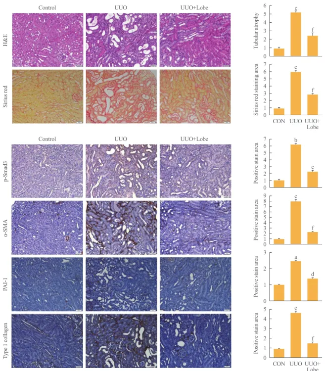
The effects of lobeglitazone on renal tubulointerstitial fibrosis were evaluated using the UUO model. As shown in Fig. 1A, he- matoxylin/eosin and sirius red staining showed that vehicle- treated UUO kidneys exhibited prominent renal tubular atrophy and tubulointerstitial fibrosis. By contrast, lobeglitazone-treated UUO kidneys showed marked attenuation of UUO-induced tu- bular atrophy and tubulointerstitial fibrosis (Fig. 1A).
Lobeglitazone suppresses the interstitial expression of profibrotic molecules
Given that TGF-β/Smad3 is a well-known mediator in the de- velopment of renal tubulointerstitial fibrosis, we examined the effects of lobeglitazone on the levels of Smad3 phosphorylation and Smad3 target genes including α-SMA, PAI-1, and type 1 collagen. The results showed that positively stained areas for phosphorylated Smad3, α-SMA, PAI-1, and type 1 collagen were evidently increased in the damaged tubules of UUO kid- neys, but these were significantly reduced by lobeglitazone treatment (Fig. 1B).
The effects of lobeglitazone on fibrotic gene expression were
6 5 4 3 2 1 0 76 54 32 10
5 4 3 2 1 0 3 2 1 0 98 76 54 32 10 76 54 32 10
Tubular atrophySirius red staining areaPositive stain areaPositive stain areaPositive stain areaPositive stain area
p-Smad3H&ESirius redα-SMAPAI-1Type 1 collagen
CON
CON UUO
UUO UUO+Lobe
UUO+Lobe
Fig. 1. Effects of lobeglitazone on unilateral ureteral obstruction (UUO)-induced renopathological changes. (A) Representative images of hematoxylin and eosin (H&E) and sirius red staining of kidney tissue sections from control (CON) mice and UUO mice with or without lobeglitazone (Lobe; 1 mg/kg) treatment. The number of atrophic tubules was determined by measuring abnormal and dilated tubular base- ment membranes in five random fields of H&E stained sections under high power magnification (×200). Areas of positive staining with sir- ius red were quantitated by computer-based morphometric analysis. All morphometric data were normalized against the corresponding val- ues in CON animals. Data in all bar graphs are expressed as fold increase relative to the CON (n=6 in each group). (B) Representative im- ages of immunohistochemical staining forp-Smad3, α-smooth muscle actin (α-SMA), plasminogen activator inhibitor 1 (PAI-1), and type I collagen in kidney tissue sections from CON mice or UUO mice with or without lobeglitazone (1 mg/kg). Areas of positive staining with p- Smad3, α-SMA, PAI-1, and type 1 collagen antibodies were quantitated by computer-based morphometric analysis. All data were expressed as the mean±SEM of five random fields from each kidney section (n=6 in each group). aP<0.05; bP<0.01; cP<0.001 vs. CON; and
dP<0.05; eP<0.01; fP<0.001 vs. UUO.
A
B Control
Control
UUO
UUO
UUO+Lobe
UUO+Lobe
c
c
c a c b
f
f
f d f e
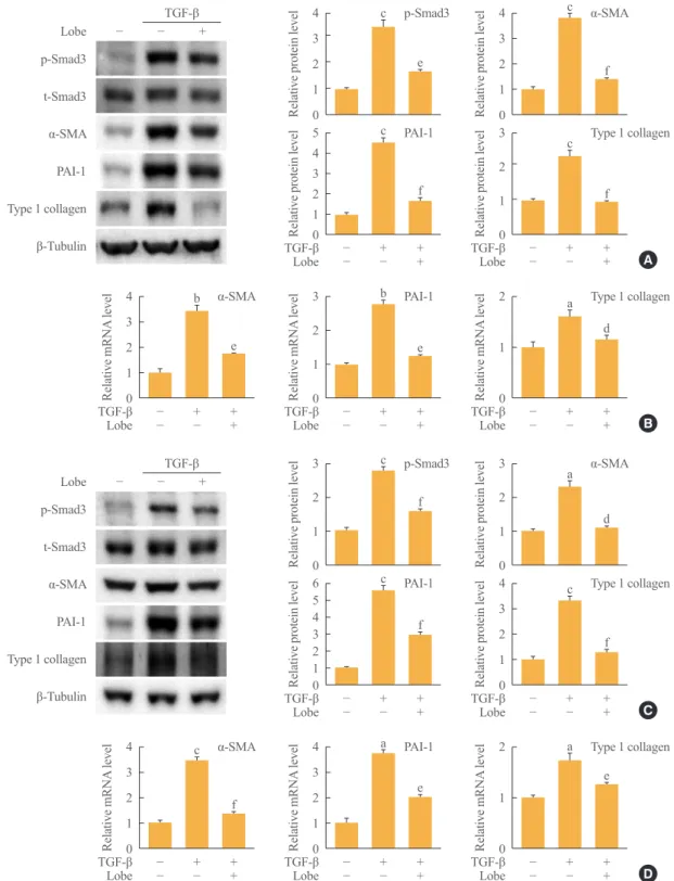
further confirmed by quantitative RT-PCR and Western blot analysis. Consistent with the immunohistochemical analysis, the protein abundance of PAI-1, α-SMA, and type 1 collagen was lower in the kidneys of mice administered lobeglitazone than in vehicle-treated kidneys (Fig. 2A). Moreover, UUO-in- duced Smad3 phosphorylation was markedly suppressed in the kidneys of lobeglitazone-treated mice (Fig. 2A). The mRNA expression levels of these genes in the kidneys of lobeglitazone- treated mice were also markedly lower than in vehicle-treated mice (Fig. 2B).
Lobeglitazone inhibits profibrotic gene expression through inhibition of TGF-β/Smad3 signaling
To examine the mechanism responsible for the antifibrotic ef- fects of lobeglitazone, we examined whether lobeglitazone in- hibits TGF-β-stimulated Smad3 signaling in cultured renal cells including NRK-49F cells and RMCs. As expected, TGF-β treat- ment increased mRNA and protein levels of PAI-1, α-SMA, and type 1 collagen, and induced Smad3 phosphorylation. Lobegli- tazone-treated NRK-49F cells showed markedly inhibited TGF- β-stimulated profibrotic gene expression and Smad3 phosphor-
Fig. 2. Effects of lobeglitazone on profibrotic gene expression in kidneys of unilateral ureteral obstruction (UUO) mice. (A) Representative Western blot analysis of p-Smad3, t-Smad3, α-smooth muscle actin (α-SMA), plasminogen activator inhibitor 1 (PAI-1), and type 1 collagen protein expression in UUO kidneys with or without lobeglitazone (Lobe; 1 mg/kg; n=6 in each group). Data are expressed as the mean±
SEM of three independent experiments. (B) Representative real-time reverse transcription polymerase chain reaction analysis of α-SMA, PAI-1, and type 1 collagen mRNA expression in UUO kidneys with or without Lobe (1 mg/kg; n=6 in each group). Data in bar graphs are mean±SEM. aP<0.05; bP<0.01; cP<0.001 vs. control (CON); and dP<0.05; eP<0.01; fP<0.001 vs. UUO.
p-Smad3 t-Smad3 α-SMA PAI-1 Type 1 collagen β-Tubulin
Control UUO UUO+Lobe
3 2 1 0
20 10 0 3.53.0
2.52.0 1.51.0 0.50
5 4 3 2 1 0
15 10 5 0
4 3 2 1 0
20 10 0
Relative protein level
Relative mRNA level
Relative protein level Relative protein level
Relative mRNA level Relative protein level
Relative mRNA level
CON
CON
CON CON
CON
CON
CON
UUO
UUO
UUO UUO
UUO
UUO
UUO
UUO+Lobe
UUO+Lobe
UUO+Lobe UUO+
Lobe
UUO+Lobe
UUO+Lobe
UUO+Lobe
A
B a
b
c c
c
c
c
e
d f
e
f
f
e
Type 1 collagen
Type 1 collagen
p-Smad3 α-SMA
α-SMA
PAI-1
PAI-1
4 3 2 1 0
3 2 1 0 3 2 1 0
4 3 2 1 0 2 1 0
2 1 0 4
3 2 1 0
3 2 1 0 5 4 3 2 1 0
65 4 3 21 0 3 2 1 0 4
3 2 1 0
4 3 2 1 0 4
3 2 1 0
Relative protein levelRelative protein levelRelative protein levelRelative protein levelRelative mRNA levelRelative mRNA level
Relative protein levelRelative protein levelRelative protein levelRelative protein levelRelative mRNA level
Relative mRNA level Relative mRNA level
Relative mRNA level
−
−
−
−
−
−
−
−
−
−
−
−
−
−
TGF-β TGF-β
TGF-β TGF-β
TGF-β TGF-β
TGF-β
TGF-β
TGF-β TGF-β
−
−
−
−
−
−
Lobe Lobe
Lobe Lobe
Lobe Lobe
Lobe
Lobe
Lobe Lobe
+
+ +
+
−
−
−
− +
+ + +
+ +
−
−
−
−
−
−
+
+ +
+ +
+ +
+ +
+ + +
+ +
+
+ + +
+ +
A
C B
D c
a c
c a
a c
c c
c b b
c a
f
d f
f d
e e
f f
f e e
e f
α-SMA
α-SMA Type 1 collagen
Type 1 collagen Type 1 collagen
Type 1 collagen p-Smad3
p-Smad3 PAI-1
PAI-1 PAI-1 α-SMA
PAI-1 α-SMA
p-Smad3
p-Smad3 t-Smad3
t-Smad3 α-SMA
α-SMA PAI-1
PAI-1 Type 1 collagen
Type 1 collagen β-Tubulin
β-Tubulin
TGF-β
TGF-β
Fig. 3. Effects of lobeglitazone (Lobe) on transforming growth factor β (TGF-β)-induced profibrotic gene expression in cultured kidney cell lines. Representative Western blot analysis (A) of p-Smad3, t-Smad3, α-smooth muscle actin (α-SMA), plasminogen activator inhibitor 1 (PAI-1), and type 1 collagen levels and representative real-time reverse transcription polymerase chain reaction (RT-PCR) analysis (B) of α-SMA, PAI-1, and type 1 collagen expression in TGF-β-stimulated NRK-49F cells. Representative Western blot analysis (C) of p-Smad3, t-Smad3, α-SMA, PAI-1, and type 1 collagen expression and representative real-time RT-PCR analysis (D) of α-SMA, PAI-1, and type 1 collagen expression in TGF-β-stimulated rat mesangial cells. Cells were co-incubated with TGF-β (5 ng/mL) and Lobe (10 μM) after 24 hours serum starvation. Data are the mean±SEM of three independent measurements (three separate experiments). aP<0.05; bP<0.01;
cP<0.001 vs. control (CON); and dP<0.05; eP<0.01; fP<0.001 vs. TGF-β alone.
−
− Lobe
Lobe
−
− +
+
ylation (Fig. 3A, B). Consistent with the effects in NRK-49F cells, lobeglitazone also suppressed TGF-β-stimulated Smad3 phosphorylation, PAI-1, α-SMA, and type 1 collagen in RMCs (Fig. 3C, D).
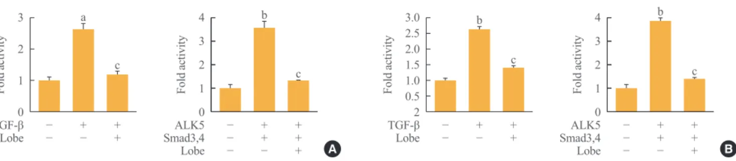
To determine whether lobeglitazone-induced suppression of profibrotic gene expression is mediated at the transcriptional level, we examined whether lobeglitazone treatment inhibits TGF-β/Smad-stimulated PAI-1 promoter activity. Indeed, lobe- glitazone treatment successfully inhibited TGF-β and ALK5/
Smad3, 4-stimulated PAI-1 promoter activity both in NRK-49F cells and RMCs (Fig. 4). These results indicate that lobegli- tazone has an antifibrotic effect through the inhibition of TGF- β-stimulated Smad3 transcriptional activity on its target genes.
DISCUSSION
The study presented here shows that lobeglitazone treatment at- tenuates renal fibrosis in UUO mice. Lobeglitazone inhibited UUO-induced profibrotic gene expression including PAI-1, α-SMA, and type 1 collagen. The antifibrotic effects of lobegli- tazone were associated with inhibition of the TGF-β/Smad3 sig- naling pathway.
PPARγ agonists are widely used as antidiabetic agents through improved insulin sensitivity and lipid metabolism [21].
On the basis of their wide range of metabolic benefits, PPARγ agonists can ameliorate diabetic nephropathy in animal and hu- man studies [22]. Recently, several lines of evidence show that PPARγ agonists can reduce acute kidney injury, indicating that their renal protective properties may be partially independent of metabolic factors. For instance, activation of PPARγ reduces glomerulosclerosis and apoptosis via regulation of inflamma-
tion in various animal models of non-diabetic nephropathy in- cluding 5/6 nephrectomy [12], passive Heymann nephritis [23], cisplatin-induced renal damage [24], and ischemia/reperfusion injury [25]. Additionally, rosiglitazone, pioglitazone, and trogli- tazone have a role in reducing renal tubulointerstitial fibrosis in the UUO model [26-29]. Rosiglitazone inhibits renal tubuloint- erstitial fibrosis through inhibiting interstitial macrophage infil- tration, downregulating the expressions of TGF-β and its down- stream target genes, and up-regulating the BMP-7 expression [26]. Several groups examined antifibrotic effects of piogli- tazone and interaction with angiotensin receptor antagonists such as L158809 and candesartan in the UUO model [27,28].
Pioglitazone and candesartan have additive protective effects on renal fibrosis, but the synergism between pioglitazone and L158809 is not clear [27]. Troglitazone attenuates renal intersti- tial fibrosis and inflammation in dose dependent manner by down regulation of TGF-β signaling pathway in the model of UUO. In accordance with these findings, our present study showed that lobeglitazone also has a protective effect on renal tubulointerstitial fibrosis and UUO-induced tubular atrophy.
Furthermore, lobeglitazone inhibited the expression of well- known TGF-β target genes, such as PAI-1, α-SMA, and type 1 collagen, as well as its major effector, phosphorylated Smad3, in the kidneys of UUO mice.
The TGF-β/Smad signaling pathway is a primary pathogenic factor that drives glomerular and tubulointerstitial fibrosis in the kidney by stimulating the synthesis of ECM molecules and by decreasing ECM degradation [4]. Although mesangial cells ex- press Smad1, 2, 3, 4, and 7, accumulating evidence demon- strates that Smad3 is mainly implicated in a pathogenic role in TGF-β-mediated renal fibrosis [30-32]. Recent studies show
4 3 2 1 0
4 3 2 1 0 3
2 1 0
3.0 2.52.0 1.5 1.0 0.52
Fold activity Fold activity
Fold activity Fold activity
− −
− −
− −
− −
− −
TGF-β TGF-β
Lobe ALK5 Lobe ALK5
Smad3,4 Smad3,4
Lobe Lobe
+ +
+ +
− −
+ +
− + − +
+ +
+ +
+ +
+ +
b b
a b
c c
c c
Fig. 4. Effects of lobeglitazone (Lobe) on the transforming growth factor β (TGF-β)/Smad3 signaling pathway in kidney cell lines. (A) Ef- fects of Lobe on plasminogen activator inhibitor 1 (PAI-1) promoter activity in NRK-49F cells. Cells were treated with TGF-β (5 ng/mL) with or without Lobe co-treatment (10 μM) for 24 hours (left panel). (B) Effects of Lobe on PAI-1 promoter activity in rat mesangial cells.
Cells were treated with TGF-β (5 ng/mL) with or without Lobe co-treatment (10 μM) for 24 hours (left panel). Cells were co-transfected with the PAI-1 promoter and expression vectors for Smad3/4 (pRK5) and ALK5 (pcDNA) with or without Lobe treatment (10 μM) for 24 hours (right panel). Data are the mean±SEM of three independent measurements. aP<0.01; bP<0.001 vs. control; and cP<0.001 vs. TGF-β alone or vs. Smad3/4 and ALK5.
A B
that several PPARγ agonists inhibit TGF-β-stimulated ECM production, indicating that PPARγ agonists attenuate renal tubu- lointerstitial fibrosis by inhibiting the TGF-β/Smad3 signaling pathway [33]. Pioglitazone inhibits renal tubulointerstitial fibro- sis and infiltration of interstitial macrophages by regulating transcription of PAI-1in UUO mice [28]. Rosiglitazone treat- ment inhibits inflammatory reactions and renal fibrosis by re- ducing the overexpression of endogenous endothelin-1, cyclo- oxygenase-2, and TGF-β in deoxycorticosterone acetate-salt hypertensive rats [34]. Our study adds evidence that another novel PPARγ agonist, lobeglitazone suppresses tubulointerstitial fibrosis after UUO in mice by inhibiting TGF-β/Smad3 signal- ing.
In conclusion, this study demonstrates that lobeglitazone has a renoprotective effect on UUO-induced renal fibrosis through inhibition of the TGF-β/Smad3 pathway. Our results suggest that lobeglitazone could play a therapeutic role in CKD, provid- ing rationale for further clinical trials to evaluate the efficacy of lobeglitazone in the treatment of CKD including renal tubuloin- terstitial fibrosis.
CONFLICTS OF INTEREST
No potential conflict of interest relevant to this article was re- ported.
ACKNOWLEDGMENTS
This work was supported by Biomedical Research Institute grant, Kyungpook National University Hospital (2016).
ORCID
Kwi-Hyun Bae http://orcid.org/0000-0002-0482-2904 Keun-Gyu Park http://orcid.org/0000-0002-8403-1298
REFERENCES
1. Meguid El Nahas A, Bello AK. Chronic kidney disease: the global challenge. Lancet 2005;365:331-40.
2. Zeisberg M, Neilson EG. Mechanisms of tubulointerstitial fibrosis. J Am Soc Nephrol 2010;21:1819-34.
3. Nangaku M. Chronic hypoxia and tubulointerstitial injury: a final common pathway to end-stage renal failure. J Am Soc Nephrol 2006;17:17-25.
4. Meng XM, Nikolic-Paterson DJ, Lan HY. TGF-beta: the
master regulator of fibrosis. Nat Rev Nephrol 2016;12:325- 38.
5. Eddy AA, Fogo AB. Plasminogen activator inhibitor-1 in chronic kidney disease: evidence and mechanisms of action.
J Am Soc Nephrol 2006;17:2999-3012.
6. Jung GS, Kim MK, Jung YA, Kim HS, Park IS, Min BH, et al. Clusterin attenuates the development of renal fibrosis. J Am Soc Nephrol 2012;23:73-85.
7. Won JC, Park CY, Oh SW, Lee ES, Youn BS, Kim MS.
Plasma clusterin (ApoJ) levels are associated with adiposity and systemic inflammation. PLoS One 2014;9:e103351.
8. Derosa G, Maffioli P. Peroxisome proliferator-activated re- ceptor-gamma (PPAR-gamma) agonists on glycemic con- trol, lipid profile and cardiovascular risk. Curr Mol Pharma- col 2012;5:272-81.
9. Cunard R, Ricote M, DiCampli D, Archer DC, Kahn DA, Glass CK, et al. Regulation of cytokine expression by li- gands of peroxisome proliferator activated receptors. J Im- munol 2002;168:2795-802.
10. Guan Y, Zhang Y, Breyer MD. The role of PPARs in the transcriptional control of cellular processes. Drug News Per- spect 2002;15:147-54.
11. Smith SA, Lister CA, Toseland CD, Buckingham RE. Rosi- glitazone prevents the onset of hyperglycaemia and protein- uria in the Zucker diabetic fatty rat. Diabetes Obes Metab 2000;2:363-72.
12. Ma LJ, Marcantoni C, Linton MF, Fazio S, Fogo AB. Per- oxisome proliferator-activated receptor-gamma agonist tro- glitazone protects against nondiabetic glomerulosclerosis in rats. Kidney Int 2001;59:1899-910.
13. Routh RE, Johnson JH, McCarthy KJ. Troglitazone sup- presses the secretion of type I collagen by mesangial cells in vitro. Kidney Int 2002;61:1365-76.
14. Guo B, Koya D, Isono M, Sugimoto T, Kashiwagi A, Hane- da M. Peroxisome proliferator-activated receptor-gamma li- gands inhibit TGF-beta 1-induced fibronectin expression in glomerular mesangial cells. Diabetes 2004;53:200-8.
15. Kim JW, Kim JR, Yi S, Shin KH, Shin HS, Yoon SH, et al.
Tolerability and pharmacokinetics of lobeglitazone (CKD- 501), a peroxisome proliferator-activated receptor-gamma agonist: a single- and multiple-dose, double-blind, random- ized control study in healthy male Korean subjects. Clin Ther 2011;33:1819-30.
16. Kim SG, Kim DM, Woo JT, Jang HC, Chung CH, Ko KS, et al. Efficacy and safety of lobeglitazone monotherapy in patients with type 2 diabetes mellitus over 24-weeks: a mul-
ticenter, randomized, double-blind, parallel-group, placebo controlled trial. PLoS One 2014;9:e92843.
17. Jin SM, Park CY, Cho YM, Ku BJ, Ahn CW, Cha BS, et al.
Lobeglitazone and pioglitazone as add-ons to metformin for patients with type 2 diabetes: a 24-week, multicentre, ran- domized, double-blind, parallel-group, active-controlled, phase III clinical trial with a 28-week extension. Diabetes Obes Metab 2015;17:599-602.
18. Lim S, Lee KS, Lee JE, Park HS, Kim KM, Moon JH, et al.
Effect of a new PPAR-gamma agonist, lobeglitazone, on neointimal formation after balloon injury in rats and the de- velopment of atherosclerosis. Atherosclerosis 2015;243:107- 19.
19. Jung GS, Jeon JH, Choe MS, Kim SW, Lee IK, Kim MK, et al. Renoprotective effect of gemigliptin, a dipeptidyl pepti- dase-4 inhibitor, in streptozotocin-induced type 1 diabetic mice. Diabetes Metab J 2016;40:211-21.
20. Jung GS, Kim MK, Choe MS, Lee KM, Kim HS, Park YJ, et al. The orphan nuclear receptor SHP attenuates renal fi- brosis. J Am Soc Nephrol 2009;20:2162-70.
21. Kim H, Haluzik M, Gavrilova O, Yakar S, Portas J, Sun H, et al. Thiazolidinediones improve insulin sensitivity in adi- pose tissue and reduce the hyperlipidaemia without affecting the hyperglycaemia in a transgenic model of type 2 diabetes.
Diabetologia 2004;47:2215-25.
22. Pistrosch F, Passauer J, Herbrig K, Schwanebeck U, Gross P, Bornstein SR. Effect of thiazolidinedione treatment on pro- teinuria and renal hemodynamic in type 2 diabetic patients with overt nephropathy. Horm Metab Res 2012;44:914-8.
23. Benigni A, Zoja C, Tomasoni S, Campana M, Corna D, Zanchi C, et al. Transcriptional regulation of nephrin gene by peroxisome proliferator-activated receptor-gamma ago- nist: molecular mechanism of the antiproteinuric effect of pioglitazone. J Am Soc Nephrol 2006;17:1624-32.
24. Jesse CR, Bortolatto CF, Wilhelm EA, Roman SS, Prigol M, Nogueira CW. The peroxisome proliferator-activated recep- tor-gamma agonist pioglitazone protects against cisplatin-
induced renal damage in mice. J Appl Toxicol 2014;34:25- 32.
25. Doi S, Masaki T, Arakawa T, Takahashi S, Kawai T, Na- kashima A, et al. Protective effects of peroxisome prolifera- tor-activated receptor gamma ligand on apoptosis and hepa- tocyte growth factor induction in renal ischemia-reperfusion injury. Transplantation 2007;84:207-13.
26. Lin Q, Gu Y, Ma J, Lin SY. Protection of rosiglitazone against renal interstitial lesion and its mechanism. Zhonghua Yi Xue Za Zhi 2005;85:1618-24.
27. Han JY, Kim YJ, Kim L, Choi SJ, Park IS, Kim JM, et al.
PPARgamma agonist and angiotensin II receptor antagonist ameliorate renal tubulointerstitial fibrosis. J Korean Med Sci 2010;25:35-41.
28. Higashi K, Oda T, Kushiyama T, Hyodo T, Yamada M, Su- zuki S, et al. Additive antifibrotic effects of pioglitazone and candesartan on experimental renal fibrosis in mice. Nephrol- ogy (Carlton) 2010;15:327-35.
29. Kawai T, Masaki T, Doi S, Arakawa T, Yokoyama Y, Doi T, et al. PPAR-gamma agonist attenuates renal interstitial fi- brosis and inflammation through reduction of TGF-beta.
Lab Invest 2009;89:47-58.
30. Lan HY, Chung AC. TGF-beta/Smad signaling in kidney disease. Semin Nephrol 2012;32:236-43.
31. Meng XM, Huang XR, Chung AC, Qin W, Shao X, Igarashi P, et al. Smad2 protects against TGF-beta/Smad3-mediated renal fibrosis. J Am Soc Nephrol 2010;21:1477-87.
32. Leask A, Abraham DJ. TGF-beta signaling and the fibrotic response. FASEB J 2004;18:816-27.
33. Ohtomo S, Izuhara Y, Takizawa S, Yamada N, Kakuta T, van Ypersele de Strihou C, et al. Thiazolidinediones provide bet- ter renoprotection than insulin in an obese, hypertensive type II diabetic rat model. Kidney Int 2007;72:1512-9.
34. Bae EH, Kim IJ, Ma SK, Kim SW. Rosiglitazone prevents the progression of renal injury in DOCA-salt hypertensive rats. Hypertens Res 2010;33:255-62.