Lumbar foraminal or extraforaminal stenosis is a common cause of radiculopathy with a frequency of approximately 8%–11% in patients with lumbar disorders and a 75%
incidence of L5 root compression in lumbar foraminal stenosis. It is caused by foraminal space narrowing with ligamentous and osseous structure hypertrophy.1,2) There are currently two major surgical treatment options for this disease: simple foraminal decompression with structural stability preservation, and wider decompression with lami-
nectomy and foraminotomy with fusion and instrumenta- tion.3,4) Microscopic decompression, designed to preserve facet joints and avoid fusion, was introduced by Wiltse and Spencer5) and modified by others with 80% patient satisfaction rates. Lumbar interbody fusion surgery was designed to expand the intervertebral space to resolve fo- raminal stenosis without a foraminotomy. However, fusion surgery was considered to be associated with potential complications, such as back muscle atrophy, adjacent seg- ment disease, and pseudoarthrosis. Thus, several authors introduced uniportal endoscopic spine surgery under docking into the Kambin’s triangle for foraminal decom- pression with preserving back muscle integrity and facet joint function. At the L5–S1 level, however, the protruding iliac crest makes it difficult to access the lumbosacral fora- men to perform proper decompression. Unilateral biportal endoscopic spinal surgery (BESS) reportedly shows good
Unilateral Biportal Endoscopic Spinal Surgery Using a 30° Arthroscope for L5–S1 Foraminal
Decompression
Ju-Eun Kim, MD
#, Dae-Jung Choi, MD*
,#Department of Orthopedic Surgery, Andong Hospital, Andong,
*Department of Spine Surgery, Barun Hospital, Jinju, Korea
Foraminal decompression using a minimally invasive technique to preserve facet joint stability and function without fusion report- edly improves the radicular symptoms in approximately 80% of patients and is considered one of the good surgical treatment choices for lumbar foraminal or extraforaminal stenosis. However, proper decompression was not possible because of the inability to access the foramen at the L5–S1 level due to prominence of the iliac crest. To overcome this challenge, endoscopy-based mini- mally invasive spine surgery has recently gained attention. Here, we report the technical skills required in unilateral extraforaminal biportal endoscopic spinal surgery using a 30° arthroscope to enable foraminal decompression at the L5–S1 level. Two 0.8-cm por- tals were created 2 cm lateral from the lateral border of the pedicles at the L5–S1 level. After sufficient working space was made, half of the superior articular process (SAP) in the hypertrophied facet joint was removed using a high-speed burr and a 5-mm wide osteotome, whereas the remaining inside part of the SAP was removed using a Kerrison punch and pituitary punch. The foraminal ligamentum flavum should be removed to inspect the conditions of the L5 exiting root and disc. Removing of the extruded disc could decompress the L5 root. The extraforaminal approach using a 30° arthroscope is considered a minimally invasive alternative technique for decompressing foraminal stenosis at the L5–S1 level that preserves facet stability and provides symptomatic relief.
Keywords: Arthroscope, Spinal stenosis, Lumbosacral region, Endoscopes
Copyright © 2018 by The Korean Orthopaedic Association
This is an Open Access article distributed under the terms of the Creative Commons Attribution Non-Commercial License (http://creativecommons.org/licenses/by-nc/4.0) which permits unrestricted non-commercial use, distribution, and reproduction in any medium, provided the original work is properly cited.
Clinics in Orthopedic Surgery • pISSN 2005-291X eISSN 2005-4408
#Current affiliation: Department of Spine Surgery, Himnaera Hospital, Busan, Korea
Received November 25, 2017; Accepted March 19, 2018 Correspondence to: Dae-Jung Choi, MD
Department of Spine Surgery, Himnaera Hospital, 85 Beomil-ro, Dong-gu, Busan 48735, Korea
Tel: +82-1644-9502, Fax: +82-51-720-1505 E-mail: djchoi9@hanmail.net
results for the treatment of degenerated lumbar spine dis- orders. However, to date there are only a few reports on the extraforaminal approach at the L5–S1 level, and an ef- fective technique has not yet been standardized.6) The aim of our study was to introduce the extraforaminal approach of BESS using a 30° arthroscope as an alternative to fusion surgery for L5–S1 foraminal stenosis.
TECHNIQUE
We conducted this study in compliance with the principles of the Declaration of Helsinki. The protocol of this study was reviewed and approved by the Institutional Review Board of Andong Hospital (IRB No. 2018-013). Written informed consents were obtained.
Basic Facilities and Instruments
A 30°, 4-mm-diameter arthroscope commonly used in joint arthroscopic surgery was prepared in an arthroscopic surgery facility. The radiofrequency (RF) wand for de- bridement and coagulation should be set on the coagula- tion rather than the ablation mode. A 4.2-mm high-speed burr for laminectomy, a shaver to perform debridement and create a working space, and basic spine instruments, such as Kerrison punch, pituitary rongeur, and curettes were used. A fluoroscope was set for level check. An irri- gation pump was optional and saline could be sufficiently infused by natural gravity.
Creating an Extraforaminal Working Space
Two portals were created as standard inlets, one for scop- ing and the other for handling of an instrument. A third portal could be added for water outflow, especially for the patients with heavier back muscle and poor water outflow.
The proximal portal should be created 2 cm away from the lateral margin of the L5 pedicle in the middle of the L5 transverse process (TP), and the distal portal should be created on the sacral ala. Each portal was 1 cm in diameter, sufficient to accommodate an arthroscope for viewing and an instrument for working. A working space surrounded by the L5 TP, L5–S1 facet joint, and ala was created using a muscle detacher to separate the muscles from the basal bony structures using the triangulation technique: the pat- tern for muscle detaching from the dorsal surface of the L5 TP via the lateral surface of the facet to the proximal surface of the ala. The rugged muscle was shaved off and the soft tissue was debrided to clear the surgical field, us- ing a shaver and an RF wand, respectively. Heavy bleeds occurred at the upper edge of the facet at the 10 and 2 o’clock positions from the dorsal branch of the lumbar
artery and lower edge of the facet just beside the muscles at center. Small bleeds occurred at the 6 o’clock position from the muscle under endoscopic view. It is helpful to orient the arthroscopic arrow in the 12 o’clock direction to control heavy bleeds and 6 o’clock direction to control small bleeds. When visual acuity deteriorates rapidly, small bleeds originate from some position just beneath the scope, and water output could become poor. Therefore, a 5.0-mm plastic cannula or an additional quarterback por- tal at the triangular point between the portals for fluent saline output could be used (Fig. 1A).
Foraminal Decompression
To decompress the exiting root, half of the superior ar- ticular process (SAP) in the hypertrophied facet joint was removed using a high-speed burr and an osteotome to create a cortical half (TP and pedicular junction of the L5) and a cancellous half (SAP from the ala) view (“half-and- half view”) (Fig. 1B). Unroofing of the foramen over the exiting root was mandatory. The arthroscopic arrow was rotated to 12 o’clock to gain a much deeper view of the fo- ramen. The comparison of a 0° and a 30° arthroscope (at the same position) revealed that the 0° arthroscope could not provide a good view because of the facet joint, but the 30° arthroscope could provide a better view of the inner foramen (Fig. 1C, D). A flavectomy was performed from the distal portion of the L5 TP using a curette. Thereafter, an annulus protrusion with fat and small vascular struc- tures was noticed at the distal half, which could easily be misunderstood as the exiting root. The root must always be located at the proximal half inferior to the TP. At the 6 o’clock position of the arthroscopic arrow, the view for dis- cectomy was better. The subpedicular cortical thickening and spur at the TP and pedicular junction could compress the root, which ran obliquely around the pedicle. A subpe- diculotomy should be performed using an angled osteo- tome to create sufficient room for a 5-mm-thick curette to freely pass under the pedicle (Fig. 2).
DISCUSSION
The L5 exiting root is one of the frequent sites for lesions, either alone or in combination with upper levels in lumbar degenerative disorders. However, surgical treatment op- tions are very limited and focused on fusion surgery with total laminectomy due to the difficulty in accessing the foramen owing to its deeper location and higher alar wing.
Open decompression surgery using the Wilte approach is a conventional challenge despite the deeper back muscle sacrifice for extraforaminal or foraminal stenosis.5) Open
microscopic foraminotomy reportedly has a success rate of 58%–80% and poor outcomes compared to other spine surgeries.7) This might be due to insufficient access to the foramen when trying to directly inspect the structure hid- den under the foramen. Excessive dissection of the para- spinal muscle in the open approach might cause back pain or muscle atrophy.
An ability of free handling and a better view with the endoscope with no need for muscle dissection to view the area beneath the isthmus and lower facet can allow
for more precise surgery around the extraforaminal and foraminal lesions and provide good results for decompres- sion surgery for foraminal or extraforaminal stenosis.1,8) The priority of endoscopy-based spine surgery is a mini- mally invasive technique that aims to preserve the back muscles and structural stability.1,8)
The recently introduced floating technique of BESS using an arthroscope has advantages over the conventional portal endoscopic surgery, even with the extraforaminal approach. Conventional one-portal endoscopic decom-
*
A B
C D
Fig. 1. (A) Creating an extraforaminal working space. A schematic drawing on the right-side approach (left). An endoscopic view (right) showing the surgical anatomy, so-called “Half-and-Half” view. Partial resection of the tip of the superior articular process (SAP) should be fully performed to expose the half view of the cortical surface of the junction of transverse process (TP) and pedicle and the other half view of the cancellous surface of the SAP.
This space of view enough to decompress the exiting root should be needed. (B) There are several bleeding foci (asterisks) at center, 10, 2, and 6 o’clock.
(C, D) The difference of 0° arthroscopic and 30° arthroscopic views. The 30° arthroscopy is more advantageous for obtaining the inside view (D) than the 0° arthroscopy (C). It is more vertically rotated to the vertebra in the 12 o’clock direction for closer approach to the pedicle.
A B
C D
Fig. 2. Subpedicular decompression and unroofing of the exiting root. (A) Subpedicular cortical, thickening (dotted line) could compress the exiting root at the transverse process (TP) and pedicular junction. (B) Subpedicular corticotomy was performed using a ventral curved pedicular chisel. (C) Unroofing of the exiting root should be checked with sufficient proximal free space between the root and distal area of the TP using a freer and medially under the TP and pedicular junction using a 5-mm-thick curette (D).
pression using the docking technique into Kambin’s tri- angle carries the potential risk of an existing nerve root injury, especially in cases with a narrowed disc space and foramen in degenerative foraminal stenosis. In severe foraminal stenosis, the floating technique of BESS being performed just outside of the foramen could prevent root irritation and using another working portal could permit the use of various surgical instruments such as a Kerrison rongeur, pituitary forceps, burr, or osteotome regardless of size. The peculiar concept of the BESS can bring about de- creasing paravertebral muscle damage and enable precise and selective foraminal decompression with the preserva- tion of structural stability, which consequently can lead to good results. Regarding the approach to the L5–S1 area, the sacral ala is a notorious barrier in the conventional en- doscopic spine surgery and offsets its other advantages due to the difficulty of access to the L5–S1. However, the por- tals used in the BESS are created inside the ala, eliminating the barrier and enabling access to the L5–S1 foramen.
To ensure successful decompression using BESS, the proximally overgrowing spur of the SAP outside the fora- men should be completely removed until the ligamentum flavum is exposed. A half-and-half view (cortical surface of the L5 at the proximal side and cancellous surface of the lower articular process of the S1 at the distal side) should then be created. The exiting root must be fully decom-
pressed from the entrance of the foramen to the extrafo- raminal area. The distal part of the L5 TP is frequently overgrown distally and ventrally, causing far-out syn- drome.9) The L5 root should be decompressed along the distal margin of the TP to the TP and Ala junction. The cortical lamina at the outside and beneath the subpedicu- lar area at the TP and Pedicle junction thickened and hy- pertrophied, which occurred due to repeated impact of the SAP and the junction, and disc height collapse could fre- quently impinge the L5 root running close to the pedicle.10) The somewhat inner side of the pedicle can be difficult to inspect. The use of a 30° arthroscope at 12 o’clock position of the arthroscopic arrow could enable clear observation of the root at the inner and distal areas of the pedicle.
Subpedicular decompression and regaining free room for the root are mandatory to eliminate radicular symptoms, such as posterior calf pain and paresthesia on the sole of the foot. Subpedicular decompression at the junction of the TP and pedicle has not been mentioned yet, because in previous studies, the surgical performance (using a naked eye or a microscopic view) focused only on the lesions in front. A notional lesion under the pedicle can be clearly visualized using the 30° arthroscope view during a BESS.
Technical Tips
Technical tips are as follows. (1) Checking fluent water
A B C
D E F
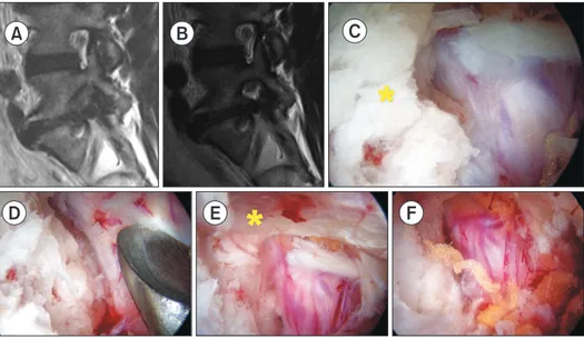
Fig. 3. A case of 53-year-old male patient with L5–S1 foraminal stenosis on the left side. (A) Preoperative T2-weighted magnetic resonance imaging (MRI) sagittal view showing grade 2 foraminal stenosis with a decreased disc height. (B) Postoperative T2-weighted MRI sagittal view showing a widened foramen after foraminotomy using biportal endoscopic spinal surgery. (C) The L5 exiting root was compressed by the distal spur (asterisk) of the transverse process (TP) at the TP and pedicular junction and the surface of the root was seen covered by fibrous degenerative tissue with embedded perineural vessels on the root. (D) The proximal surface of the root should be fully decompressed after resection of the spur of the distal TP and the root should be in unroofed state. (E) The 0° arthroscopic view of the outlet of the root (asterisk) did not show the proximal curved part of the root under the pedicle. (F) The 30° arthroscopic view showed the deeper inner side for inspection of the corner under the pedicle.
output is important when creating a portal. The subcuta- neous fascia should be released or cross-cut to enable the flow. (2) An accessory portal or cannula could help pre- vent poor output of the irrigation saline. Water congestion could create a poor visual field, a longer operation time, and consequent muscle edema. (3) The 6 o’clock rotation of the arthroscopic arrow could provide a better view of the working space on the TP, while the 12 o’clock view could enable the foraminotomy. (4) The radicular artery ran through the middle of the view overlapping the root and was easily damaged by the instruments and shaver.
This caused the visual field to deteriorate rapidly because of heavy blood flow. A small-tipped RF wand, which was generally used for wrist arthroscopy, was used to control the bleeding at the foramen. (5) Subpedicular decompres- sion is mandatory to eliminate the cortical thickening of the TP and pedicle junction at the foraminal surface. The
L5 root runs obliquely along the pedicle and can become impinged at the junction (Fig. 3).
We described a biportal endoscopic decompression technique for foraminal stenosis at the L5–S1 level using a 30° arthroscope to suggest a standard endoscopy-based approach. The extraforaminal approach using BESS with a 30° arthroscope could allow for a better inspection of the extraforaminal and intraforaminal areas and promote complete decompression of the foraminal lesion. BESS could be an option for patients with L5–S1 foraminal ste- nosis who do not want fusion surgery.
CONFLICT OF INTEREST
No potential conflict of interest relevant to this article was reported.
REFERENCES
1. Ahn Y, Oh HK, Kim H, Lee SH, Lee HN. Percutaneous en- doscopic lumbar foraminotomy: an advanced surgical tech- nique and clinical outcomes. Neurosurgery. 2014;75(2):124- 33.
2. Jenis LG, An HS. Spine update: lumbar foraminal stenosis.
Spine (Phila Pa 1976). 2000;25(3):389-94.
3. Chang SB, Lee SH, Ahn Y, Kim JM. Risk factor for unsatis- factory outcome after lumbar foraminal and far lateral mi- crodecompression. Spine (Phila Pa 1976). 2006;31(10):1163- 7.
4. Kang K, Rodriguez-Olaverri JC, Schwab F, Hashem J, Razi A, Farcy JP. Partial facetectomy for lumbar foraminal stenosis.
Adv Orthop. 2014;2014:534658.
5. Wiltse LL, Spencer CW. New uses and refinements of the paraspinal approach to the lumbar spine. Spine (Phila Pa 1976). 1988;13(6):696-706.
6. Choi DJ, Jung JT, Lee SJ, Kim YS, Jang HJ, Yoo B. Biportal endoscopic spinal surgery for recurrent lumbar disc hernia-
tions. Clin Orthop Surg. 2016;8(3):325-9.
7. Cho SI, Chough CK, Choi SC, Chun JY. Microsurgical fo- raminotomy via Wiltse paraspinal approach for foraminal or extraforaminal stenosis at L5-S1 Level: risk factor analysis for poor outcome. J Korean Neurosurg Soc. 2016;59(6):610- 4.
8. Ruetten S, Komp M, Merk H, Godolias G. Full-endoscopic interlaminar and transforaminal lumbar discectomy versus conventional microsurgical technique: a prospec- tive, randomized, controlled study. Spine (Phila Pa 1976).
2008;33(9):931-9.
9. Yamada K, Matsuda H, Nabeta M, Habunaga H, Suzuki A, Nakamura H. Clinical outcomes of microscopic decompres- sion for degenerative lumbar foraminal stenosis: a compari- son between patients with and without degenerative lumbar scoliosis. Eur Spine J. 2011;20(6):947-53.
10. Orita S, Inage K, Eguchi Y, et al. Lumbar foraminal stenosis, the hidden stenosis including at L5/S1. Eur J Orthop Surg Traumatol. 2016;26(7):685-93.