CONTENTS
Ⅰ. INTRODUCTION
Ⅱ. MATERIALS AND METHODS
Ⅲ. RESULTS
Ⅳ. DISCUSSION
Ⅴ. CONCLUSION REFERENCES KOREAN ABSTRACT
Ⅰ. INTRODUCTION
Myofascial pain is a regional myogenous pain condition characterized by local areas of firm hypersensitive bands of muscle tissue known as trigger points. A trigger point is a focus of hyperirritability in a tissue that, when compressed, is locally tender and, if sufficiently hypersensitive, gives rise to referred pain and tenderness and sometimes to referred autonomic phenomena and distortion of proprioception. A trigger point may present in either active or latent. When active, trigger points are painful to palpation and refer pain, tenderness, and autonomic symptoms such as redness, swelling and sweating to remote structures in predictable and reproducible patterns characteristic for each muscle. These referred symptoms often occur in otherwise normal
structures. When latent, trigger points are still locally tender and may show a twitch response but do not produce any referred phenomena
1). Inactivation of the trigger points with injection of local anesthetics, ice, or vapocoolant spray followed by stretch or transcutaneous electrical nerve stimulation(TENS) relieves the larger area of pain
2). Medical lasers can be divided into two main types;
the high-intensity laser is now commonly used in the orofacial region for soft tissue excision(CO
2,Argon and Nd:YAG) and experimentally for hard tissue applications(Erbium:YAG, TEA CO
2and Excimer) while photodynamic therapy is finding increasing application in such contexts as the management of oral neoplasms and destruction of periodontal pathogens(Tuneable Dye and Helium Neon)
3). On the other hand, the low-intensity laser has been advocated for pain control and promotion of healing in this anatomical region(Gallium Aluminium Arsenide, Gallium Arsenide, Helium Neon)
4).
Low level laser therapy(LLLT) has been applied to many musculoskeletal pain syndromes in clinical trials since the work of Mester on the biological and medical effect of LLLT in the early seventies.
The studies were reviewed by Mester et al.
5). They, using 15 experimental biological models, found that in the 0.5∼5J/cm power range, laser radiation
Therapeutic Effect of Low Level Laser Therapy on the Trigger Points
Soo-Hyun Cho, D.D.S., June-Sang Park, D.D.S., M.S.D., Ph.D., Myung-Yun Ko, D.D.S., M.S.D., Ph.D.
Department of Oral Medicine, College of Dentistry, Pusan National University
stimulates cellular function of the whole of the tissue irradiated : at higher powers, while not damaging the tissue, it reduces or even stops biological functions.
The effect of LLLT for various pain conditions has been researched on craniomandibular disorders by Bezuur et al.
6), chronic orofacial pain by Hansen and Thoroe
7), chronic lower back pain by Klein and Eek
8), trigger point by Olavie et al.
9)and Snyder- Mackler et al.
10), chronic myofascial pain by Waylonis et al.
11)and by Thorsen et al.
12). In the conclusion of two meta-analysis, while Thorsen et al.
13)concluded in meta-analysis of 40 trials that LLLT had no effect on musculoskeletal pain, Beckerman et al.
14)assessed the results of 36 trials and suggested that the efficacy of laser therapy for musculoskeletal disorders seemed, on average, to be larger than the efficacy of a placebo treatment. However, many researchers and clinicians have questioned the biological and medical benefits of LLLT.
Tenderness upon muscle palpation, which indicates a decreased pressure pain threshold(PPT)
15,16)
, is a common clinical sign in myofascial pain
17). Pressure algometers enable the quantification of local muscle tenderness in patients with musculoskeletal disorders
18,19), and in asymptomatic subjects
20,21). Jensen et al.
16), Chung et al.
20), Fischer
22), and Reeves et al.
23)revealed a good reliability and a validity of algometer measurements in the masticatory muscles as a quantitative evaluation of PPT.
The ability to reliably measure trigger point sensitivity has important clinical and research implications. Since trigger point sensitivity is a clinical sign that changes with treatment, the pressure algometer may provide a useful tool to quantify the clinical outcome of treatment modalities.
The purpose of this double-blind study is 1) to ascertain the clinical effect of GaAlAs diode laser therapy on the trigger points, which may alter the pain threshold measured by pressure algometer, and 2) to compare actual laser-induced effect with placebo.
Ⅱ. MATERIAL AND METHODS 1. Subjects
We performed two steps in this double-blind study. The first study was to evaluate the effect of LLLT within patient groups, and the second study was mainly to compare the effects of actual laser therapy with placebo treatment between the patient and control groups. In the first study, 69 dental students, 39 males and 30 females, at Pusan National University, were studied by pressure algometer after laser application. The mean age of male subjects was 23.7years, ranging from 22 to 29years, and that of female subjects was 23.3years, ranging from 22 to 25years. They were randomly assigned to either a LLLT group(n=37) or a sham LLLT group(n=32).
To clarify the reliability of LLLT, 39 subjects, 19 patients and 20 controls, took part in the second study. All of the subjects were randomly classified into four various groups; 10 in the LLLT-patient group, 9 in the sham LLLT-patient group, 10 in the LLLT-control group and 10 in the sham LLLT-control group. We used the same apparatus and the method was identical to the previous study.
The patients have not experienced physical therapy prior to inclusion in these studies.
2. Apparatus
The electronic algometer type I used in this study consists of a gun shaped application handle with a round rubber tip (diameter = 11mm) and a main body that has a digital display panel, calibration knob, control knob of application rate slope, and a patient-operated switch(Fig. 1).
PPT was measured in Kpa by algometer. The algometer handle was applied perpendicularly to the masseter, temporalis and trapezius and maintained at 30 Kpa/sec.
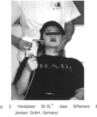
The laser apparatus is a handylaser 50-SL
Ⓡclass
B which is fitted with a 820nm, 50mW, GaAlAs
diode(Fig. 2). The power output can be regulated
Fig. 1. Electronic algometer type I(Somedic Produc- tion, Stockholm, Sweden)
Fig. 2. Handylaser 50-SLⓇ class B(Reimers &
Janssen GmbH, Germany)
with ease and precision using the handylaser; In 40 seconds, 2 Joules are emitted with continuous beam and when the frequency modulation is in operation, 1 Joule is emitted. The laser is activated for 40 seconds and is automatically turned off. The handylaser 50-SL is also available without timer.
The red pilot diode at the head of the laser indicates the direction of the visible laser beam. The GaAlAs diode laser is ideally suited for this double blind
Fig. 3. The electronic algometer was held perpendi- cularly to the superficial masseter.
study since the laser light is invisible and emits no heat, sound or other physically detectable indication when it is activated. Therefore, it is possible to randomly receive placebo treatment and actual laser treatment.
3. PROCEDURE
Before the physical examination, all of the subjects were instructed to push the button on the patient-operated switch as soon as they experienced pain. As the subject feels pain, he or she pushes the button on the patient-operated switch, the digital display stops immediately for about five seconds, and the red light turns on so that the operator can record the value easily.
During this test, he or she who made the measurements could not see the values of the measurement.
All tests were performed with the subjects in a reclined position with the neck supported. During the measurement on the masticatory muscles, the investigator applied manual counter-pressure contralaterally to stabilize the head(Fig. 3).
For reliability, the measurements were taken
three times at each marked trigger point and the
mean value of the three measurements was
accepted. Before treatment, on the 2nd week and
after treatment, the PPT was taken on both the
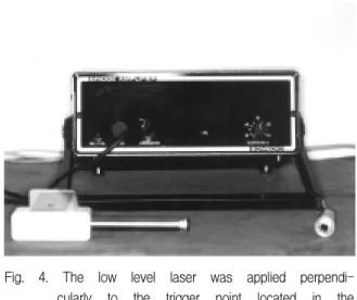
Fig. 4. The low level laser was applied perpendi- cularly to the trigger point located in the superficial masseter.
LLLT and the sham LLLT group, both two patient and two control groups.
All of the subjects received either a LLLT or a sham LLLT(Fig. 4). The laser was set to deliver a pulse energy at 1J per square centimeter of tissue for 40 seconds. Each marked trigger point was either irradiated with 2J per square centimeter of repetitively pulsed GaAlAs laser or received placebo application for 80 seconds. The total energy emitted at each session was 2J and each subject was treated with 5 sessions. The probe was in contact with the skin at a right angle. To preserve the double-blind study, the subjects were positioned sitting or semi-supine so as to obscure viewing of the laser beam. The same unit was used for the placebo treatment, for which no laser beam was emitted. Each subject was treated twice at the first weeks and once a week during the following three weeks. A total treatment was 5 times during 4weeks.
4. STATISTICAL ANALYSIS
All measurements in each group were averaged.
Statistical analysis was performed to evaluate the increase of the PPT values. Differences in the PPT between the LLLT and sham LLLT group, patient and control groups, inter-patient and inter-control
groups before treatment were analyzed with the unpaired t-test. The repeated-measure ANOVA was performed to analyze the treatment and time effect for the PPT changes within each muscle.
The statistical comparison of P-values of the LLLT and sham LLLT group, inter-patient and inter-control groups was carried out with the paired t-test separately at each session. P-values of less than 0.05 were interpreted as significant, and the level in confidence intervals was 95%. All statistical analyses were used with Statview
TMII software.
Ⅲ. RESULTS
The results are presented in table 1∼13. The mean and standard deviations of the PPT values in both the LLLT and sham LLLT group before treatment are shown in table 1. There were no statistically significant differences between the LLLT and sham LLLT group. In general, the PPT values of the sham LLLT group, except for the masseter, were higher than those of the LLLT group before treatment(Table 1).
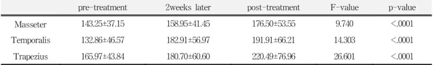
The comparison of the PPT values measured from individual muscles and the significant differences between the LLLT and sham LLLT group are shown as seen in Table 2 and 3. Results of the ANOVA indicated a significant increase of the PPT values in the LLLT group(P<0.001), but not in the sham LLLT group.
Comparing the p-value of the PPT values in the LLLT group with that in the sham LLLT group, it was found that on the third irradiation a statistically significant increase was more prominent in the
Table 1. Pressure pain threshold of the LLLT group and sham LLLT group before treatment.
LLLT group sham LLLT group p-value Masseter 143.25±37.15 142.04±21.64 .8771 Temporalis 132.86±46.57 137.69±33.12 .7269 Trapezius 165.97±43.84 178.07±48.12 .1333
Table 5. Pressure pain threshold of the patient group and control group before treatment.
patient group control group p-value Masseter 150.03±30.03 216.89±38.92 <.0001 Temporalis 181.45±38.77 253.97±52.90 <.0001 Trapezius 159.48±27.53 232.49±41.70 <.0001
Table 6. Pressure pain threshold of the inter-patient groups before treatment.
LLLT group sham LLLT group p-value Masseter 156.29±31.87 141.09±25.08 .4744 Temporalis 178.75±34.02 186.22±46.50 .0390 Trapezius 160.50±30.57 158.13±23.43 .7526 Table 2. Pressure pain threshold of the LLLT group in each muscle with time.
pre-treatment 2weeks later post-treatment F-value p-value
Masseter 143.25±37.15 158.95±41.45 176.50±53.55 9.740 <.0001
Temporalis 132.86±46.57 182.91±56.97 191.91±66.21 14.303 <.0001
Trapezius 165.97±43.84 180.70±60.60 220.49±76.96 26.601 <.0001
Table 3. Pressure pain threshold of the sham LLLT group in each muscle with time.
pre-treatment 2weeks later post-treatment F-value p-value
Masseter 142.04±21.64 130.68±20.72 128.79±34.81 1.829 .1485
Temporalis 137.69±33.12 150.25±22.75 153.75±31.53 2.631 .0615
Trapezius 178.07±48.12 168.61±49.64 175.07±62.77 2.362 .0729
Table 4. Comparison of the p-value of the LLLT group and sham LLLT group in each muscle with time.
site LLLT group
sham LLLT group pre-treatment 2weeks later post-treatment
Masseter
pre-treatment .0221 .0001
2weeks later .0188 .0062
post-treatment .0767 .7798
Temporalis
pre-treatment .0002 .0002
2weeks later .0831 .2938
post-treatment .1467 .7240
Trapezius
pre-treatment .0247 <.0001
2weeks later .0573 <.0001
post-treatment .6363 .2647
LLLT group(P<0.05). This difference increased gradually with following treatment session (P<0.001), but no significant increase was found within the sham LLLT group(Table 4).
The mean and standard deviations of the PPT values in both patient and control groups before treatment are shown in table 5. The PPT values of all patient groups were significantly lower than those of all control groups(p<0.0001).
Table 7. Pressure pain threshold of the inter-control group before treatment.
LLLT group sham LLLT group p-value Masseter 218.96±29.11 215.38±45.07 .7048 Temporalis 256.71±50.36 251.98±55.21 .7166 Trapezius 229.15±34.52 234.75±46.18 .5803
Table 8. Pressure pain threshold of the LLLT-patient group in each muscle with time.
pre-treatment 2weeks later post-treatment F-value p-value
Masseter 156.29±31.87 174.79±57.75 175.28±30.01 4.726 .0116
Temporalis 178.75±34.02 206.83±40.07 210.64±29.43 21.432 <.0001
Trapezius 160.50±30.57 167.84±39.20 179.42±33.33 5.101 .0089
Table 9. Pressure pain threshold of the sham LLLT-patient group in each muscle with time.
pre-treatment 2weeks later post-treatment F-value p-value
Masseter 141.09±25.08 145.76±26.66 147.08±23.56 1.190 .3120
Temporalis 186.22±46.50 196.44±37.20 193.71±39.43 1.315 .2792
Trapezius 158.13±23.43 152.23±25.50 159.65±25.86 1.200 .3103
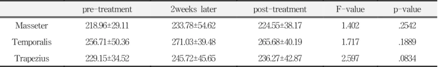
Table 10. Pressure pain threshold of the LLLT-control group in each muscle with time.
pre-treatment 2weeks later post-treatment F-value p-value
Masseter 218.96±29.11 233.78±54.62 224.55±38.17 1.402 .2542
Temporalis 256.71±50.36 271.03±39.48 265.68±40.19 1.717 .1889
Trapezius 229.15±34.52 245.72±45.65 236.27±42.87 2.597 .0834
Table 6 and 7 show the comparison of the PPT values measured from the inter-patient and inter-control groups before treatment. There were no statistically significant differences in the inter-patient groups, except for the temporalis, and in the inter-control groups in each muscle.
The change of the PPT values measured from each muscle and the significant differences both the inter-patient and inter-control groups with treatment sessions are shown as seen in Table 8∼
11. Table 8 clearly shows the positive effect of
laser therapy on increase of the PPT values found
in the LLLT-patient group and all muscles showed
a significant difference(P<0.05). The PPT values in
the sham LLLT-patient group were increased but
no statistical change was found between them for
each session(Table 9). Though all of the subjects
received each actual laser and sham laser
irradiation, we found little change of the PPT
values in both control groups (Table 10 and 11).
Comparing the p-value of the PPT values in the LLLT-patient and the LLLT-control group with
that in the sham LLLT-patient group and the sham LLLT-control group, it was found that on the third irradiation a statistically significant increase was
Table 11. Pressure pain threshold of the sham LLLT-control group in each muscle with time.pre-treatment 2weeks later post-treatment F-value p-value
Masseter 215.38±45.07 222.95±45.25 224.86±33.96 1.117 .3324
Temporalis 251.98±55.21 259.39±48.42 268.64±34.54 2.024 .1390
Trapezius 234.75±46.18 239.37±46.00 244.19±29.25 .908 .4075
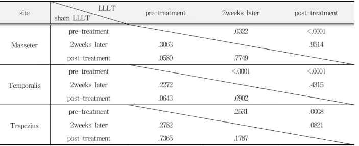
Table 12. Comparison of the p-value of the inter-patient group in each muscle with time.
site LLLT
sham LLLT pre-treatment 2weeks later post-treatment
Masseter
pre-treatment .0322 <.0001
2weeks later .3063 .9514
post-treatment .0580 .7749
Temporalis
pre-treatment <.0001 <.0001
2weeks later .2272 .4315
post-treatment .0643 .6902
Trapezius
pre-treatment .2531 .0008
2weeks later .2782 .0821
post-treatment .7365 .1787
Table 13. Comparison of the p-value of the inter-control group in each muscle with time.
site LLLT
sham LLLT pre-treatment 2weeks later post-treatment
Masseter
pre-treatment .1433 .3238
2weeks later .3201 .3895
post-treatment .1882 .7226
Temporalis
pre-treatment .0216 .3857
2weeks later .4599 .4306
post-treatment .0577 .1277
Trapezius
pre-treatment .0315 .3444
2weeks later .5599 .1976
post-treatment .1759 .4418
more prominent in the LLLT-patient than in the LLLT-control group(P<0.05). In the LLLT-patient group, this difference increased from on the third to on the fifth irradiation(P<0.001). However, no significant increase was found within the sham LLLT-patient group and both control groups(Table 12 and 13).
Ⅳ. DISCUSSION
The exact nature of a trigger point is not known.
It has been suggested that certain nerve endings in the muscle tissues may become sensitized by algogenic substances that create a localized zone of hypersensitivity
24-26). There may be a local tempe- rature rise at the site of the trigger point, suggesting an increase in metabolic demand or reduction of blood flow to these tissues
1,27). The importance of pain and dysfunction originating from myofascial trigger points is gaining increased recognition by clinicians. Since there are no laboratory or radiographic changes associated with myofascial pain and trigger points sensitivity, diagnosis and treatment evaluation depends on an accurate hands on examination of the muscle to locate focal tenderness in palpable muscle bands.
Precise information on predetermined trigger point locations and examination techniques can be obtained from Travell and Simmons
1). Once located, quantification of the tenderness of a trigger point is impressive with manual palpation alone. Both in clinical practice and experimentally it would be of great value to have a reliable, yet simple method to quantify trigger point sensitivities once they have been manually located. The pressure algometer may be suited for this purpose
23).
The first generation of pressure algometers in the 1930s to 1960s, were rather than crude instruments working on a spring load principle
18). More recent models working on mechanical force gauges include those of Fischer
22), and Tunks et al.
28)Electronic pressure algometers working in strain gauge principle have also been developed
16). Many researcher reported about the validity and reliability
of PPT measurement using algometer
18,20-23). Chung et al.
20)reported that there was high intraexaminer and interexaminer reliability in a study on PPT measurement of head and neck muscles of 40 normal subjects, and concluded that the algometer can be very useful to investigate the head and neck muscles tenderness for clinical practice and research. Fischer
22)suggested that PPT measurement was excellent in reproducibility and validity. Reeves et al.
23)demonstrated a high degree of validity and reliability of the instrument in the detection of myofascial trigger points in temporomandibular muscles.
The reliability of PPT measurements can be affected by several factors. These factors include the size of contact area and the rate of application.
The PPT increases as the area of contact decreases and increases with the increasing rate of application
16,29). The intermuscle regional differences in both temporalis and masseter muscles have been reported
21). Therefore, this study was designed to obtain the reliability and reproducibility of the algometer by using specific marked trigger point detected from Travell and Simmons' technique.
In this study, the application rate was 30Kpa/sec
recommended by manufacturer. List et al.
29)and
Doland and Keefe
30)emphasized that a constant
pressure rate is necessary to obtain a good
reliability with the algometer. To apply the
pressure with a uniform rate, a visual signal was
given to the investigator. The rate of application,
30Kpa/sec used in this study, was chosen to avoid
prolonged pressure application, which may in itself
affect the PPT by traumatizing the tissues and may
certainly make it difficult for the investigator to
maintain a constant rate of application throughout
the measurement due to fatigue in his arm. On the
other hand, the pressure application rate should be
slow enough to allow the subject to signal at the
PPT pressure to reduce overestimation of the PPT
because of the reaction time. In addition, at very
fast application rates, it is difficult to ensure a
constant rate of pressure application by visual
feedback from the digital display panel.
The use of laser on abnormal pain producing tissue can cause an almost immediate relief of spasm. This may be related to depolarization and repolarization of abnormally contracted muscle fibers, relief of arteriolar muscle spasms in the affected areas with reactive vasodilation, or electron excitation in the mitochondrial membranes with changes of transport and metabolic processes
31,32). Moreno
33)postulated that low intensity laser can stimulate the energy processes via ATP formation and activation of enzyme activity leading to restoration of normal properties on the cell- organ-organism levels.
The low level laser is widely recommended for routine clinical use, especially in the treatment of ulcerative or inflammatory disease, functional disorders, and chronic pain conditions. The recommendations are primarily based on positive clinical experience rather than classical placebo controlled clinical trials. Only a limited number of controlled studies have been performed. Of these, few indicate positive results in pain treatment
6,9,10,34), whereas several recent studies fail to show any difference between laser and placebo treatment
7,8,11,12). Bezuur et al.
6)found that the infrared laser irradiated the TMJ area was effective and significantly increased the maximum mouth opening in the arthrogenous patients but no significant increase of the maximum mouth opening was found in the myogenous patients. Olavi et al.
9)suggested that low power 904nm IR laser treatment may have a beneficial effect on the pain threshold when it is low, as is usually the case in the trigger points during myofascial pain syndromes. On the other hand, the same laser therapy had no effect on the pain threshold in normal subjects
35,36)and on normal pain threshold
37). A double-blind controlled study by Snyder-Mackler et al.
10)found that there were both a statistically significant reduction of pain and increased skin resistance at the trigger points in the group that was irradiated with a 0.95mW He-Ne CW laser. Walker
34)reported that laser irradiation may have an effect on serotonin metabolism, thereby serving as a mechanism of
pain relief.
A double-blind controlled trial by Klein and Eek
8)showed that there were significant improvement in objective parameters in both the laser and placebo group, but no relative advantage accrued to either group. Under the short-term conditions, low- energy laser stimulation plus exercise did not provide a significant advantage over exercise alone.
The report of Waylonis et al.
11)suggested that no difference in pain response and treatment effectiveness was noted in the treated and placebo groups that were irradiated with low output He-Ne laser. A double blind, cross-over study by Thorsen et al.
12)reported that a 830nm GaAlAs diode laser had no beneficial effects between LLLT and placebo for myofascial pain.
In this double blind study, one of the popular 820nm GaAlAs diode laser was used at the sensitive trigger points. The therapeutic effect in both the LLLT and sham LLLT group and the comparison between laser treatment and placebo in both patient and control groups were assessed by pressure pain threshold measurements. In the first study, A significant increase of the PPT values was found in the LLLT group from the third day of laser exposure (P<0.05), increasing even more until the last laser session(P<0.001). In the sham LLLT group, there was in fact no significant increase of the PPT values. By contraries, the PPT values measured from masseter and trapezius were decreased with treatment. In the temporalis, the increase of the PPT values was slightly observed on the third sham irradiation although it might be a temporary phenomena without any statistical significance. Our findings are in accordance with the results of several studies
3,6,9,10,34).
In the second study, we found that although the PPT values of all groups were slightly increased with treatment, a statistically significant increase was more prominent in the LLLT-patient group than in the sham LLLT-patient group and both control groups. Moreover, this difference was increasingly larger during treatment sessions.
Therefore, this result was identical to the result of
the first study. In the sham LLLT-patient group, there was increase of the PPT values in the contrast to the first study, but the difference of PPT values between pre-treatment and post- treatment was no more than 7Kpa. The PPT values in both control groups were significantly higher than those in both patient groups. In the LLLT-control group, it was found that a significant difference was observed on the third irradiation, except for the masseter, but the PPT values on the fifth irradiation were decreased. There were no statistical differences within each session but the PPT values were increased gradually with treatment in the sham LLLT-control group. Hansen and Thoroe
7)have suggested that no statistically significant difference between the analgesic effect of the laser and placebo irradiation was found and placebo was superior to laser stimulation. Also, the placebo response is supposed to be determined by several independent factors as the setting of the study, the doctor-patient relationship and the belief and anticipation of the patients. In this study, the PPT values in all four groups were increased with treatment sessions. Therefore, this results indicated that a slight placebo response was found in the sham LLLT-patient group and both control groups.
In the LLLT-control group, Our results are similar to the results of the previous mentioned studies
35-37). The increase of the PPT values in placebo treatment groups with mock irradiation is explained by the belief that the sham laser is actually activated on laser probe contact surface and by the anticipation that there is a therapeutic effect during sham laser irradiation. However, the increase rate in the sham LLLT-patient group was lower than that in the sham LLLT-control group.
From that result, it is believed that abnormal skin overlaying a trigger point is more sensitive to the pressure than normal one. Hence, In this study, our results showed that actual laser-induced effect is superior to placebo response.
We conclude that the low level laser has an effective modality of treatment for the trigger points and the efficacy of laser is superior to
placebo response. A minimum of the three times treatments has been suggested for assessing the efficacy of laser treatment, and a 5-session course has been recommended.
There is a need for further study to establish the effect of various frequency, energy dosage and irradiation schedules in reaction to pain conditions of any kind. Also, the long-term laser effects need to be evaluated.
Ⅴ. CONCLUSION
To evaluate the effect of LLLT, 69 dental students at Pusan National University, were studied by pressure algometer after laser application. They were randomly assigned to either a LLLT group or a sham LLLT group. To clarify the reliability of LLLT, 39 subjects, 19 patients and 20 controls subjects, were randomly classified into four various groups; 10 in the LLLT-patient group, 9 in the sham LLLT-patient group, 10 in the LLLT-control group and 10 in the sham LLLT-control group.
Measurement of pressure pain threshold over trigger points irradiated with GaAlAs diode laser displayed a significant increase in the pain threshold after treatment.
The obtained results are as follows:
1. A significant increase of the PPT values was found from the third day of laser exposure in the LLLT group(P<0.05), increasing even more until the last laser session(P<0.001). In the sham LLLT group, there was no significant increase of the PPT values.
2. The significant increase of the PPT values was higher in the LLLT-patient group than in the sham LLLT-patient group(P<0.05). In the LLLT and sham LLLT-control group, there was no statistical difference after laser irradiation.
3. Even if a slight placebo response was found in
the sham LLLT-patient group and both control
groups, actual laser-induced effect in the LLLT-
patient group was superior to placebo response.
REFERENCE
1. Travell J.G., Simmons D.G. : Myofascial pain and dysfunction, the trigger point manual. Baltimore, Williams & Wilkins Co., 13-37, 1983.
2. Okeson J.P. : Orofacial pain : Guidelines for assess- ment, diagnosis and management. Qunitessence Co., 139, 1996.
3. Bradley P.F. : Thermal surgery in the management of maxillofacial malignancy, In; Malignant tumors of the maxillofacial region. Oral & Maxillofacial Surgery Clinics of North America, 1993.
4. Kert J. and Rose M. ; Clinical laser therapy-Low level laser therapy. 2nd ed., Copenhagen, Scandinavian Medical Laser Technology, 1989.
5. Mester E., Mester A.F. and Mester A. : The biomedical effect of laser application lasers. Surg.
Med., 5:31-39, 1988.
6. Bezzur N.J., Habets L.M.H., and Hansson T.L. : The effect of therapeutic laser treatment in patients with craniomandibular disorders. J. Craniomandib. Disord., 2:83-86, 1988.
7. Hansen H.J., and Thoroe U. : Low power laser biosti- mulation of chronic orofacial pain, A double-blind placebo controlled cross-over study in 40 patients.
Pain, 43:169-179, 1990.
8. Klein, R.G., Eek, B.C. : Low-energy laser treatment and exercise for chronic low back pain : double-blind controlled trial. Arch. Phys. Med. Rehabil., 71:34-37, 1990.
9. Olavie A., Pekka R., Pertti K. : Effects of the infrared laser therapy at treated and non-treated trigger points. Acupuncture electro-therapeutics Res. Int. J., 14:9-14, 1989.
10. Snyder-Mackler L., Barry A.J., Perkins A.I., Soucek M.D. : Effects of Helium-Neon laser irradiation on skin resistance and pain in patients with trigger points in the neck or back. Physical therapy, 69:336-341, 1989.
11. Waylonis G.W., Wilke S., O'Toole D., Waylonis D.B.
: Chronic myofascial pain : management by low output helium-neon laser therapy. Arch. Phys. Med.
Rehabil., 69:1017-1020, 1988.
12. Thorsen H. et al : Low level laser therapy for myofascial pain in the neck and shoulder girdle, A double blind, cross-over study. Scan. J. Rheumatol., 21:139-142, 1992.
13. Thorsen, H., Gam, A.N., Lonnberg, F. : The effect of low-level laser therapy on musculoskeletal pain : a
meta analysis. Pain, 52:63-66, 1993.
14. Berkerman, H., de Bie, R.A., Bouter, L.M., De Cuyper H.J., Oostendorp, R.AB. : The efficacy of laser therapy for musculoskeletal and skin disorders : A criteria-based meta-analysis of randomized clinical trials. Phys. Ther., 72:483-491, 1992.
15. Helkimo M.: Studies on function and dysfunction of the masticatory system, II, index for anamnestic and clinical dysfunction and occlusal state. Swed. Dent. J., 67:101-121, 1974.
16. Jensen K., Andersen H.O., Olesen J., Lindblom, U. : Pressure pain threshold in human temporal region, evaluation of the new pressure algometer. Pain, 25:313-323, 1986.
17. McNeil C(ed). : Temporomandibular disorders;
Guidelines for classification, assessment and management. Chicago, Quintessence, 65, 1993.
18. Jensen K. : Quantification of tenderness by palpation and use of pressure algometers. In: Fricton J.R, Awad E(eds). : Advances in Pain Research and Therapy, New York : Raven, 165-181, 1990.
19. Ohrbach R., Gale E.N. : Pressure pain thresholds clinical assessment and differential diagnosis : Reliability and validity in patients with myogenic pain. Pain, 39:157-169, 1989.
20. Chung S.C., UM B.Y., Kim H.S. : Evaluation of pressure pain threshold in head and neck muscles by electronic algometer : intrarater and interrater reliability. J. Craniomand. Pract, 10:28-34, 1992.
21. Ohrbach R., Gale E.N. : Pressure pain thresholds in normal muscles : Reliability, measurement effects and topographic differences. Pain, 37:257-263, 1989.
22. Fischer A.A. : Pressure algometry over normal muscles, standard values, validity and reproducibility of pressure threshold. Pain, 30;115-126, 1987.
23. Reeves J. L., Jaeger B., Graff-Radford S. B. : Reliability of the pressure algometer as a measure of myofascial trigger point sensitivity. Pain, 24:313-321, 1986.
24. Mense S., Meyer H. : Bradykinin-induced sensitiza- tion of high-threshold muscle receptors with slowly conducting afferent fibers. Pain(supp 1), S204, 1981.
25. Simons D.G., Travell J. : Myofascial trigger points, a possible explanation. Pain, 10(1):106-109, 1981.
26. McMillan A.S., Blasberg B. : Pain-pressure threshold in painful jaw muscles following trigger point injection. J. Orofac. Pain, 8(4):384-390, 1994.
27. Travell J. : Introductory comments. In Ragan C, editor : Connective tissues: Transactions of the fifth
conference. New York, 12-22, 1954.
28. Tunks E., Crook J., Norman G., Kalaher S. : Tender points in fibromyalgia. Pain, 34:11-19, 1988.
29. List T., Helkimo M, : Method for measuring muscle pain with the aid of algometer, [abstract No. 69].
Swed. Dent. J., 11:294, 1987.
30. Doland E.A., Keefe F.J.: Muscle activity in myofascial pain dysfunction syndrome patients, a structured clinical evaluation. J. Craniomandib. Disord., 2:101-105, 1988.
31. Kryspin, J. : Laser stimulation in chronic pain.
Manuscript submitted for publication, 1981.
32. Brucini, M., Duranti, R., Galletti, R., Pantaleo, T., Zucchi, P.L. : Pain thresholds and electromyographic features of periarticular muscles in patients with osteoarthritis of the knee. Pain, 10:57-66, 1981.
33. Moreno, C. : Ultrastructure of mitochrions after laser microirradiation. Journal de Microscopic, 16(3):269- 279, 1973.
34. Walker J.B, : Relief from chronic pain by low power laser irradiation. Neurosci. Lett., 43:339-344, 1983.
35. Fischer, A.A. : Diagnosis and management of chronic pain in physical medicine and rehabilitation. In Ruskin, A.P.(ed) : Current therapy in psychiatry.
W.B.Saunders, Philadelphia, 1984.
36. Fischer, A.A. : Pressure threshold meter: Its use for quantification of tender spots. Arch. Phys. Med.
Rehabil., 67:838, 1986.
37. Kolari, P.J., Hietanen, M., Van Nondelstrand, P., Airaksinen, O., Pontinen, P. : Characteristics of mid lasers for physical therapy. Scand. J. Acup.
Electrother. In press. 1988.
국문초록
발통점에 대한 저출력 레이저의 치료효과