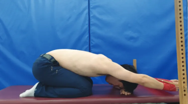
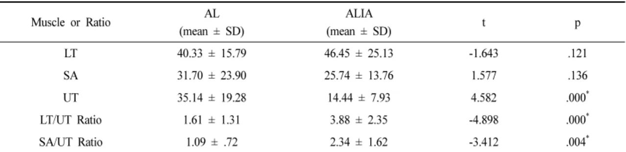
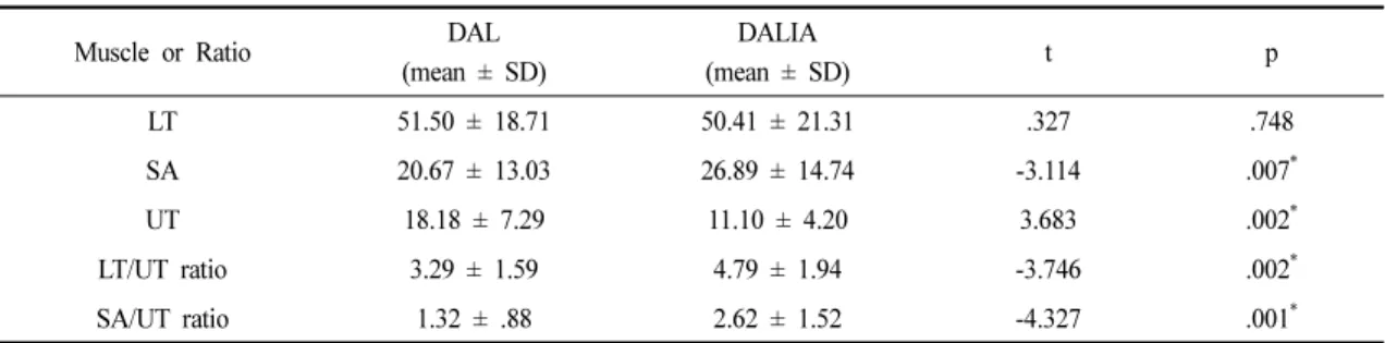
Effects of Reciprocal Inhibition Using Thera-band on Scapular Muscle Activities During Arm-lifting Exercises in Subjects with Rounded Shoulder Posture
10
0
0
전체 글
(3)
(4)
(5)
(6)
(7)
(8)
(9)
(10)
수치
관련 문서
This study investigated the effects of university Taekwondo coaches' types of leadership on players' anxiety about intense competition. The study subjects were
In an effort to develop a new method to remove nitrogen, this study examined the effects of C/N ratio, carbon source and nitrogen concentration on
This study was to do a comparative analysis on kinematic differences and differences in muscle activity between the skilled and the unskilled in windsurfing
This study attempts to quantify the economic effects of increases in international oil prices on Korea’s energy and biodiesel industry by using a small open computable
In future studies focused on antioxidant effect and active oxygen reduction in resistance exercise using thera-band will be conducted in-depth research that
Effects of Group Art Therapy by Using Hanji on Spontaneity of the Osteoporosis Eldery
In conclusion, the result that the walk-using safety educational activities designed for young children had effects on their safety problem solving ability
Conclusion: In conclusion, the relaxant effects of neuromuscular blockers on the uterine smooth muscle may be transmitted via nicotinic acetylcholine receptors
