134
Clinical Application of Amniotic Membrane and Freeze-dried Amniotic Membrane in Management of Partial-thickness Burn Wounds
Junhyung Kim, M.D., Ph.D., Hyunseok Jin, M.D., Hyun Ji Kim, M.D.
Dae Gu Son, M.D., Ph.D. and Kihwan Han, M.D., Ph.D.
Department of Plastic and Reconstructive Surgery, Keimyung University Dongsan Medical Center, Daegu, Korea
Dressing materials for wound management of the patients with partial thickness burn must be able to shorten the healing time with epithelialization, reduce the pain for management, and minimize scar formation after the healing process. For this purpose, the authors conducted a comparative study of the application of a preveously used, fresh amniotic membrane and a freeze-dried amniotic membrane for the management of partial thickness burn wounds in our hospital.
We treated four patients with a biologic dressing for partial thickness burn wound on the face, using the fresh amniotic membrane on one side of the face and the freeze-dried amniotic membrane on the other side. Fresh amniotic membrane underwent sterilization after delivery. Freeze-dried amniotic membrane (Bioland Ltd., Cheonan Chungnam, Korea) was prepared from fresh amniotic membrane with an additional process consisting of refrigeration at -70
oC, drying for more than 48 hours, and sterilized from radiation. All of the patients were injured in a flame burn. There age range was 28∼53 years, with an average age of 38.7 years. The patients were compared for a mean follow-up of five months with infection, time of peeling off, flushing on the face, and formation of the hypertrophic scar evaluated with chromometry.
After the management with amniotic membrane, all patients were treated without infection or hypertrophic scar formation. After an average of 7 days, the amniotic membrane was peeled off from the margin, and after an average of 12 days we could inspect complete peeling off. From the comparison of the two membranes, no clear difference was evident and the healing time was the same. On chromometry, the healed skin color was brighter and reddish.
Amniotic membrane was demonstrated to be an effective dressing material. However Its use is subject to restriction due to the awkwardness of donor security, the difficulty of custody, and the risk
부분층 화상 치료에서 생양막과 동결건조양막의 임상적 비교적용
계명대학교 의과대학 성형외과학교실
김준형․진현석․김현지․손대구․한기환
ꠏꠏꠏꠏꠏꠏꠏꠏꠏꠏꠏꠏꠏꠏꠏꠏꠏꠏꠏꠏꠏꠏꠏꠏꠏꠏꠏꠏꠏꠏꠏꠏꠏꠏꠏꠏꠏꠏꠏꠏꠏꠏꠏꠏꠏ
책임저자:김준형, 대구광역시 중구 동산동 194번지, 우편번호: 700-712, 계명대학교 의과대학 성형외과학교실 Tel: 053-250-7635, Fax: 053-255-0632, E-mail: [email protected]
서 론
부분층 화상은 진피의 유두층(papillary layer)까지 만 화상을 입은 것으로서 표피 재생이 가능하여 식 피술을 하지 않고도 2∼3주 내에 최소한의 반흔만을 남기고 치유된다. 그러나 부분층 화상도 적절한 치 료를 받지 못하거나, 감염이 동반되면 전층 화상으 로 쉽게 변화하기 때문에 이러한 부분층 화상의 국 소적 치료는 매우 중요하다. 기술의 발전과 함께 삼 출물의 흡수가 뛰어나고 치료가 간편한 새롭고, 다 양한 드레싱재료들이 많이 이용되고 있지만, 교환 시 출혈과 통증이 있고 고가인 문제점이 있다.
생물성 드레싱의 일종인 양막은 1910년 Davis가 피 부를 대체하기 위하여 처음 사용한 후 여러 방법과 목 적으로 이용되어 왔으나,
1)공여자 확보의 어려움, 보관 의 불편함, 수직감염의 위험 등 여러 단점들이 있어 최 근에는 그 사용이 많이 감소하였다. 저자들은 이런 단 점들을 보완하고 쉽게 이용할 수 있게 무균적인 조작 후 동결건조시켜 상품화한 양막(Bioland Ltd., Cheonan Chungnam, Korea)을 부분층 화상 환자의 안면부에서 생양막과 함께 적용하여 차이점을 알아보았다.
대상 및 방법
2002년 3월부터 9월까지 본원에 내원한 안면부 부분층 화상 환자 4예를 대상으로 하였다. 화상의 깊 이는 임상적으로 판단하였고, 모든 환자는 화상 후 24시간 안에 내원하여 입원치료 하였다. 모두 남성 이었고 화염화상이었으며 28∼53세(평균 38.7세)였 고 추적관찰기간은 13∼19개월(평균 15개월)이었다.
과거병력이나 가족력에서 특이 사항은 없었으며, 기
저질환도 없었다.
치료용 생양막은 간염, 매독, 결핵, 후천성면역결 핍증 등 전염병의 병력이나 혈액학적 검사 상 이상 소견이 없고 조기파수나 자궁내막증의 병력이 없는 건강한 산모로부터 얻어 수술실에서 분리하였다. 태 반과 융모막으로 부터 양막을 박리하여 5회에 걸쳐 0.9% 생리식염수에 씻어 묻어 있는 혈액이나 태변을 완전히 제거하고, 0.025% 염화차아연소산염(sodium hypochlorite) 용액에 담아 1회 소독한 뒤, 5회에 걸쳐 생리식염수에 다시 헹구었다. 세척과 소독을 한 양 막을 넓게 펴고 거즈에 싼 뒤 소독된 용기에 넣고 냉 장고에서 4
oC 온도로 보관하였고 제조한 뒤 5일 이 내에 사용하였다. 동결건조양막은 생양막과 유사한 조건의 양막을 세척과 소독과정을 거친 후 냉동실에 서 -80°C로 24시간 이상 동결 후 진공상태의 동결 건조기(freezing dry chamber)에 넣어 48시간 이상 건 조시켰다. 동결건조된 양막은 진공상태에서 개별적 으로 포장 후 방사선을 조사하여 멸균시켰으며 냉장 실에서 4°C 온도로 보관하였다.
양막을 도포할 때에 필요에 따라 환자의 통증을 감소하기 위하여 일반적으로 진통제 meperidine (De- merol
Ⓡ) 50∼100 mg을 사용하였다. 화상부위를 1%
zephiran 용액으로 소독한 다음 0.9% 생리식염수로 소독액이 남지 않도록 깨끗하게 씻은 후 양막을 도 포하였다. 안면부에서 반쪽은 생양막으로 다른 쪽은 동결건조양막을 이용하였다. 준비된 양막을 구별하 여 안면부의 화상부위가 충분히 덮이도록 잘 고정하 였으며 기포나 삼출액이 없도록 밀착시켰다. 도포한 양막위에 마른 거즈와 솜붕대를 이용하여 가볍게 압 박하였으며 24시간이 지난 후에 고정한 양막이 떨어 지지 않도록 조심스럽게 거즈와 솜붕대를 제거하였 고, 기포나 삼출액이 생긴 경우에는 작은 절개를 통 of direct infection. Nevertheless, from this study, we confirmed the absence of the any difference in terms of effective management between fresh amniotic membrane and freeze-dried amniotic membrane.
ꠏꠏꠏꠏꠏꠏꠏꠏꠏꠏꠏꠏꠏꠏꠏꠏꠏꠏꠏꠏꠏꠏꠏꠏꠏꠏꠏꠏꠏꠏꠏꠏꠏꠏꠏꠏꠏꠏꠏꠏꠏꠏꠏꠏꠏꠏꠏꠏꠏꠏꠏꠏꠏꠏꠏꠏꠏꠏꠏꠏꠏꠏꠏꠏꠏꠏꠏꠏꠏꠏꠏꠏꠏꠏꠏꠏꠏꠏꠏꠏꠏꠏ
Key Words: Fresh amniotic membrane, Freeze-dried amniotic membrane, No difference
하여 배출한 후 건조시켰다.
임상적으로 서로 다른 두 양막이 건조되는 정도 및 환자가 통증을 느끼는 정도와 삼출액이 고이는 정도, 완전히 탈락되는 시기를 관찰하였고, 완치 후 양막을 도포하였던 부위와 화상을 입지 않았던 정상피부에서 비색계(chromometry)를 이용하여 안면홍조의 지속과 과색소침착과 같은 피부색 변화를 비교하여 보았다.
결 과
4예 모두 감염이나 다른 합병증 없이 단 1회의 양 막을 이용한 생물성 드레싱으로 10일에서 14일에 화 상부위가 상피로 덮이면서 반흔 없이 치유되었다.
양막드레싱을 후 5일째 주변부에서부터 탈락이 되었 고 평균 12일째 완전히 탈락하였다. 1예에서 도포한 양막 밑에 부분적으로 삼출액이 고였으나 5 mm 정 도의 절개를 가하여 배액 후 압박하였다.
양막드레싱 후 24시간이 지나 거즈를 제거하였을 때 동결건조양막을 도포한 부위가 더 빨리 건조되는 것이 관찰되었으며 그 뒤에는 양쪽 반안면 모두 같 은 양상을 보였고 육안적으로 뚜렷한 차이점을 찾을 수 없었다. 양막의 탈락되기 시작한 시기와, 환자의 통증, 삼출액이 고이는 정도, 완전히 탈락된 날짜 등 양쪽 양막 모두 동일하였다.
장기추적관찰에서 안면부 비후성 반흔이나 홍조 는 관찰되지 않았다. 화상을 입은 후 평균 15개월에 비색계(Minolta chromameter CR-300, Minolta co., Osaka, Japan)를 이용하여, 양막을 도포 후 치유된 양 쪽 안면부와 화상을 입지 않은 안면부 세 부분의 피 부색을 측정하여 비교해 보았다. 비색계로 측정한 값은 L, a, b값으로, L값은 피부의 밝기(brightness) 정 도를 나타내며, 그 범위는 0 (흑색)에서 부터 100 (백 색)까지이다. a값은 피부의 적색도(redness)를 의미하 며, 적색(+)에서 부터 녹색(-)까지의 분포로 나타낸 다. b값은 피부의 황색도(yellowness)로 황색(+)에서 부터 청색(-)까지의 분포를 보인다. L값의 측정결과 화상을 입지 않은 안면부의 평균값은 47.33이었고 생양막을 부착한 부위는 51.71, 동결건조양막을 부착 한 부위는 49.62로 나타나 화상으로 치유된 부위의
피부색이 더 밝게 나타났다. a값은 화상을 입지 않은 안면부의 평균값이 15.83이었고, 생양막을 부착한 부 위는 16.43, 동결건조양막을 부착한 부위는 16.83으 로 화상으로 치유된 부위의 피부색이 더 적색에 가 깝게 나타났다. b값은 화상을 입지 않은 안면부의 평 균값이 19.04이었고, 생양막을 부착한 부위는 15.92, 동결건조양막을 부착한 부위는 17.18로 나타나 화상 을 입지 않은 안면부가 더 황색에 가깝게 나타났다.
그러나 화상을 입지 않은 안면부와 화상으로 치유된 부위가 육안적으로 뚜렷하게 구별되지는 않았다.
1. 증례 1
28세 남자로 안면부와 우측 상완부에 전체 체표면 적의 6%의 화염화상을 입고 응급실로 내원하였다.
내원당일 안면부는 0.9% 생리식염수와 1% zephiran 으로 세척과 소독을 한 뒤 습식드레싱(wet dressing) 을 유지하였고, 우측 상완부는 세척과 소독을 하고 기존의 1% silver sulfadiazine (silvadene
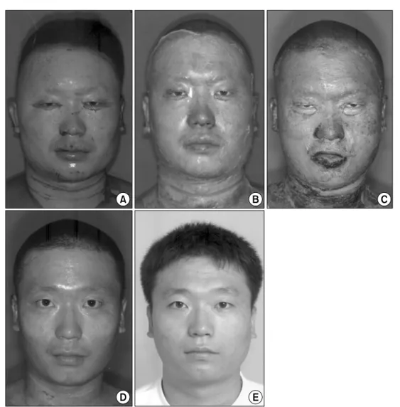
®) 크림으로 드레싱하였다. 내원 다음날 전체 체표면적의 4%의 부분층 화상을 입은 안면부에 생양막과 동결건조양 막을 도포하였고, 24시간 후에 거즈를 조심스럽게 제거하고 잘 건조시켰다. 6일 만에 주변부부터 탈락 하기 시작하였고 13일째 완전히 탈락되었다. 양쪽 반안면부에서 뚜렷한 차이점을 찾을 수 없었으며 세 균의 감염이나 동통 없이 완쾌되었다. 우측 상완부 는 내원 5일째 분층식피술을 하였고 다른 합병증 없 이 잘 치유되었다(Fig. 1).
2. 증례 2
37세 남자로 전안면부와 경부에 전체 체표면적의 5%의 부분층 화염화상을 입고 당일 내원하였다.
0.9% 생리식염수와 1% zephiran으로 세척과 소독을 한 뒤 습식드레싱으로 하루를 유지한 후 생양막과 동결건조양막을 역시 같은 방법으로 도포하였다.
24시간이 지난 뒤 거즈를 제거하였을 때 양쪽 협부
에 부분적으로 삼출액이 고였으나 최소한의 절개를
통하여 배출하였으며 감염이나 다른 합병증 없이
14일째 완전히 탈락되었다. 양쪽 반안면부에서 탈락
되기 시작한 날짜, 완전히 탈락된 날짜 등 뚜렷한 차
이점이 나타나지 않았다(Fig. 2).
고 찰
양막을 이용한 생물성 드레싱은 조기에 화상부위 를 덮어줌으로써 항균작용과 함께 체액누출을 방지 하고 상피화(epithelialization)의 촉진 및 조기재활치
료가 가능한 장점이 있다. 그러나 간염, 매독 등과 같은 전염병의 전파가능성이 있으며, 공여자 확보가 어렵고, 제조과정에서 시간과 채취기술의 습득이 필 요하며, 장기간 보관이 어려운점 등의 단점이 있다.
특히 화상 환자가 내원하였을 때마다 기존의 드레싱 재료들처럼 쉽게 사용할 수 없어 그 사용이 많이 제 한된 것이 사실이다.
Fig. 1. A 28-year-old male patient with superficial second degree burn on the face and deep second degree burn on the right forearm, 6% of total body surface area, (A) Immediate appearance of the face with burn wound. At the day of burns, after cleansing and sterilizing with normal saline and zephiran, wet dressing was performed for 24 hours, (B) At postburn 1 day, fresh amniotic membrane on right hemiface and freeze-dried amniotic membrane on left side was applied each other, (C) Postburn 7 days, some of the dried amniotic membrane were peeled off after the healing of the wound, (D) Post burn 13 days, all of the dried amniotic membrane were peeled off, (E) Appearance of the face, one year after burn following application of the amniotic membrane.
생양막의 이런 단점을 보완한 동결건조양막은, 먼 저 공여자의 철저한 병력검사와 혈청학적 검사 후, 분만이 이루어지고 있는 수술실에서 무균적 조작을 통하여 양막을 분리해서, 4°C의 보관용기에 담아 24 시간 내에 세척과 소독을 한다. 항생제가 첨가된 생 리식염수에 24시간 처리하여 양막조직으로부터 기 질(stroma)층을 제거한 뒤 멸균된 거즈에 양막을 접
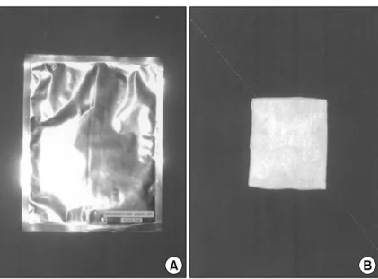
히지 않게 올려놓고 -80°C의 냉동실에서 동결시킨 다. 진공상태의 동결건조기 내에서 48시간 건조시킨 다음 수분흡수를 최소화하기 위하여 진공상태를 유 지하면서 알루미늄 재질의 포장지로 이중 포장한다.
개별 포장된 양막은 25 KGy로 약 1주일 동안 방사
선을 조사한 뒤 냉장보관한다. 이상 제조된 동결건
조양막은 진공의 상태에서 철저하게 포장되어 있으
Fig. 2. A 37-year-old male patient with superficial second degree burn on the face and neck, 5% of total body surface area. (A) Immediate appearance of the face with burn wound. (B) At postburn 1 day, fresh amniotic membrane on right hemiface and freeze- dried amniotic membrane on left side was applied. (C) Postburn 5 days, some of the dried amniotic membrane were peeled off partially. (D) The amniotic membrane was completely peeled off from the burn areas, after 14 days of application. (E) Fifteen months follow up, note a absence of the hypertrophic scars and discoloration on the face.므로 평균 6개월 이상의 4°C 냉장보관으로 내용물의 변화가 없어 예기할 수 없는 화상환자의 내원에도 상기의 단점들을 보완할 수 있다(Fig. 3). Thomson 등 은 -80°C의 온도로 양막을 동결시킨 후에 300일 이
상 보관이 가능하며 양막의 효과에 변화가 없다고 하였다.
9)환자에게 적용 시 생양막은 크기와 모양이 일정하 지 않아 도포할 양을 미리 예측할 수 없고, 수축력이
Fig. 3. (A) A freeze-dried amniotic membrane (Bioland Ltd., Chonan Chungnam, Korea) is well packed with a aluminium packing material after the radiation. (B) When the package is unpacked, it is taken a unfold membrane with a layer of gauze.Fig. 4. (A) The freeze-dried amniotic membrane was applied on the partial skin defect area after the split thickness skin graft on the back. (B) It was also applied on the split thickness skin defect as the donor site for the split thickness skin graft on the upper arm.
커서 서로 겹쳐지는 현상이 자주 나타났으며 밀착시 키기 위해 힘을 주어 펴게 되므로 찢어지는 경우가 자주 발생하였다. 그러나 동결건조양막은 크기가 다 양하게 제품화되어 있고 수축력이 적어 도포 시 겹 쳐지지 않아 얼굴에 쉽게 잘 밀착되어 도포하는 시 간 및 환자의 고통도 줄일 수 있었다. 또 식피술 후 부분적으로 피부결손이 생긴 경우와 분층식피술의 공여부에도 효과적으로 사용할 수 있었다(Fig. 4).
10)양막드레싱을 한 후 1일째 생양막보다 동결건조양 막을 도포한 부위가 더 빨리 건조되는 것을 관찰할 수 있었는데 이는 저자들이 생양막을 제조하는 과정 에서 기질층을 완전히 제거하지 않아 부분적으로 두 껍게 만들어 졌고 이에 비해 동결건조양막은 더 얇 게 제작되어 빨리 건조된 것으로 생각된다.
양막드레싱 후 피부색의 변화를 알아보기 위해 비 색계를 이용해 피부색을 측정했을 때 화상을 입지 않은 안면부가 대구광역시민의 안면부의 피부색과 비교하였을 때, L값이 더 낮고, a값이 더 높아 비교 적 어둡고 붉은 피부색으로 나타났는데 이는 4예 모 두 옥외에서 일하는 직업을 가진 남자여서 평소에 태양광선에 자주 노출되어 왔음을 추측할 수 있었 다.
11)화상을 입은 뒤 치유된 부위가 화상을 입지 않 은 부위보다 L값이 더 높고, a값이 더 높아 비교적 밝고 붉은 피부색으로 나타났는데 이는 아직도 육안 으로 뚜렷하지 않은 홍반이 남아 있고, 표피에 드러 나는 혈관화의 정도가 더 발달되어 있음을 알 수 있 었다. 또한 화상을 입지 않은 부위에서 b값이 더 높 게 나타나 좀더 황색에 더 가까웠는데 이는 화상을 입은 뒤 치유된 부위에서 아직 표피층과 진피층에 있는 멜라닌의 양이 화상을 입지 않은 원래의 피부 색깔의 양에 도달하지 않았음을 간접적으로 알 수 있었다.
12)그러나 증례의 수가 적고 추적기간이 평균 15개월로 다소 단기간이어서 추후 피부색의 변화에 대한 장기적인 추적관찰이 필요할 것으로 생각된다.
결 론
양막은 드레싱 재료로써 유용함이 밝혀졌지만 공 여자의 확보, 보관의 어려움, 수직감염의 위험 등으
로 사용에 제한이 있었다. 그러나 이 연구를 통 하여 동결건조양막이 생양막과 비교하였을 때 치 료효과의 차이가 없고 보다 쉽게 환자에게 적용 할 수 있음을 알 수 있었다. 그 뿐 아니라 자가식 피술 후 부분적으로 피부결손이 생긴 부분이나, 분층식피술의 공여부 치료에도 유용하게 사용될 수 있음을 알 수 있었다.
참 고 문 헌
1) Davis JS: Skin transplantation with review of 550 cases at the Johns Hopkins Hospital. Johns Hopkins Hosp Rep 15: 307, 1910
2) Sabella N: Use of fetal membranes in skin grafting. Med Rec NY 83: 478, 1913
3) Brown JB, McDowell F: Massive repairs of burns with thick split-skin grafts. Ann Surg 115: 658, 1942 4) Brown JB, Fryer MP, Randall P, Lu M: Postmortem
homografts as “biological dressings” for extensive burns and denuded areas. Ann Surg 138: 618, 1953 5) Ramakrishnan KM, Jayaraman V: Management of par-
tial-thickness burn wounds by amniotic membrane: a cost-effective treatment in developing countries. Burns 23: 33, 1997
6) William CQ, Herbert CH, Michael S: Clinical trials of amniotic membranes in burn wound care. Plast Reconstr Surg 70: 711, 1982
7) Robson MC, Krizek TJ, Koss N, Smburg JL: Human amniotic membrane as a temporary wound dressing.
Surg Gynecol Obstet 136: 904, 1973
8) Robson MC, Krizek TJ: The effect of human amniotic membranes on the bacteria population of infected rat burns. Ann Surg 177: 144, 1972
9) Thomson PD, Parks DH: Monitoring, banking, and clinical use of amnion as a burn wound dressing. Ann Plast Surg 7: 354, 1981
10) Saranatra W, Kanyika C, Siripong S, Yongyuth V: Ap- plication of freeze-dried amniotic membrane: a control trial at the donor site of split-thickness skin grafing.
Bulletin Hosp Joint disease Ortho Instit 50: 27 1990 11) Choi TH, Han KH: Chromometric analysis of the skin
colors of Daegu citizen. Keimyung Med J. In press 12) Lim JJ, Son DG, Choi DW, Han KH: Measurement of
the skin color changes of free flaps by chromometry. J Korean Soc Plast Reconstr Surg 28: 255, 2001