여성에서 골 성장과 보존
인제대학교 의과대학 상계백병원 산부인과학교실 최 훈
Development and Conservation of the Female Skeleton
Hoon Choi
Department of Obstetrics & Gynecology, Inje University Sanggyepaik Hospital, Seoul, Korea
Estrogen is the key hormone for maintaining bone mass and estrogen deficiency is the major cause of age related bone loss in female. Estrogen together with growth hormone and IGF-1 initiate a 3- to 4- pubertal growth spurt that doubles skeletal mass and is also required for the attainment of maximal peak bone mass.
Aging women undergo two phases of bone loss. The menopause initiates an accelerated phase of predominantly cancellous bone loss that declines rapidly over 4~8 yr to become asymptotic with a subsequent slow phase that continues indefinitely. The accelerated phase results from the loss of the direct restraining effects of estrogen on bone turnover, an action mediated by estrogen receptors in both osteoblasts and osteoclasts. In ensuing slow phase, the rate of cancellous bone loss is reduced, but the rate of cortical bone loss is unchanged or increased. This phase is mediated largely by secondary hyperparathyroidism that result from the loss of estrogen actions on extraskeletal calcium metabolism. Impaired osteoblast function due to estrogen deficiency, aging, or both also contributes to the slow phase of bone loss.
Key Words: Female skeleton, Bone development, Bone loss
여성에서 골 성장
1) 성 스테로이드는 골격의 성숙과 성적 차이에 관여 한다. 골격 크기와 용적골밀도는 사춘기 전 소년과 소녀에서 비슷하다. 사춘기시작과 청장년 사이에 근 육량은 배로 증가한다1. 신장의 성장 속도와 골 재형 성 율은 사춘기 초기에 가장 빠르다1-5. 이에 반해 용 적 골밀도의 최대 증가는 2년 후 (소년은 초경시기;
접수일: 2007년 12월 20일, 승인일: 2008년 4월 7일 책임저자: 최 훈, 인제대학교 상계백병원 산부인과
Tel: 02)950-1990/1991, Fax: 02)938-4109/4108 E-mail: [email protected]
소년은 사춘기 후반)에 일어난다. 소년의 성장 양상 은 소녀와 2가지 면에서 다르다. 소년은 사춘기가 늦기 때문에 사춘기 전 성장기간이 2년 이상 더 길 다. 사춘기 성장가속은 소녀의 3년에 비해 4년 동안 지속된다. 이러한 차이는 남성에서 여성보다 신장이 10% 더 크고 최대 골량이 25% 더 많다는 것을 의미 한다. 대부분의 경우 남성의 골량이 많은 것은 뼈 크 기가 크기 때문이다.
골 성장은 형성에 의해 뼈의 크기와 형태가 증가 하는 데 기인한다. 직선적인 골 성장은 연골내질 (endochondral) 성장판의 골화 (ossification)에 의한다.
방사 골 (radial bone) 성장은 골막의 두께 (periosteal
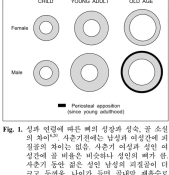
CHILD YOUNG ADULT OLD AGE
Female
Male
Periosteal apposition (since young adulthood)
Fig. 1. 성과 연령에 따른 뼈의 성장과 성숙, 골 소실 의 차이6,20. 사춘기전에는 남성과 여성간에 피 질골의 차이는 없음. 사춘기 여성과 성인 여 성간에 골 비율은 비슷하나 성인의 뼈가 큼.
사춘기 동안 젊은 성인 남성의 피질골이 더 크고 두꺼움. 나이가 들면 골내막 재흡수로 인하여 피질골의 과도한 손실이 남성과 여성 에서 발생하나 여성은 폐경으로 인하여 남성 보다 피질골 손실이 더 많음. 남성은 여성보 다 골막이 3배 두꺼워지므로 연령에 의한 골 내막 재흡수에 의한 골 소실을 상쇄할 수 있 음. 피질골질의 공증 (porosity)은 남성과 여성 에서 모두 발생하나 여성에서 폐경으로 인하 여 더 많이 생김.
apposition)에 의한다. 골수강 크기는 골내막 재흡수 로 증가한다. 사춘기 전 성장은 다리의 성장이 비례 적으로 커며 사춘기 성장은 몸통의 성장이 비례적으 로 크다6. 사춘기 성장 가속기 동안 골내막의 골흡수 보다 골막이 두꺼워지는데 이로써 사지의 크기 및 용적 골밀도 (골 부피에 포함된 골량)가 증가한다7. 사춘기는 성장·판이 닫힘으로써 끝나게 되며 이 시 기에 용적 골밀도는 최대 골량의 90~95%에 이르게 된다. 이러한 과정을 강화 (consolidation)라고 하며 골 격은 지속적인 골막 병치와 소주의 조밀화로 인하여 최고치에 달하게 된다. 또한 강화 과정 동안 골밀도 의 증가는 피질골내 공간의 감소와 연관이 있다. 강 화란 과정이 얼마나 오래 지속되는가에 관해서는 논 란이 있다. 일부는2 20대 후반까지만 지속된다고 주 장하는 반면에 일부는 척추 골밀도는 30대 후반까지 지속될 것이라고 주장하고 있다8.
사춘기 전에는 성장호르몬 (GH)과 Insulin like growth factor (IGF-1)축의 기본적인 수치가 뼈성장을 천천히 지속적으로 유지한다. 시상하부에서 성선자극호르몬 분비호르몬 (GnRH)의 파동형 분비의 증가로 혈중내 성선자극호르몬 수치가 증가하고 따라서 성선에서 성호르몬의 분비가 증가되면 사춘기는 촉발된다9. 혈 청 에스트로겐의 증가는 성장호르몬의 파동형 분비 를 1.5~3.0배 증가시킨다10,11. 성장호르몬, IGF-1과 에스트로겐의 증가는 성장가속기가 진행되는데 도 움이 된다. 성장호르몬과 IGF-1는 성장가속기 3~4 년 동안 최고치를 유지한 후 수년에 걸쳐 서서히 저 하하면서 사춘기 전 수준으로 돌아가지만 혈중 성 스 테로이드 수치는 성인 수준으로 유지된다10.
혈중 에스트로겐 증가가 사춘기 성장가속에 중요 하다. 에스트로겐 수용체 α 유전자 또는 방향화 효 소 유전자 변이가 동종접합체인 남성에서는 혈청 테 스토스테론 수치가 정상 또는 증가되어 있어도 성장 가속이 진행되지 않는다11-15. 혈청 에스트로겐 수치 가 지속적으로 증가하면 남성과 여성 모두에서 성장 판이 닫히게 되지만 에스트로겐 수용체 α 유전자12 또는 방향화 효소 유전자 변이13-15가 동종접합체인 남성에서는 골단 (epiphyses)이 열려있다. 안드로겐 수용체 변이에 의한 고환 여성화 남성은 성장판이 닫힌다16,17.
성스테로이드는 혈중내 성장호르몬과 IGF-1 수치 의 영향과 관계없이 골성숙 동안 골량을 증가시킨 다. 사춘기가 지나면 소년이 소녀보다 골량이 25%
더 많은데 이는 사춘기 동안 혈청 테스토스테론의 증가에 기인한다. 성장호르몬 분비와 IGF-1 생산의 증가는 소년보다 소녀에서 크거나 비슷하다. 에스트 로겐은 용적 골밀도와 연관이 있다. 방향화 효소의 유전적 문제로 인하여 에스트로겐을 생성할 수 없는 청장년의 골밀도는 각 뼈 부위에서 예측치보다 25~
40% 감소한다15.
골의 각 부분인 골막, 골 내막, 피질골질 (intracor- tical)은 골 성장과 성숙 동안 그리고 골 소실 동안 변할 수 있다7. 각 부분의 변화는 골밀도를 측정하는 이중 에너지 X-ray 흡수 계측법 (dual-energy X-ray absorptiometry: DXA)으로 측정할 수 없다. DXA는 면적 골밀도 (g/cm2)의 정보를 제공해 주는데 깊이를
Decr. sensing of strain by mechanostat
Incr. bone
tumover Rapid transient bone loss (mainly cancellous bone)
Resetting of mechanostat
Remodeling
imbalance Decr. PTH
secretion Decr. 1.25(OH)2D production
Decr. Ca absorption Decr. renal Ca
conservation Rapid bone loss ceases
(after 4~8 yrs) E-deficiency
(rapid onset)
A. MECHANISM OF EARLY, ACCELERATED BONE LOSS IN WOMEN
B. MECHANISM OF LATE, SLOW BONE LOSS IN WOMEN E-deficiency
(rapid onset)
Decr. Ca absorption Decr. TRCa
Decr. OB function
External Ca wasting
Incr. dietary Ca requirement
2o hyper-
parathyroidism Slow bone loss (greater cortical than
cancellous losses) Parathyroid
gland semi- autonomy
Fig. 2. 폐경 여성에서 골 소실의 2가지 유형24.
제대로 측정할 수 없기 때문에 뼈가 클 경우 용적 골밀도 (g/cm3)를 과도하게 측정하게 된다. 따라서 같 은 골량에서는 뼈가 클수록 더 강하다는 생체 역학적 자료에도 불구하고 통상적인 검사방법으로는 골 크 기는 무시된다18,19. 나이와 성에 따른 뼈의 크기와 형 태의 차이는 Fig. 1과 같다.
여성에서 골소실
여성에서 성 스테로이드가 감소하기 시작하는 폐 경전 5년 동안 골 소실이 있는데 대퇴골 근위부부터 시작된다21. 여성에서 다른 부위에 실질적으로 폐경 전 골소실이 있는지 또는 젊은 성인 남자에서 골소 실이 일어나는지에 대해서는 논란이 많다. 일부 연 구에 의하면 여성에서 폐경전 골소실이 척추와8,22 대퇴부에서23 일어난다고 하였다. 이러한 결과가 옳 다면 여성에서 재형성 불균형은 골의 성장과 성숙이 중단 후에 시작되며 폐경에 의해 증폭된다고 여길 수도 있다.
여성에서 골소실은 두 가지 양상을 거치게 된다 (Fig. 2)24. 초기의 일시적인 가속기로 폐경에서 시작 되는 것과 느리게 지속되는 시기로 구분된다. 초기
의 골 소실은 4~8년에 걸쳐 기하급수적으로 진행되 다가 느리게 꾸준히 진행된다. 초기에 해면골의 20~
30%가 골 소실이 되지만 피질골은 5~10%에서 골 소실이 발생된다. 자연폐경은 나이가 다르므로 폐경 에 따른 골변화는 난소절제술후에 분명하다. 난소절 제술을 받은 중년여성을 대상으로 2년 동안 시행한 종단연구에 의하면25 해면골은 18%의 골소실이 있 었으나 피질골에서는 4%만 골소실이 있었다. 이에 반하여 자연폐경 여성을 대상으로 한 cohort 연구들 에 의하면 폐경전 3년 이내에 요추와 대퇴골 근위부 에서 4%와26 1~2%의27 골소실이 각각 있었다고 하 였다. 한 연구에서는26 폐경 중단후 3년까지 골소실 이 약 7% 있은 후 느리게 진행하였으며 다른 연구에 서는27 골소실이 14% 있었으며 8~10년까지 진행되 었다고 하였다. 난소절제술을 받은 폐경 여성에서25 자연폐경 여성보다26,27 골소실이 높은 율로 진행된 이유는 난소절제술후 혈중 에스트로겐 농도가 급작 스럽게 감소하였으며 혈중 테스토스테론 농도도 감 소하였으므로 골소실이 신속하게 진행되었다. 또한 난소절제술후 폐경 여성에서25 quantitative CT를 이 용하여 골소실을 측정하였으므로 이중 에너지 X- ray 흡수 계측법으로 측정한 경우보다 해면골의 골
소실이 분명하게 나타나기 때문이다.
골소실의 초기 가속기는 폐경에서 시작되는데 이 는 에스트로겐 투여로 예방될 수 있으므로25,28 난소 기능의 손실에 의해 초래되는 것이 분명하다. 폐경 이행기의 2~4년 동안 혈청 에스트론 농도는 폐경 전보다 25~35% 감소하지만 혈청 에스트라디올 농 도는 폐경 전보다 10~15% 감소한다29. 폐경후 난소 에서 테스토스테론의 생산 감소로 인하여 혈청 테스 토스테론 농도도 감소하지만30 부신피질과 난소 간 질에서 테스토스테론이 생산되므로 그 정도는 심하 지 않다. 생화학 표지자의 측정에 의하면 폐경시기 에 골 흡수는 90% 정도 증가한 반면에 골 형성은 45%만 증가한다고 한다31. 골교체율의 증가와 골재 형성의 불균형은 골소실을 가속화하는 데 특히 골 내피에서 그러하다. 폐경 동안 골소실은 신속하게 일어나지만 골밀도측정기에 의한 골밀도 감소의 일 부는 BMU 수의 증가로 발생한 재형성 공간의 증가 와 연관이 있다32. 이 시기 동안 신속한 골소실은 골 에서 칼슘을 세포 밖으로 유출시키는데 이에 따른 고 칼슘혈증은 요 칼슘 배설의 증가33, 장에서 칼슘 흡수 감소34, 부갑상선호르몬의 부분적 분비 억제로 예방된다35. 폐경 초기에 골소실이 가속되는 것은 급 작스러운 에스트로겐 결핍에 따른 골세포의 화학적 감각 저하에 의해 초래된다는 설이 있으나 아직 확 실하지 않다.
과거에는 노령 여성에서 골소실이 느리게 지속되 는 시기는 연령에 의한 골외 칼슘대사의 장애로 기인 한 것이며 에스트로겐 결핍은 거의 영향이 없다고 하 였으나 약 10년 전부터 실제로 에스트로겐 결핍이 골 외 칼슘대사 이상과 2차적 고 부갑상선호르몬증의 주 요원인이라는 가설이 제기되었다36. 이 가설은 일부
연구에29,37 의해 입증되었는데 노령의 폐경 여성이 장
기간 에스트로겐 치료를 받는 경우 혈청 부갑상선호 르몬 농도와 골 교체 표지자의 농도가 폐경전 여성의 수준으로 감소되지만 치료를 받지 않은 여성에서는 이들 농도가 증가된 상태로 남아 있다고 하였다.
에스트로겐 결핍이 골소실에서 두 가지 양상을 보 이는 것은 에스트로겐이 골에 대한 작용에 두 가지 형태가 있다고 생각할 수 있다. 즉 에스트로겐이 골 세포에 직접 작용하는 것과 골외 칼슘대사에 대한
영향으로 부갑상선호르몬 분비의 변화에 의한 것이 다. 에스트로겐은 장에서 칼슘의 흡수를 증가시키며
35,38, 신장에서 칼슘의 배설을 억제한다39,40. 이러한
장과 신장에 대한 에스트로겐의 직접적인 작용이 없 다면 칼슘은 지속으로 소모되게 된다. 이러한 손실 은 다량의 칼슘 복용으로 보상되지 않으면 2차적 고 부갑상선호르몬증을 유발하게 되고 골소실이 느리 게 지속적으로 진행된다.
골소실의 초기 가속기의 특징은 제한적이며 해면 골 소실이 불균형적인데 반해 느리게 지속되는 시기 의 특징은 2차적 고 부갑상선호르몬증이 지속되는 한 골소실이 끝없이 지속되며 해면골과 피질골이 비 슷하거나 피질골에서 더 심해지기도 한다41-43. 폐경 여성에서 골 소실의 주된 원인은 골 흡수의 증가이지만 골형성의 감소도 원인으로 관여한다. 폐 경 여성에서 골소실의 두 시기에서 골흡수가 골형성 보다 과다한데 이는 보상장애를 의미한다29,31. 이러 한 이상은 나이에 연관된 요인인 성장인자의 생산 감소44, 혈액내 성장호르몬10,45과 IGF-1 수치 감소46-48 등에 의한다고 하였다.
에스트로겐은 실험에 의하면 조골세포에서 IGF-1, TGF-beta, procollagen 합성을 증가시키며49 조골세포 괴사를 방해하면서50,51 조골세포의 수명을 증가시킨 다고 한다. 한 연구에 의하면52 고용량의 에스트로겐 을 노년 폐경 여성 (평균나이, 65세)에게 피하이식 시킨 후 6년 후에 골 생검을 하여 이식전과 비교한 바 해면골양이 22% 증가하였고 소주골 벽 두께가 12% 증가하였다고 하였는데 이는 고용량의 에스트 로겐이 골 형성을 촉진한다는 것을 시사한다.
그러나 아직 에스트로겐이 조골세포 기능을 촉진 하는지에 대한 일치된 견해는 없으며 촉진한다면 증 식을 촉진하는지 또는 괴사를 억제하는지에 대해서 도 잘 밝혀져 있지 않다.
참 고 문 헌
1. Katzman DK, Bachrach LK, Carter DR, Marcus R.
Clinical and anthropometric correlates of bone mineral acquisition in healthy adolescent girl. J Clin Endocrinol Metab 1991;73:1332-9.
2. Theintz G, Buchs B, Rizzoli R, Slosman D, Clavien H, Sizonenko PC, et al. Longitudinal monitoring of bone mass accumulation in healthy adolescencts: evidence for a marked reduction after 16 years of age at the levels of lumbar spine and femoral neck in female subjects. J Clin Endocrinol Metab 1992;75:1060-5.
3. Lu P, Cowell CT, Lloyd-Jones SA, Briody JN, Howman-Giles R. Volumetric bone mineral density in normal subjects, aged 3-27 years. J Clin Endo- crinol Metab 1996;81:1586-90.
4. Gilsanz V, Roe TF, Mora S, Costin G, Goodman WG. Changes in vertebral bone density in black girls and white girls during childhood and puberty.
N Engl J Med 1991;352:1597-600.
5. Cadogan J, Blumsolin A, Barker ME, Eastell R. A longitudinal study of bone gain in pubertal girls, anthropometric and biochemical correlates. J Bone Miner Res 1998;13:1602-12.
6. Bass S, Delmas PD, Pearce G, Hendrich E, Tabensky A, Seeman E. The differing tempo of growth in bone size, mass, and density in girls is region specific. J Clin Invest 1999;104:795-804.
7. Seeman E. From density to structure: growing up and growing old on the surfaces of bone. J Bone Miner Res 1997;12:509-21.
8. Matkovic V, Jelic T, Wardlaw GM, Ilch JZ, Goel PK, Wright JK, et al. Timing of peak bone mass in Caucasian females and its implication for the pre- vention of osteoporosis. J Clin Invest 1994;93:799- 808.
9. Terasawa T, Fermandez DI. Neurobiological mecha- nisms of the onset of puberty in primates. Endocr Rev 2001;22:111-51.
10. Giustina A, Veldhuis JD. Pathophysiology of the neyroregulation of growth hormone secretion in experimental animals and the human. Endocr Rev 1998;19:717-97.
11. Grumbach MM. Estrogen, bone, growth and sex. a sea change in conventional wisdom. J Pediatr
Endocrinol Metab 2000;13:1439-55.
12. Smith EP, Boyd J, Frank GR, Takahashi H, Cohen RM, Specker B, et al. Estrogen resistance caused by a mutation in the estrogen-receptor gene in a man. N Engl J Med 1994;331:1056-61.
13. Morishima A, Grumbach MM, Simpson ER, Fisher C, Qin K. Aromatase deficiency in male and female siblings caused by a novel mutation and the physiological role of estrogens. J Clin Endocrinol Metab 1995;80:3689-98.
14. Carani C, Qin K, Simoni M, Faustini-Fustini M, Serpente S, Boyd J, et al. Effect of testosterone and estradiol in a man with aromatase deficiency.
N Engl J Med 1997;337:91-5.
15. Bilezikian JP, Morishima A, Bell J, Grumbach MM. Increased bone mass as a result of estrogen therapy in a man with aromatase deficiency. N Engl J Med 1998;339:599-603.
16. Zachman M, Prader A, Sobel EH, Crigler Jr JF, Ritzen EM, Atares M, et al. Pubertal growth in patients with androgen insensitivity: indirect evi- dence for the importance of estrogen in pubertal growth in girls. J Pediatr 1986;108:694-7.
17. Marcus R, Leary D, Schneider DT, Shane E, Favus M, Quigley CA. The contribution of testosterone to skeletal development and maintenance: lessons from the androgen insensitivity syndrome. J Clin Endocrinol Metab 2000;85:1032-7.
18. Beck TJ, Looker AC, Ruft CB, Sievanen H, Wahner HW. Structural trends in the aging femoral neck and proximal shaft: analysis of the third national health and nutrition examination survey dual-energy x-ray absorptiometry data. J Bone Miner Res 2000;15:2297-304.
19. Bouxsein MT. Application of biomechanics to the aging human skeleton. In: Glowacki J, Rosen CJ, Bilazikian JP, eds. The aging skeleton. San Diego:
Academic Press; 1999;chap 27:315-30.
20. Garn SM. The course of bone gain and the phases of bone loss. Orthop Clin North Am 1972;3:503-20.
21. Siemenda C, Hui SI, Longcope C, Johnston CC.
Sex steroids and bone mass. a study of changes about the time of menopause. J Clin Invest 1987;
80:1261-9.
22. Riggs RL, Wahner HW, Melton III LJ, Richelson LS, Judd HL, Offord KP. Rates of bone loss in the appendicular and axial skeletons of women: evi- dence of substantial vertebral bone loss before menopause. J Clin Invest 1986;77:1487-91.
23. Melton III LJ, Atkinson EJ, CyConner MK, O'Fallon WM, Riggs BI. Determinants of bone loss from the femoral neck in women of different ages.
J Bone Miner Res 2000;15:24-31.
24. Riggs BL, Khosla S, Melton LJ. Sex steroids and the construction and conservation of the adult skeleton. Endocr Rev 2002;23:279-302.
25. Genant HK, Cann CE, Ettinger B, Gordan GS.
Quantitative computed tomography of vertebral spongiosa: a sensitive method for detecting early bone loss after oophorectomy. Ann Intern Med 1982;97:699-705.
26. Recker R, Lappe J, Davis K, Heaney R. Charac- terization of perimenopausal bone loss: a prospec- tive study. J Bone Miner Res 2000;15;1965-73.
27. Guthrie JR, Ebeling PR, Hopper JL, Barrett-Connor E, Dennerstein L, Dudley EC, et al. A prospective study of bone loss in menopausal Australian-born women. Osteoporos Int 1998;8:282-90.
28. Linsay R, Aitken JM, Anderson JB, Hart DM, MacDonald EB, Clarks AC. Long term prevention of postmenopausal osteoporosis by oestrogen: evi- dence for an increased bone mass after delayed onset of oestrogen treatment. Lancet 1979;1:1038-41.
29. Khosia SK, Atkinson EJ, Melton III LJ, Riggs BT.
Effects of age and estrogen status on serum para- thyroid hormone levels and biochemical markers of bone turnover in women: a population study. J Clin Endocrinol Metab 1997;82:1522-7.
30. Horton R, Roamnoff E, Walker J. Androstenen- dione and testosterone in ovarian venous and peri-
pheral plasma during ovariectomy for breast cancer.
J Clin Endocrinol Metab 1966;26:1267-9.
31. Gamero P, Sornay-Rendu E, Chapuy M, Delmas PD. Increased bone turnover in late postmenopausal women is a major determinant of osteoporosis. J Bone Miner Res 1996;11:337-49.
32. Heaney RP. The bone-remodeling transient: implica- tion for the interpretation of clinical studies of bone mass change. J Bone Miner Res 1994;9:1515-23.
33. Young MM, Nordin BEC. Effects of natural and artificial menopause on plasma and urinary calcium and phosphorus. Lancet 1967;2:118-20.
34. Gennari C, Agnusdei D, Nardi P, Civitelli R. Estro- gen preserves a normal intestinal responsiveness to 1,25-dihydroxyvitamin D3 in oophorectomized women.
J Clin Endocrinol Metab 1990;71:1288-93.
35. Cosman F, Shen V, Xie F, Seibel M, Ratcliffe A, Linsay R. Estrogen protection against bone resor- bing effects of parathyroid hormone infusion. Ann Intern Med 1993;118:337-43.
36. Riggs BL, Khosia S, Melton III LJ. A unitary model for involutional osteoporosis: estrogen defi- ciency causes both type I and type II osteoporosis in postmenopausal women and contributes to bone loss in aging men. J Bone Miner Res 1998;13:
763-73.
37. McKane RW, Khosla S, Risteli J, Robins SP, Muhs JM, Riggs BI. Role of estrogen deficiency in pathogenesis of secondary hyperparathyroidism and incrased bone resorption in elderly women. Proc Assoc Am Physicians 1997;109:174-80.
38. Gallagher JC, Riggs BL, DeLuca HF. Effect of estrogen on calcium absorption and serum vitamin D metabolites in postmenopausal osteoporosis. J Clin Endocrinol Metab 1980;51:1359-64.
39. Nordin BEC, Need AG, Morris HA, Horowitz M, Robertson WG. Evidence for a renal calcium leak in postmenopausal women. J Clin Endocrinol Metab 1991;72:401-7.
40. McKane WR, Khosia S, Burritt MF, Kan PC,
Wilson DM, Ory SJ, et al. Mechanisms of renal calcium conservation with estrogen replacement therapy in women in early postmenopause-a clinical research center study. J Clin Endocrinol Metab 1995;80:3458-64.
41. Parisien M, Melish RWE, Silverberg SJ, Shane E, Lindsay R, Bilezikian JP, et al. Maintenance of cancellous bone connectivity in primary hyperpara- thyroidism: trabecular strut analysis. J Bone Miner Res 1992;7:913-9.
42. Silverberg SJ, Gartenberg F, Jacobs TP, Shane E, Siris E, Starum RB, et al. Longitudinal measur- ments of bone density and biochemical indices in untreated primary hyperparathyroidism. J Clin Endocrinol Metab 1995;80:723-8.
43. Dempster DW, Cosman F, Parisien M, Shen V, Lindsay R. Anabolic actions of parathyroid hor- mone on bone. Endocr Rev 1993;14:690-709.
44. Marie PJ, Hott M, Launay JM, Graulet AM, Gueris J. In vitro production of cytokines by bone surface- derived osteoblastic cells in normal and osteoporotic postmenopausal women: relationship with cell pro- liferation. J Clin Endocrinol Metab 1993;77:824-30.
45. Ho KY, Evans WS, Blizzard RM, Veidhuid JD, Merriam GR, Samojlik E, et al. Effects of sex and age on the 24-hour profile of growth hormone secretion in man: importance of endogenous estra- diol concentrations. J Clin Endocrinol Metab 1997;
64:51-8.
46. Bennet A, Wahner HW, Riggs BL, Hintz RL.
Insulin-like growth factors I and II, aging and bone density in women. J Clin Endocrinol Metab 1984;
59:701-4.
47. Boonen S, Mohan S, Dequeker J, Aerssens J, Vanderschueren D, Verbeke G, et al. Dwon-regu- lation of the serum stimulatory components of the insulin-like growth factor (IGF) system (IGF-I, IGF- II, IGF binding protein[BP]-3, and IGFBP-5) in age- related (type II) femoral neck osteoporosis. J Bone Miner Res 1999;14:2150-8.
48. Pfeilschifter J, Diel I, Kloppinger T, Bismar H, Schuster EM, Balbach S, et al. Concentrations of insulin-like growth factor (IGF)-I, IGF-II, IGF bin- ding protein-4 and -5 in human bone cell condi- tioned media do not change with age. Mech Ageing Dev 2000;117:109-14.
49. Ernst M, Heath JK, Rodan GA. Estradiol effects on proliferation, messenger ribonucleic acid for colla- gen and insulin-like growth factor-I, and para- thyroid hormone-stimulated adenylate cyclase acti- vity in osteoblastic cells from calvariae and long bones. Endocrinology 1989;125:825-33.
50. Manolagas SC. Birth and death of bone cells: basic regulatory mechanisma and implications for the pathogenesis and treatment of osteoporosis. Endocr Rev 2000;21:115-37.
51. Gohel A, McCarthy MB, Gronowicz G. Estrogen prevents glucocorticoid-induced apoptosis in osteo- blasts in vivo and in vitro. Endocrinology 1999;
140:5339-7.
52. Khastgir G, Studd J, Holland N, Alaghband-Zadeh J, Fox S, Chow J. Anabolic effect of estrogen replacement on bone in postmenopausal women with osteoporosis histomorphometric evidence in a longitudinal study. J Clin Endocrinol Metab 2001;
86:289-95.