J. Exp. Biomed. Sci. 10 (2004) 467–472
Cerebral Blood Flow Velocity of Young People in Their Twenties
Seok-Cheol Choi
†, Heun-Young Kwon, Tae-Un Kim and Jai-Young Kim
Department of Clinical Laboratory Science, College of Health Sciences, Catholic University of Pusan, Busan 609-757, Korea
Transcranial Doppler sonography (TCD) is a useful diagnostic method to measure cerebral blood flow velocity in various cerebral disorders. However, we haven't data enough to be available for young persons, especially in the twenties in Korea. This study was performed to collect the basic data of the cerebral blood flow velocity and to understand the cerebral physiology in the twenties. We determined the mean velocities of middle, anterior, and posterior cerebral artery, and vertebral and basilar artery (MCA-V, ACA-V, PCA-V, VA-V, and BA-V, respectively) in eighty-two healthy volunteers. For evaluating cerebral autoregulation, only the MCA-V was measured under various conditions such as stable, apnea, and hyperventilation state. Right and left MCA-V were 80.66±14.03 and 83.22±14.40 cm/sec at stable state, 90.13±17.47 and 90.26±16.38 cm/sec at apnea, and 54.83±11.09 and 55.33±10.74 cm/sec at hyperventilation.
Right and left ACA-V were 49.11±15.71 and 48.19±13.75 cm/sec. Right and left PCA-V were 39.44±9.12 and 37.91
±6.74 cm/sec. Right and left VA-V were 33.65±9.26 and 36.18±10.39 cm/sec. BA-V was 48.49±11.16 cm/sec. Right and left MCA-V, VA-V, and right ACA-V and PCA-V in women were higher than those of men (P<0.05). No significant differences were found between men and women in the others. These findings indicate that cerebral hemodynamics and autoregulation were normal in young people in their twenties. The velocities of MCA, ACA, PCA, and BA were high values in women as compared with men.
Key Words: Transcranial Doppler (TCD), Young people, Cerebral blood flow velocity, Cerebral autoregulation
서 론
초음파를 이용한 말초혈관 내 혈류속도 측정은 1959년 Sa- tomura에 의해 최초로 시행된 이래 경동맥 혈류 평가검사에 널리 이용되어 뇌혈관 관련 질환의 진단에 유용하게 사용되 었다. 그러나 이 방법은 5~10 MHz의 탐촉자를 사용하기 때 문에 두개강 내 뇌혈관의 혈류속도 측정은 불가능하여 그 적 용범위에 한계가 있었다. 1982년 Aaslid (Aaslid et al., 1982)가 뼈와 연조직 투과시 감쇠가 적은 저주파수 (2 MHz)의 탐촉 자를 개발함으로써 두개골 중 가장 얇은 측두골 부분을 통해 Willis환 동맥들의 혈류속도 측정이 가능해졌다. 경두개 초음 파 검사 (Transcranial Doppler, TCD)라 불리우는 이 검사법은 측정의 용이함과 함께 비혈관적, 비침습적, 반복적인 실시간 값의 측정이라는 매우 큰 장점을 가지고 있어 현재 임상에서
널리 이용되고 있는 유용한 방법이다 (Lindegaard et al., 1985;
Seiler et al., 1986). 최근 식생활의 서구화와 다양한 사회적 요 인에 의해 한국인의 뇌혈관 질환 발생률이 급격히 증가함에 따라 TCD의 임상적 유용성은 더욱 강조되고 있다. 그러나 연구보고의 대부분은 임상적 사례에 관한 내용이기 때문에 실제 건강인에 대한 TCD자료가 상대적으로 축적되어있지 않고 특히 젊은 연령층에 대해 참고할 만한 국내자료가 절 대 부족한 실정이다. 저자들은 20대 연령층을 대상으로 TCD 를 이용한 뇌혈류 속도 측정을 통해 젊은이들의 뇌혈류역 학 및 생리에 대한 이해와 향후 관련 연구를 위한 기초 자료 로 활용하기 위해 이 연구를 시행하게 되었다.
대상 및 방법
1. 대 상
저자들은 부산시 소재 P대학교에 재학 중인 20대 연령층 (범위 20~24세, 평균 22.0±1.75세) 대학생들 중 뇌혈관질환, 심혈관질환, 고혈압, 당뇨병 등과 같은 질환의 병력이 없고 신경학적 이상소견이 없는 건강한 젊은이들을 대상으로 연 구하였다. 대상자들 중 특정 약물을 복용중인 사람들은 연구
*논 문 접 수: 2004년 9월 9일 수정재접수 : 2004년 10월 26일
†교신저자: 최석철, (우) 609-757, 부산광역시 금정구 부곡3동 9, 부산가톨릭대학교 보건과학대학 임상병리학과
Tel: 051-510-0564, Fax: 051-510-0568 e-mail: [email protected]
에서 제외시켰다. 각 측정값들은 전체 대상자들에 대한 평 균값을 구하여 분석하였고 남녀 성별로 분류하여 주요 측정 변수들을 비교분석하였다 (Table 1, 2).
2. 방 법 1) 뇌혈류 속도
뇌혈류 속도는 Remed TCD (ReMed Co., Israel)와 2 MHz pulsed probe를 이용하여 아래의 부위와 조건에서 측정하였 고 각 값은 평균수치 (mean velocity)를 기록하였다.
2) 측정부위 및 조건
모든 대상자들에 대해 앙와위의 안정상태에서 좌측 및 우
측의 전대뇌동맥, 중대뇌동맥, 후대뇌동맥, 추골동맥의 평균 혈류속도를 각각 측정하였고 기저동맥의 평균 혈류속도도 함 께 측정하였다 (이하 Rt or Lt ACA-V, MCA-V, PCA-V, VA-V 그리고 BA-V로 각각 표시). ACA-V, MCA-V, PCA-V는 관골 궁 바로 위쪽의 측두창 (temporal window)을 통해 탐촉자의 각 도를 변화시켜 가면서 각 해당 혈관의 혈류속도를 측정하였 고 (Fig. 1) BA-V와 VA-V는 후두와창 (suboccipital middle line) 과 유양돌기의 2 cm뒷쪽 (paramedian line)에서 각각 측정하였 다 (Fig. 2). 각 측정부위의 초음파 발사 깊이는 ACA의 경우 60~70 mm, MCA의 경우 50~60 mm, PCA의 경우 60~75 mm, Table 1. Demographic characteristics in study population
Variable Mean SD
Age (year) 21.02 1.75
Height (cm) 164.4 9.35
Weight (kg) 55.0 9.35
BP (mmHg)a 118.48/77.34 11.42/9.55 Mean BP (mmHg) 91.05 9.17
HR (beat/min)b 77.67 11.78
Data were expressed as mean ± standard deviation (SD).
a, systolic blood pressure/diastolic blood pressure; b, heart rate
Table 2. Demographic characteristics of male and female groups Gender
Variable
Male Female Sample size (number) 30 52
Age (year) 22.27±2.74 21.45±2.54 Height (cm) 173.70±5.28* 161.47±3.86 Weight (kg) 67.0±6.35* 51.09±5.71 S-BP (mmHg)a 128.67±12.64* 115.37±10.82 D-BP (mmHg)b 79.22±9.62 76.76±10.93 M-BP (mmHg)c 95.70±11.58* 89.63±10.27 HR (beat/min)d 75±12.53 79±11.84 Data were expressed as mean ± SD.
*, P<0.05 (compared with female group). a, systolic blood pressure;
b, diastolic blood pressure; c, mean blood pressure; d, heart rate
Fig. 1. The temporal window (A, B) allows the insonation of the anterior (ACA, C), middle (MCA, D), and posterior (PCA, E) cerebral arteries through the thin temporal bone. The diagram depicts the sampling volume For Doppler recording (cylinder).
(BA=basilar artery; VA=vertebral artery). Fig. 2. The suboccipital window (A, B) allows the insonation of the vertebral (VA, C) and basilar (BA, D) arteries through the foramen magnum.
A
B C
D
E
A
B C
D
ACA
MCA PCA
BA VA
ACA at a depth of insonation of 70 mm
MCA at a depth of insonation of 50 mm
PCA at a depth of insonation of 65 mm
VA at a depth of insonation of 65 mm
BA at a depth of insonation of 100 mm
VA의 경우 60~70 mm, BA의 경우 80~110 mm였다. 또한 환경 변화에 따른 뇌의 자동조절기전의 변화유무를 조사하기 위해 30초간의 무호흡 상태에서, 그리고 30초간 과호흡 상태 (50~
55회/분)에서 각각 MCA-V를 측정하여 안정상태의 측정치와 비교하였다.
3) 혈압 및 심장박동수
자동혈압 측정기를 이용하여 전체 대상자들의 수축기 혈 압, 확장기 혈압, 평균혈압, 심장박동수를 각각 측정하였다.
4) 자료분석 및 통계처리
모든 연구대상자들의 안정상태의 각 부위별 뇌혈류속도의 좌우 비교와 혈압 및 심장박동수의 각 측정치의 남녀간 비교 에는 unpaired t-test를 적용하였고, 안정상태의 MCA-V와 무호 흡 혹은 과호흡 상태의 MCA-V 간의 비교에는 paired t-test를 적용하였다. 가능한 모든 값은 평균 ± 표준편차로 나타내었다.
자료처리에 사용한 통계분석 프로그램은 SPSS 10.0이었고 P<
0.05일 때 통계적으로 유의한 차이가 있는 것으로 판단하였다.
결 과
1. 안정상태의 평균 뇌혈류속도
도플러상 뇌혈류의 방향은 중대뇌동맥과 후대뇌동맥은 상향, 전대뇌동맥, 뇌기저동맥, 추골동맥은 하향을 보였다 (Fig. 3).
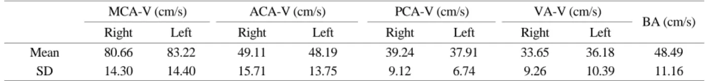
안정상태의 평균 뇌혈류속도는 중대뇌동맥의 경우 상향파로 서 80.66±14.30 및 83.22±14.40 cm/sec (우측과 좌측), 전대 뇌동맥의 경우 하향파로서 49.11±15.71 및 48.19±13.75 cm/sec, 후대뇌동맥의 경우 상향파로서 39.24±9.12 및 37.91±6.74 cm/
sec, 추골동맥의 경우 하향파로서 33.65±9.26 및 36.18±10.39 cm/sec, 뇌기저동맥의 경우 하향파로서 48.49±11.16 cm/sec였 고 우측과 좌측동맥 간 유의한 차이는 없었다 (P>0.05, Table 3). 뇌혈관별 평균 혈류속도는 중대뇌동맥, 전대뇌동맥, 뇌기저
동맥, 후대뇌동맥, 추골동맥의 순으로 점차 낮았다.
2. 무호흡 및 과호흡 상태의 뇌혈류속도
우측 및 좌측의 중대뇌동맥 평균 뇌혈류속도는 안정상태에 비해 무호흡 시 유의하게 상승하였으나 (P=0.0006, P=0.011) 과 호흡 시에는 유의하게 감소하였다 (P=0.007, P=0.0033, Table 4).
3. 성별에 따른 차이
안정상태에서 각 혈관부위별 평균혈류속도는 남녀간 전 반적으로 차이를 보였는데, 여자군의 좌측 및 우측 중대뇌동 맥, 우측 전대뇌동맥, 우측 후대뇌동맥, 좌측 및 우측 추골동 맥의 혈류속도가 남자군의 경우 보다 유의하게 더 높았다 (P<0.05, Table 5). 나머지 혈관의 평균 뇌혈류속도는 남녀간 에 유의한 차이가 없었다. 혈압의 경우 남자군이 여자군 보 다 수축기 및 평균 혈압은 유의하게 더 높았고 확장기 혈 압과 심장박동수는 차이가 없었다 (Table 2).
고 찰
1843년 Christian Doppler에 의해 도플러 효과 (Doppler effect) 가 발표된 이래 1959년 Satomura가 도플러 방법을 이용하여 말초혈관의 혈류속도를 비침습적이고 경피적으로 측정함으 로써 도플러의 임상적 이용시대를 열었다. 그 후 1982년 노 르웨이의 물리학자이자 인공두뇌 학자인 Aaslid가 저주파 (2 MHz)의 탐촉자를 사용하여 측두골을 통한 두개강 내 혈관 의 혈류속도를 측정하는 방법을 개발함으로써 폐쇄성혈관 질환뿐만 아니라 지주막하 출혈 후 혈관연축의 확인에 유용 한 검사로 제시되었다. 오늘날 TCD는 뇌혈류역학 변화를 평 가하기 위한 필수장비로 활용되고 있다.
한편 그 동안 국내에서도 몇몇 연구자들에 의해 TCD를 이 용하여 경두개 뇌혈관 혈류속도의 정상치에 대한 보고가 있 었으나 연구대상이 20대에서 70대 사이의 광범위한 연령층 이였던 까닭에 특정 연령대에 대한 뇌혈류역학의 이해자료 로는 다소 부족함이 있었다.
저자들은 이 연구에서 20대 연령층의 젊은이들만을 연구 대상으로 한정시켜 이 연령대의 뇌혈류역학에 대한 좀더 깊 은 이해와 자료의 축적을 시도하였다. 우리의 연구에서 뇌혈관 별 평균 혈류속도의 빠르기는 중대뇌동맥, 전대뇌동맥, 후대뇌 Table 4. Comparison of mean velocity of MCA at stable state to
those at apnea and hyperventilation condition Condition Position
Stable Apnea Hyperventilation Rt-MCA (cm/s) 80.66±4.30 90.13±17.4* 54.83±11.09* Lt-MCA (cm/s) 83.22±14.40 90.26±17.4* 55.33±10.74* Data were expressed as mean ± SD.
*, P<0.01 (compared with stable condition)
Table 3. Mean velocity in the intracranial vessels at stable condition
MCA-V (cm/s) ACA-V (cm/s) PCA-V (cm/s) VA-V (cm/s) Right Left Right Left Right Left Right Left BA (cm/s)
Mean 80.66 83.22 49.11 48.19 39.24 37.91 33.65 36.18 48.49
SD 14.30 14.40 15.71 13.75 9.12 6.74 9.26 10.39 11.16
Data were expressed as the mean ± SD
동맥의 순서로서 선행 연구보고들 (Kwun et al., 1989; Ahn et al., 1991; Kim et al., 1995; Krejza et al., 1999; Szydlik et al., 2000) 과 일치하였고 전체 연구대상자들 중 혈관별 뇌혈류 속도가 이 순서를 벗어나는 사례는 거의 없었다. 또한 각 혈관별 좌 측과 우측의 값에 의미있는 차이를 보이는 경우가 없으므 로 연구 대상자 모두의 뇌혈류 역학이 정상 상태를 유지함을 알 수 있었다. 중대뇌동맥은 뇌분포 혈관 중 가장 내경이 크 고 대뇌반구로 공급하는 전체 뇌혈류량의 80%를 담당하는 혈관인 까닭에 평균 뇌혈류 속도가 가장 높다.
따라서 저자들의 연구에서 측정 조건의 변화에 따른 뇌자 동조절기전의 정상적 반응을 조사하기 위해 중대뇌동맥의 평 균 뇌혈류 속도만을 측정하여 평가지표로 활용하였다. 뇌자동 조절 기전의 정상적 반응을 평가하기 위해 이 연구에서 채택 한 방법은 연구대상자로 하여금 무호흡 상태와 빈호흡 상태를 일정시간 동안 유지하여 동맥혈액의 이산화탄소분압 (PCO2) 레벨을 변화시키는 방법이었다. 동맥혈액의 PCO2 레벨 변화
는 뇌혈류에 영향을 미치게 된다. 뇌혈관의 CO2 반응성은 혈중 이산화탄소가 혈관내피세포하의 뇌-혈관문 (brain-vessel barrier)을 통과하여 세포 간 산성도를 저하시켜 뇌혈관의 근섬 유를 수축시킴으로써 나타나게 된다 (Wade et al, 1988; Widder, 1989). 동맥혈액의 PCO2가 변화하면 뇌의 연막혈관은 20초 후에 변화가 일어나는데 (Auer, 1978), 과호흡에 따른 저탄산 혈증에 대한 변화는 상대적으로 빨리 나타나고 (Severinghaus.
et al., 1967) 무호흡으로 인한 고탄산혈증 시에는 상대적으로 느리게 나타나며 만성적인 노출시 그 반응성은 저하된다 (Le- vasseur et al., 1979). PCO2 반응성은 외상 (Saunders et al., 1979), 두개 내 수술 (Haggendal et al., 1965), 동맥경화 (Lindegaard et al., 1987), 뇌지주막하 출혈 (Jakubowski et al., 1982) 등의 경 우 저하되고 특히 뇌대사가 저하된 혼수상태의 환자에서 현 저히 저하된다 (Ingvar et al., 1964). 대개 동맥혈액의 PCO2가 25~57 mmHg 사이에서는 PCO2가 1 mmHg 감소 또는 증가 되면 뇌혈류는 2~6% (평균 4%) 감소 또는 증가되며 이때 변 Table 5. Comparison between male and female group in the velocity at stable condition
MCA-V (cm/s) ACA-V (cm/s) PCA-V (cm/s) VA-V (cm/s) Right Left Right Left Right Left Right Left BA (cm/s)
Ma 73.79 69.73 39.64 44.57 34.50 36.29 30.46 32.38 39.54
Fb 82.37* 87.0* 51.48* 49.11 40.43* 38.33 34.40* 37.07 50.60*
*, P<0.05 (compared with male group). a, male group; b, female group
Fig. 3. Summary figure displays the arterial segments investigated and wave forms recorded in a normal transcranial Doppler ultrasono- graphic examination. LMCA=left middle cerebral artery; RMCA=right middle cerebral artery; LACA=anterior cerebral artery; RACA=
right anterior cerebral artery; LPCA=left posterior cerebral artery; RPCA=right posterior cerebral artery; LVA=left vertebral artery; RVA=
right vertebral artery; BA=basilar artery.
화된 PCO2 레벨에 대해 뇌기저 큰 동맥들의 반경은 변화가 없고 미세동맥의 반경 변화로 뇌혈류의 변화가 일어난다고 한 다 (Kety et al., 1948; Huber et al., 1967).
혈중 PCO2 변화에 따라 연막의 소동맥들의 내경이 변화되 면 그 부위를 공급하는 뇌기저 동맥에 이 변화가 반영되며 이 는 TCD를 이용하여 시간에 따른 평균뇌혈류속도의 변화로 환 산할 수 있다. 한편 Markwalder 등 (Markwalder et al., 1984)은 정상적인 경우 동맥혈액의 PCO2 1 mmHg 변화 시 중대뇌동 맥의 혈류 속도는 3~4% 변화된다고 보고한 바 있다.
저자들의 연구에서도 모든 연구대상자들이 무호흡 상태에 서는 중대뇌동맥의 평균 뇌혈류 속도가 안정 시보다 유의하 게 증가하였고 과호흡 시에는 유의한 감소를 보였으며, 그 변 화 양상이 과호흡의 경우가 무호흡의 경우보다 빠른 변화를 보임으로써 기존의 연구저자들이 보고한 결과들과 거의 일치 하였다. 이러한 결과들은 저자들의 연구에서 대상이 된 20대 연령층의 젊은이들이 지극히 정상적인 뇌혈류 역학 및 대사 를 유지하고 있음을 보여주는 증거라 할 수 있다. 그러나 저 자들의 연구에서 측정된 중대뇌동맥의 평균혈류 속도와 국 내의 선행 연구결과 사이에는 차이를 보였다. 저자들의 결과 는 80.66 cm/sec (우측)와 83.22 cm/sec (좌측)로써 권 등 (Kwun et al., 1989)의 61 cm/sec, 안 등 (Ahn et al., 1991)의 55.9 cm/sec, 김 등 (Kim et al., 1995)의 49 cm/sec (남자군)와 57 cm/sec (여 자군)와는 현저한 차이가 있었다. 연구 간의 이러한 차이가 무엇 때문에 발생하였는지에 관해 향후 지속적 연구를 통한 규명이 필요하겠지만 현재의 관점에서는 연구대상간의 특성 적 차이에 기인한 것으로 판단된다. 저자들의 경우 20대 연령 층의 젊은이들만을 연구대상화 한데 비해 전술한 국내 타 연 구저자들은 20대뿐만 아니라 30대에서 70대에 걸친 고 연령 층이 다수 포함된 대상을 연구함으로써 중대뇌동맥의 평균 혈류 속도 결과가 상대적으로 낮은 값으로 통계처리된 것으 로 생각된다. 연령의 증가는 뇌혈류 속도의 감소를 가져오는 데 20대에서 70대 사이의 연령에서 평균 뇌혈류 속도는 약 15~24% 정도 감소된다 (Arnolds et al., 1986; Vriens et al., 1989).
Grolimund 등 (Grolimund et al., 1988)은 연령 1세의 증가 시 중대뇌동맥의 평균혈류 속도는 약 0.51% 감소한다고 보고하였 다. 연령의 증가에 따른 뇌혈류 속도의 저하는 큰 뇌동맥의 직 경 변화보다는 작은 뇌혈관의 저항 증가와 더 관련이 있을 것 으로 생각된다 (Kim et al., 1995). 최근 Krejza 등 (Kre- jza et al., 1999)과 Szydlik 등 (Szydlik et al., 2000)은 20대 연령층의 중대 뇌동맥의 평균 혈류 속도가 약 81 cm/sec라고 보고함으로써 저자들의 연구결과와 거의 일치하였다. 그 외 나머지 뇌혈관별 혈류속도는 타 연구들과 유의한 차이를 보이지 않았다.
한편 성별에 따른 안정 상태 시 뇌혈류 속도의 비교 결과 는 전반적으로 여자군이 남자군 보다 높은 것으로 나타났 다. 이러한 성별에 따른 뇌혈류 속도의 차이에 대해 이 등
(Lee et al., 2001)은 여성의 경우 estrogen 호르몬이 분비됨으 로써 남자보다 뇌혈류 속도가 약 3~5% 정도 빠르다고 설명 한 바 있고, Grolimund 등 (Grolimund et al., 1988)과 Vriens 등 (Vriens et al., 1989)은 헤마토크리트 (hematocrit)의 차이에 기 인한 것으로 설명하고 있다. 이들은 여성이 남성 보다 헤마 토크리트 수치가 낮으므로 더 높은 뇌혈류 속도를 보였다고 주장하였다. Brass 등 (Brass et al., 1988)은 헤마토크리트 수치 와 뇌혈류 속도 사이에 유의하게 높은 음의 상관성이 있다 고 설명한 바 있다. 저자들의 연구에서도 비록 결과에서 제시 하지는 않았지만 뇌혈류 검사 시 채혈을 통해 측정한 헤마토 크리트치가 여자군의 경우 평균 35.89%, 남자군의 경우 평균 44.37%를 보임으로써 여자군이 유의하게 더 낮았다. TCD검사 시 함께 측정한 수축기 혈압과 평균 혈압 역시 여자군이 남 자군보다 유의하게 낮았는데 이러한 혈압의 차이가 남녀간의 뇌혈류 속도의 차이에 영향을 미친 것으로는 판단되지 않는 다. 그 이유는 고혈압이나 뇌혈관 질환이 없는 사람의 경우 평 균혈압이 50~150 mmHg 사이로 유지되면 뇌혈류는 특별한 변 화 없이 일정한 상태를 유지할 수 있기 때문이다 (Schell et al., 1993). 결국 저자들의 연구에서 나타난 여자군의 상대적으로 더 높은 뇌혈류 속도는 전술한 에스트로겐 및 헤마토크리트 의 차이에 기인한 것으로 사료된다.
그러나 권 등 (Kwun et al., 1989), 안 등 (Ahn et al., 1991), 김 등 (Kim et al., 1995)의 경우 남녀간의 중대뇌동맥의 뇌혈류 속 도에 유의한 차이가 없다고 보고함으로써 저자들의 연구결과 와 상치되었다. 이러한 차이는 이미 전술한 연구대상의 차이 에 따른 결과로 판단된다. Vriens 등 (Vriens et al., 1989)과 Bode 등 (Bode et al., 1988)은 성별에 따른 뇌혈류 속도의 차이는 20~60대 연령층과 어린아이 군에서는 유의한 차이를 보이지 만 70대 이상의 연령층에서는 남녀간의 차이가 유의하지 않 다고 보고한 바 있다. 따라서 국내 타 연구들의 경우 60대 이 상의 고 연령층이 연구대상에 포함됨으로써 남녀간의 차이를 상쇄시킨 것으로 사료되나 향후 좀 더 큰 연구 집단을 대상 으로 한 지속적 연구가 필요할 것으로 판단된다.
결론적으로, 저자들의 연구에 있어 기왕의 질병이 없는 20 대 연령층의 뇌혈류역학 및 자동조절기전은 정상적이었고 여 자군이 남자군에 비해 뇌혈류 속도가 상대적으로 더 높았다.
향후 더 많은 건강인을 대상으로 한 다양한 조건 (예를 들어 음주 전후, 흡연, 자세의 다양한 변화)에서의 뇌혈역학 및 뇌 생리에 대한 연구를 실시하여 좀더 폭 넓고 깊이 있는 데이터 수집이 지속되어야 할 것으로 생각된다.
REFERENCES
Aaslid R, Markwalder TM, Nornes H. Noninvasive transcranial Doppler ultrasound recording of flow velocity in basal cere-
bral arteries. J Neurosurg. 1982. 57: 769-774.
Ahn GB, Chi CS, Chung CS. Recording of cerebral blood flow velocity transcranial Doppler ultrasound in normal subjects. J Kor Neurol. 1991. 9: 37-45.
Arnolks BJ, von Reutern GM. Transcranial Doppler sonography.
Examination technique and normal reference values. Ultraso- und Med Biol. 1986. 12: 115-123.
Auer L. Pial arterial reactions to hyper-and hypocapnia: A dynamic experimental study in cars. Eur Neurol. 1978. 17: 351-362.
Bode H, Wais U. Age dependence of flow velocities in basal cere- bral arteries. Arch Dis Child. 1988. 63: 606-611.
Brass LM, Pavlakis SG, De Vivo D. Transcranial Doppler measur- ements of the middle cerebral artery. Effect of hematocrit.
Stroke. 1988. 19: 1466-1469.
Grolimund P, Seiler RW. Age dependence of the flow velocity in the basal cerebral arteries-a transcranial Doppler ultrasound study.
Ultrasound Med Biol. 1988. 14: 191-198.
Haggendal E, Ingvar DH, Lassen NA. Pre-and postoperative mea- surements of regional cerebral blood flow in three cases of intr- acranial arteriovenous aneurysms. J Neurosurg. 1965. 22: 1-6.
Ingvar DH, Haggendal E, Nilsson NJ. Cerebral circulation and me- tabolism in a comatous patient. Arch Neurol. 1964. 11: 13-21.
Jakubowski J, Bell BA, Symon L. A primate model of subarachn- oid haemorrhage: Change in regional cerebral blood flow, au- toregulation, carbon dioxide reactivity and central conduction time. Stroke 1982. 13: 601-611.
Kety SS, Schmidt CF. The effects of altered tensions of carbon dio- xide and oxygen on cerebral blood flow and cerebral oxygen consumption of normal young men. J Clin Invest. 1948. 27:
484-492.
Kim GW, Sohn YH, Lee SM, Lee JH, Kim DS, Kim JY, Kim JS.
The reference values and influencing factors of TCD measur- ements in 200 normal korean adults. J Kor Neurosurg Soc.
1995. 13: 1-10.
Krejza J, Mariak Z, Walecki J, Szydlik P, Lewko J, Ustymowicz A.
Transcranial color Doppler sonography of basal cerebral ar- teries in 182 healthy subjects: Age and sex variability and no- rmal reference values for blood flow parameters. Am J Roen- tgenol. 1999. 172: 213-218.
Kwun BD, Kwon Y, Rhim SC, Whang CJ. Transcranial Doppler ultrasound recording of flow velocity in basal cerebral arte- ries: Examination technique and normal values in Korean. J Kor Neurosurg Soc. 1989. 18: 379-388.
Lee TG, Yoon SS. Normal findings of transcranial Doppler. Kor J Stroke. 2001. 3: 7-13.
Levasseur JE, Wei EP, Kontos HA. Responses of pial arterioles after prolonged hypercapnia and hypoxia in the awake rabbit.
J Appl Physiol. 1979. 46: 89-95.
Lindegaard KF, Bakke SJ, Grolimund P. Assessment of intracra- nial hemodynamics in carotid artery disease by transcranial doppler ultrasound. J Neurosurg. 1985. 63: 890-898.
Linderaard KF, Grolimund P, Aaslid R, Nornes H. Evaluation of cerebral AVM's using transcranial doppler ultrasound. J Neu- rosurg. 1986. 65: 335-344.
Lindegaard KF, Lundar T, Wiberg J. Variations in middle cerebral artery blood flow investigated with noninvasive transcranial blood flow measurements. Stroke. 1987. 18: 1025-1030.
Markwalder TM, Grolimund P, Seiler RW. Dependency of blood flow velocity in the middle cerebral artery on end-tidal carbon dioxide partial pressure - A transcranial ultrasound Doppler study. J Cereb Blood Flow Metab. 1984. 4: 368-372.
Saunders MI, Miller JD, Stablein D. The effects of graded experi- mental trauma on cerebral blood flow and responsiveness to CO2. J Neurosurg. 1979. 51: 18-26.
Schell RM, Kern FH, Greeley WJ, Schulman SR, Frasco PE, Cro- ughwell ND, Newman M, Reves JG. Cerebral blood flow and metabolism during cardiopulmonary bypass. Anesth Analg.
1993. 76: 849-865.
Seiler RW, Grolimund P, Aaslid R. Cerebral vasospasm evaluated by transcranial ultrasound correlated with clinical grade and CT-visualized subarachnoid hemorrhage. J Neurosurg. 1986.
64: 594-596.
Severinghaus JW, Lassen N. Step hypocapnia to separate from tissue PCO2 in the regulation of cerebral blood flow. Circ Res. 1967. 20: 272-278.
Szydlik P, Mariak Z, Krejza J, Swiercz M, Keller A. Transcranial color Doppler estimation of blood flow parameters in respe- ctive basal cerebral arteries in healthy subjects. Neurol Neur- ochir Pol. 2000. 34: 523-536.
Vriens EM, Kraaier V, Musbach M. Transcranial pulsed Doppler measurements of blood velocity in the middle cerebral artery:
Reference values at rest and during hyperventilation in hea- lthy volunteers in relation to age and sex. Ultrasound Med Biol. 1989. 15: 1-8.
Wade JPH, Brown MM. Handbook of regional cerebral blood flow (Knezevic S, Maximilian VA, Mubrin Z. Eds). 1988. pp255- 282. Hillsdale Lawrence Erlbaum. USA.
Widder B. The Doppler CO2 test to exclude patients not in need of ECC bypass surgery. J Neurol Neurosurg Psychiatr. 1989. 52:
38-42.