Evaluation of marginal fit of 2 CAD-CAM
anatomic contour zirconia crown systems and lithium disilicate glass-ceramic crown
Min-Kyung Ji1, Ji-Hee Park1, Sang-Won Park1,2, Kwi-Dug Yun1, Gye-Jeong Oh2, Hyun-Pil Lim1*
1Department of Prosthodontics, School of Dentistry, Chonnam National University, Gwangju, Republic of Korea
2RIS Foundation for Advanced Biomaterials, Chonnam National University, Gwangju, Republic of Korea
PURPOSE. This study was to evaluate the marginal fit of two CAD-CAM anatomic contour zirconia crown systems compared to lithium disilicate glass-ceramic crowns. MATERIALS AND METHODS. Shoulder and deep chamfer margin were formed on each acrylic resin tooth model of a maxillary first premolar. Two CAD-CAM systems (Prettau®Zirconia and ZENOSTAR®ZR translucent) and lithium disilicate glass ceramic (IPS e.max®press) crowns were made (n=16). Each crown was bonded to stone dies with resin cement (Rely X Unicem). Marginal gap and absolute marginal discrepancy of crowns were measured using a light microscope equipped with a digital camera (Leica DFC295) magnified by a factor of 100. Two-way analysis of variance (ANOVA) and post- hoc Tukey’s HSD test were conducted to analyze the significance of crown marginal fit regarding the finish line configuration and the fabrication system. RESULTS. The mean marginal gap of lithium disilicate glass ceramic crowns (IPS e.max®press) was significantly lower than that of the CAD-CAM anatomic contour zirconia crown system (Prettau®Zirconia) (P<.05). Both fabrication systems and finish line configurations significantly influenced the absolute marginal discrepancy (P<.05). CONCLUSION. The lithium disilicate glass ceramic crown (IPS e.max®press) had significantly smaller marginal gap than the CAD-CAM anatomic contour zirconia crown system (Prettau®Zirconia). In terms of absolute marginal discrepancy, the CAD-CAM anatomic contour zirconia crown system (ZENOSTAR®ZR translucent) had under-extended margin, whereas the CAD-CAM anatomic contour zirconia crown system (Prettau®Zirconia) and lithium disilicate glass ceramic crowns (IPS e.max®press) had over- extended margins. [J Adv Prosthodont 2015;7:271-7]
KEY WORDS: Anatomic contour zirconia crown; CAD-CAM; Lithium disilicate glass ceramic crown; Marginal gap; Absolute marginal discrepancy; Marginal fit
INTRODUCTION
Zirconia has excellent aesthetic quality, biocompatibility, and mechanical property. In addition, the price of zirconia is inexpensive compared to gold. Thus, zirconia is getting attention as a proper material for posterior teeth restoration to replace the existing ceramic.1-3 Commercialization of zir- conia is closely linked to the development of CAD/CAM introduced to the dental industry 20 years ago.4-6 Recently, the introduction of new CAD/CAM milling technology and new zirconia made it possible to manufacture anatomic contour zirconia crown, enabling the forming of occlusal surface anatomically instead of in the form of porcelain veneer.7,8 Anatomic contour zirconia crowns have excellent fracture resistance property because it does not have super-
Corresponding author:
Hyun-Pil Lim
Department of Prosthodontics, School of Dentistry, Chonnam National University, 42, Jebong-ro, Dong-gu, Gwangju 61469, Republic of Korea Tel. 82 62 530 5638: e-mail, [email protected]
Received September 17, 2014 / Last Revision January 7, 2015 / Accepted January 19, 2015
© 2015 The Korean Academy of Prosthodontics
This is an Open Access article distributed under the terms of the Creative Commons Attribution Non-Commercial License (http://creativecommons.
org/licenses/by-nc/3.0) which permits unrestricted non-commercial use, distribution, and reproduction in any medium, provided the original work is properly cited.
This research was supported by the Basic Science Research Program through the National Research Foundation of Korea (NRF) funded by the Ministry of Science, ICT & Future Planning (2013R1A1A1010115).
structure. In addition, it has higher strength even when the volume of tooth preparation is small during the manufac- ture of a crown.9 However, long-term clinical success of dental restoration is influenced not only by mechanical property, aesthetic quality, and biocompatibility, but also by marginal fit. Large marginal gap causes failure of a crown by dissolving dental cement so quickly that plaques easily accumulate, leading to marginal leakage and secondary car- ies.10,11 Through in vitro and in vivo studies on marginal fit of dental restorations, marginal fit has been proven to be one of the major factors causing secondary caries and peri- odontal diseases.12-16 It has been proven by McLean in 1971 that clinically allowable marginal fit is within 120 μm range when fabricating a dental restoration.17 Martínez-Rus has reported that CAD/CAM ceramic crown has clinically allowable marginal fit within the range of 17 to 118 μm.18 Chamfer margins and shoulder margins were recommended to fabricate the precise anatomic contour crowns.19
Although computer-controlled technique is used in pro- ducing dental restoration in order to improve the accuracy during the manufacturing process, not enough studies have been conducted on the marginal fit of anatomic contour zirconia crowns fabricated using CAD/CAM system.
Therefore, the objective of this study was to compare the marginal fit of anatomic contour zirconia crown to that of lithium disilicate glass ceramic crown regarding the finish line configuration and fabrication systems.
Null hypotheses for this experiment are as follows: (1) Finish line configuration has no influence on the marginal fit of anatomic contour crowns; (2) Fabrication systems do not affect the marginal fit of anatomic contour crown.
MATERIALS AND METHODS
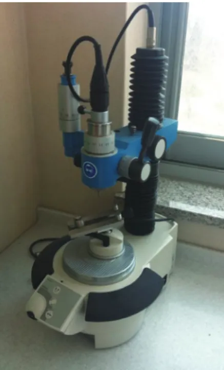
An acrylic resin model of maxillary first premolar (Columbia Dentiform Corp, Long Island City, NY, USA) was prepared. Tooth reduction was uniformly conducted with hand piece mounted in a milling machine (F4 basic, DentsplyDeguDent, Germany, Fig. 1). Total occlusal con- vergence angle was 12 degrees. An occlusal reduction of 1.5 mm at the center of the occlusal surface was executed.
The definitive die had a 1 mm shoulder and deep chamfer margin on each mesiodistal and buccolingual marginal sur- face (Fig. 2). Four reference points were formed at the mid- dle of the buccal, palatal, mesial, and distal root surface of tooth margin for later measurements. The width of each margin was managed by keeping it no wider than half of the diamond tip used for preparation. A flat-end diamond bur with 2.3 mm in diameter (Komet 6848.314.023; Gebr.
Brasseler GmbH & Co KG) was used for the shoulder mar- gin. A tapered diamond bur with 2.1 mm in diameter (Komet 6856.314.021; Gebr.Brasseler GmbH & Co KG) was used for the chamfer margin. The widths of the mar- gins were measured with a 3-dimensional measuring micro- scope (Measuring microscope; Mitutoyo, Kawasaki, Japan) to determine whether planned dimensions (1 mm shoulder and chamfer) were achieved. Forty eight impressions of the
resin tooth were obtained from the definitive die by using a light viscosity and putty vinyl polysiloxane (Exaflex and Exafine; GC Corporation, Tokyo, Japan). Forty eight impres- sions were poured with type IV die stone (suprastone, Kerr Corporation, Orange, CA, USA). Each stone die was care- fully removed from the impression and examined for the presence of air bubbles or other defects.
Fig. 1. Photograpic view of the handpiece mounted milling machine.
Fig. 2. Proximal view of tooth preparation on the die and finish line configuration. (a) total occlusal convergence angle; (b) occlusal reduction; (c) margin width; (d) shoulder margin; (e) deep chamfer margin.
(a) 12 degrees
(d) shoulder (b) 1.5 mm
(e) deep chamfer (c)
1 mm 1 mm
1 mm
1 mm
Two CAD/CAM system (Prettau®Zirconia; Zirkonzahn GmbH; Gais, Italy/ ZENOSTAR®ZR translucent; Wieland Dental GmbH, Pforzheim, Germany) zirconia crowns and IPS e.max®press (IvoclarVivadent AG, Schaan, Liechtenstein) lithium disilicate glass ceramic crowns were divided into three groups according to fabrication methods (n=16). Each group was sub-categorized into two groups (shoulder mar- gin, chamfer margin) based on the finish line configuration.
Therefore, a total of 6 groups were used in this study (n=8, Table 1).
All crowns were fabricated with anatomical shape according to the directions of manufacturing companies for each fabrication system (Fig. 3). The 5-TEC CAD/
CAM system (Zirkonzahn GmbH; Gais, Italy) was used to fabricate the Prettau®Zirconia crowns. The dies were scanned sequentially with a SCANNER S600 ARTI (Zirkonzahn GmbH;
Gais, Italy). ZirkonzahnArchiv software (Zirkonzahn GmbH;
Gais, Italy) was used to design anatomic contour zirconia crowns. The simulated die spacer was programmed at 30 μm, starting at 1 mm ahead from the margin. The Prettau® Zirconia blocks were milled with M5 milling machine (Zirkonzahn GmbH; Gais, Italy). All specimens were fired in a furnace (Zirkonofen600; Zirkonzahn GmbH; Gais, Italy) at 1600ºC. The ZENO TEC®System (Zenotec;
Wieland Dental GmbH, Pforzheim, Germany) was used to fabricate the ZENOSTAR®ZR translucent crowns. The dies were scanned sequentially with 3shape D250 (Wieland Dental GmbH, Pforzheim, Germany). Anatomic contour
zirconia crowns were designed with design software (Dental Designer; Wieland Dental GmbH, Pforzheim, Germany).
This information was transmitted to a milling machine (Zenotec T1; Wieland Dental GmbH, Pforzheim, Germany).
The crowns were produced by milling the ZENOSTAR® ZR translucent block and firing (Zenotec Fire; Wieland Dental GmbH, Pforzheim, Germany) at 1450ºC. The IPS e.max®Press lithium disilicate glass ceramic crowns were fabricated using the lost wax technique. Two coats of die spacer were applied to the stone dies20 1 mm above the cer- vical end of the preparation to ensure good marginal fit.
The crowns were fabricated by pressure injection of ceramic ingots in the Programat®EP5000 furnace (IvoclarVivadent AG, Schaan, Liechtenstein) following the manufacturer’s rec- ommendations.
Forty eight crowns were luted with resin cement (Rely X Unicem, 3M ESPE, Seefeld, Germany) on the stone dies, respectively. Finger pressure was then applied for ten min- utes as in clinical situation. All crowns that were completely bonded to stone dies were embedded with acrylic resin (Caulk, Dentsply, Milford, DE, USA) for observation.
Cured resin blocks were cut in the middle of buccopalatal and mesiodistal directions using linear precision saw (Isomet 5000; Buehler Ltd., Lake Bluff, IL, USA) referring to four reference points. Each reference point of every sample was measured three times using a light microscope equipped with a digital camera (Leica DFC295; Leica Microsystems Ltd., Heerbrugg, Switzerland) and magnified
Table 1. Experimental groups depending on the fabrication system and finish line configuration used in this study
Fabrication system Composition Finish line configuration
IPS e.max®press
(IvoclarVivadent AG,Schaan, Liechtenstein) Lithium disilicate glass-ceramic Deep chamfer (n=8) Shoulder (n=8) Prettau®Zirconia
(Zirkonzahn GmbH; Gais, Italy)
Presintered yttrium stabilized zirconium dioxide
Deep chamfer (n=8) Shoulder (n=8) ZENOSTAR®ZR translucent
(Wieland Dental GmbH, Pforzheim, Germany)
Deep chamfer (n=8) Shoulder (n=8)
Fig. 3. Photograpic view of the three types of crown restoration: (A) Prettau®Zirconia; (B) ZENOSTAR®ZR translucent;
(C) IPS e.max®press lithium disilicate glass ceramic.
A B C
by a factor of 100.
The criterion proposed by Holmes et al.21 was used to measure the marginal fit. The marginal gap and the absolute marginal discrepancy were measured. The perpendicular measurement from internal surface of the crown to the edge of the tooth finish line was defined as marginal gap.
The distance from the edge of the crown to the edge of the tooth finish line was defined as absolute marginal dis- crepancy (Fig. 4).
IBM SPSS Statistics 20 (IBM SPSS Inc., Chicago, IL, USA) was used for statistical analysis. Two-way analysis of variance (ANOVA) and post-hoc Tukey’s HSD test were conducted to analyze significance of crown marginal fit regarding the finish line configuration and the fabrication system.
RESULTS
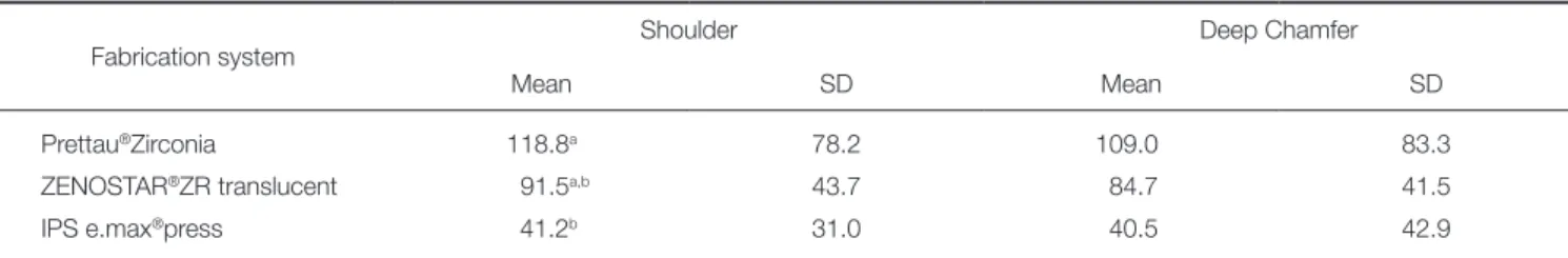
The mean marginal gap of shoulder and deep chamfer fin- ish line configurations was 118.8 ± 78.2 μm and 109.0 ± 83.3 μm in the Prettau®Zirconiagroup, 91.5 ± 43.7 μm and 84.7 ± 41.5 μm in the ZENOSTAR®ZR translucent group, 41.2 ± 31.0 μm and 40.5 ± 42.9 μm in the IPS e.max®press group, respectively (Table 2). The two-way ANOVA showed that the fabrication system significantly influenced the mar- ginal gap (P<.05). There was no significant difference in marginal gap depending on the finish line configuration, and in the interaction between the fabrication system and the finish line configuration (P>.05)(Table 3). According to Tukey honestly significant difference (HSD) test results, the marginal gap of IPS e.max®press was significantly smaller than Prettau®Zirconia (P<.05). The mean absolute marginal discrepancy of shoulder and deep chamfer finish line con- figurations was 73.9 ± 32.9 μm and 37.8 ± 37.1 μm in the Prettau®Zirconia group, -14.3 ± 61.2 μm and -51.3 ± 66.9 μm in the ZENOSTAR®ZR translucent group, 29.4 ± 19.1 Fig. 4. Schematic diagram of crown and tooth with the
following measurement part: A, over-extended margin;
and B, under-extended margin. (m): marginal gap; (a):
absolute marginal discrepancy.
Crown Crown
Tooth Tooth
a m a m
A B
Table 2. The mean and standard deviations of marginal gap depending on fabrication system in each finish line configu ration (unit: μm)
Fabrication system Shoulder Deep Chamfer
Mean SD Mean SD
Prettau®Zirconia 118.8a 78.2 109.0 83.3
ZENOSTAR®ZR translucent 91.5a,b 43.7 84.7 41.5
IPS e.max®press 41.2b 31.0 40.5 42.9
Different superscripted lowercase letters in column indicate significant differences (P<.05).
Table 3. Two-way ANOVA results for effect of fabrication system, finish line configuration, and their interactions on mean marginal gap
Source Sum of squares Degree of freedom Mean squares F-statistics P vlaue
Corrected Model 44492.075a 5 8898.415 2.742 .031
Intercept 314466.469 1 314466.469 96.902 .000
Fabrication system 43922.085 2 21961.043 6.767 .003
Finish line configuration 396.175 1 396.175 .122 .729
Finish line configuration
×Fabrication system 173.814 2 86.907 .027 .974
Error 136298.839 42 3245.210
Total 495257.383 48
Corrected Total 180790.914 47
a. R Squared = .246 (Adjusted R Squared = .156).
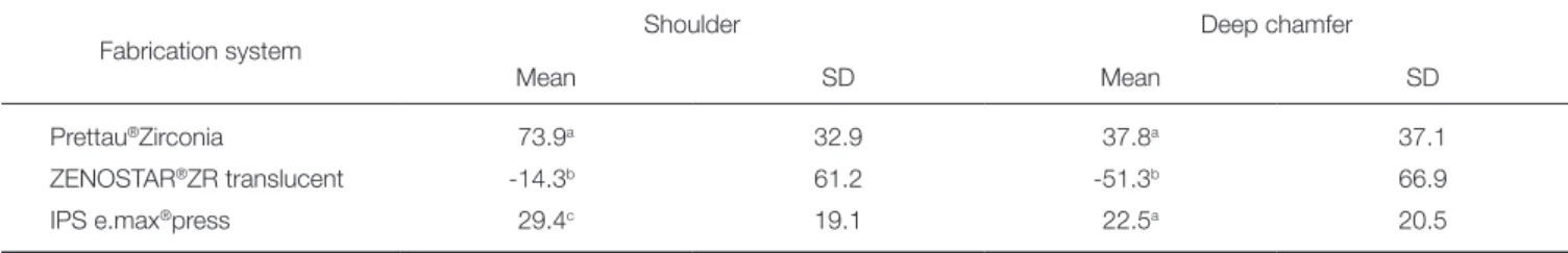
μm and 22.5 ± 20.5 μm in the IPS e max press group, respectively (Table 4). ZENOSTAR®ZR translucent had under-extended margin, whereas Prettau®Zirconia and IPS e.max®press had over-extended margin. The two-way ANOVA analysis results showed that both the fabrication system and the finish line configuration significantly influenced the absolute marginal discrepancy (P<.05). There was no signif- icant interaction between the 2 variables (P>.05)(Table 5).
Tukey honestly significant difference (HSD) test indicated a significant difference between IPS e.max®press and ZENO- STAR®ZR translucent, and also between Prettau® Zirconia and ZENOSTAR®ZR translucent (P<.05).
DISCUSSION
This study examined the marginal fit of two CAD/CAM anatomic contour zirconia crowns (Prettau®Zirconia of ZikozahnandZENOSTAR®ZR translucent of Wieland Dental) and lithium disilicate glass ceramic crowns (IPS e.
max®press of IvoclarVivadent) which are widely used clini- cally. The null hypothesis that the fabrication system has no influence on marginal fit of anatomic contour crowns was rejected, whereas the other null hypothesis that the finish
line configuration has no influence on marginal fit of ana- tomic contour crown was accepted.
Acrylic resin tooth was used in this study to simulate clinical situation. Tooth preparation was conducted uni- formly using surveyor milling machine. Each crown was sectioned at the same position with four reference points.
To simulate the cementation of the clinical situation, finger pressure was applied to each of the crowns for ten minutes.
In general, sectioning embedded specimens 22-25 and direct visualization26,27 are the two methods used to measure mar- ginal fit. Direct visualization has an advantage that it is measurable without destroying specimens, making it appli- cable under clinical situation. However, it is hard to obtain precise measurements by direct visualization.24 The section- ing method was used in this study in order to measure mar- ginal fit precisely.
Our results revealed that the maximum marginal gap was 118.8 ± 78.2 μm when shoulder margin was formed on crowns using Prettau®Zirconia. The amount of marginal gap was in the clinically acceptable range according to Martínez-Rus.18 The marginal fit of restoration manufac- tured by CAD/CAM system was increased compared to the IPS e.max®press system. Procedures added on computers Table 4. The mean and standard deviations of absolute marginal discrepancy depending on fabrication system in each finish line configuration (unit: μm)
Fabrication system Shoulder Deep chamfer
Mean SD Mean SD
Prettau®Zirconia 73.9a 32.9 37.8a 37.1
ZENOSTAR®ZR translucent -14.3b 61.2 -51.3b 66.9
IPS e.max®press 29.4c 19.1 22.5a 20.5
Different superscripted lowercase letters in column indicate significant differences (P<.05).
“-” indicate the under-extended margin.
Table 5. Two-way ANOVA results for effect of fabrication system, finish line configuration, and their interactions on mean absolute marginal discrepancy
Source Sum of squares Degree of freedom Mean squares F-statistics P vlaue
Corrected Model 75951.430a 5 15190.286 7.955 .000
Intercept 12776.765 1 12776.766 6.691 .013
Fabrication system ×
Finish line configuration 2350.913 2 1175.456 .616 .545
Fabrication system 65075.848 2 32537.924 17.040 .000
Finish line configuration 8524.668 1 8524.668 4.464 .041
Error 80197.311 42 1909.459
Total 168925.508 48
Corrected Total 156148.742 47
a. R Squared = .486 (Adjusted R Squared = .425)
caused some errors, although handwork errors were reduced by the CAD/CAM system.28 The large marginal gap com- pared to other studies are due to shrinkage error that could increase before and after sintering due to the larger material volume of anatomic zirconia crowns than zirconia copings.
The differences in marginal fit between each system result from zirconia blocks with various shrinkage rates depend- ing on manufacturing company, scanning process, size of milling bur used, and the number of milling axis.
This study has some limitations. Finger pressure used clinically to lute crowns on stone dies was not standardized.
Therefore, the use of a loading device is needed to apply uniform load on all crowns. In addition, stone dies instead of teeth were used to evaluate marginal fit of anatomic contour crown. Although stone dies or resin dies have been allowed to be used for evaluating marginal fit,24,29 the use of human tooth would be ideal to simulate clinical procedure.
The results of this study suggest that IPS e.max®press is better than Prettau®Zirconia regarding the marginal gap when fabricating an anatomic contour crown. Further study and clinical applications should be performed to compen- sate for the shrinkage of zirconia that might happen during the fabricating procedure. The development of CAD/CAM technology is needed to produce more accurate milling pro- cess.
CONCLUSION
Within the limitations of this study, we have the following conclusions: 1) IPS e.max®press had significantly smaller mar- ginal gap than Prettau®Zirconia; 2) Finish line configuration did not show significant influence on the marginal gap; 3) In terms of absolute marginal discrepancy, ZENOSTAR®ZR translucent had under-extended margins, whereas Prettau® Zirconia and IPS e.max®press had over-extended margins;
4) The measured marginal gaps of the two CAD/CAM anatomic contour zirconia crown and lithium disilicate glass ceramic crown were within the range of clinical acceptance.
ORCID
Min-Kyung Ji http://orcid.org/0000-0002-3525-644X Sang-Won park http://orcid.org/0000-0002-9376-9104 Kwi-Dug Yun http://orcid.org/0000-0002-2965-3967 Gye-Jeong Oh http://orcid.org/0000-0001-5628-0505 Hyun-Pil Lim http://orcid.org/0000-0001-5586-1404 REFERENCES
1. Filser F, Kocher P, Weibel F, Lüthy H, Schärer P, Gauckler LJ.
Reliability and strength of all-ceramic dental restorations fab- ricated by direct ceramic machining (DCM). Int J Comput Dent 2001;4:89-106.
2. Conrad HJ, Seong WJ, Pesun IJ. Current ceramic materials and systems with clinical recommendations: a systematic re- view. J Prosthet Dent 2007;98:389-404.
3. Christel P, Meunier A, Heller M, Torre JP, Peille CN. Mechanical
properties and short-term in-vivo evaluation of yttrium-ox- ide-partially-stabilized zirconia. J Biomed Mater Res 1989;23:
45-61.
4. Kugel G, Perry RD, Aboushala A. Restoring anterior maxil- lary dentition using alumina- and zirconia-based CAD/CAM restorations. Compend Contin Educ Dent 2003;24:569-72, 574, 576.
5. Rekow D. Computer-aided design and manufacturing in den- tistry: a review of the state of the art. J Prosthet Dent 1987;
58:512-6.
6. Yin L, Song XF, Song YL, Huang T, Li J. An overview of in vitro abrasive finishing & CAD/CAM of bioceramics in re- storative dentistry. Int J Mach Tool Manuf 2006;46:1013-26.
7. Baig MR, Tan KB, Nicholls JI. Evaluation of the marginal fit of a zirconia ceramic computer-aided machined (CAM) crown system. J Prosthet Dent 2010;104:216-27.
8. Preis V, Behr M, Kolbeck C, Hahnel S, Handel G, Rosentritt M. Wear performance of substructure ceramics and veneer- ing porcelains. Dent Mater 2011;27:796-804.
9. Jung YS, Lee JW, Choi YJ, Ahn JS, Shin SW, Huh JB. A study on the in-vitro wear of the natural tooth structure by oppos- ing zirconia or dental porcelain. J Adv Prosthodont 2010;2:
111-5.
10. Felton DA, Kanoy BE, Bayne SC, Wirthman GP. Effect of in vivo crown margin discrepancies on periodontal health. J Prosthet Dent 1991;65:357-64.
11. Knoernschild KL, Campbell SD. Periodontal tissue responses after insertion of artificial crowns and fixed partial dentures.
J Prosthet Dent 2000;84:492-8.
12. Kokubo Y, Ohkubo C, Tsumita M, Miyashita A, Vult von Steyern P, Fukushima S. Clinical marginal and internal gaps of Procera AllCeram crowns. J Oral Rehabil 2005;32:526-30.
13. Boening KW, Wolf BH, Schmidt AE, Kästner K, Walter MH.
Clinical fit of Procera AllCeram crowns. J Prosthet Dent 2000;84:419-24.
14. May KB, Russell MM, Razzoog ME, Lang BR. Precision of fit: the Procera AllCeram crown. J Prosthet Dent 1998;80:
394-404.
15. Weaver JD, Johnson GH, Bales DJ. Marginal adaptation of castable ceramic crowns. J Prosthet Dent 1991;66:747-53.
16. Behr M, Proff P, Kolbeck C, Langrieger S, Kunze J, Handel G, Rosentritt M. The bond strength of the resin-to-zirconia interface using different bonding concepts. J Mech Behav Biomed Mater 2011;4:2-8.
17. McLean JW, von Fraunhofer JA. The estimation of cement film thickness by an in vivo technique. Br Dent J 1971;131:
107-11.
18. Martínez-Rus F, Suárez MJ, Rivera B, Pradíes G. Evaluation of the absolute marginal discrepancy of zirconia-based ce- ramic copings. J Prosthet Dent 2011;105:108-14.
19. Pera P, Gilodi S, Bassi F, Carossa S. In vitro marginal adapta- tion of alumina porcelain ceramic crowns. J Prosthet Dent 1994;72:585-90.
20. Cho SH, Chang WG, Lim BS, Lee YK. Effect of die spacer thickness on shear bond strength of porcelain laminate ve- neers. J Prosthet Dent 2006;95:201-8.
21. Holmes JR, Bayne SC, Holland GA, Sulik WD. Considerations
in measurement of marginal fit. J Prosthet Dent 1989;62:
405-8.
22. Blackman R, Baez R, Barghi N. Marginal accuracy and geom- etry of cast titanium copings. J Prosthet Dent 1992;67:435- 40.
23. Oruç S, Tulunoglu Y. Fit of titanium and a base metal alloy metal-ceramic crown. J Prosthet Dent 2000;83:314-8.
24. Gavelis JR, Morency JD, Riley ED, Sozio RB. The effect of various finish line preparations on the marginal seal and oc- clusal seat of full crown preparations. J Prosthet Dent 1981;
45:138-45.
25. Han HS, Yang HS, Lim HP, Park YJ. Marginal accuracy and internal fit of machine-milled and cast titanium crowns. J Prosthet Dent 2011;106:191-7.
26. Witkowski S, Komine F, Gerds T. Marginal accuracy of tita- nium copings fabricated by casting and CAD/CAM tech- niques. J Prosthet Dent 2006;96:47-52.
27. Tan PL, Gratton DG, Diaz-Arnold AM, Holmes DC. An in vitro comparison of vertical marginal gaps of CAD/CAM ti- tanium and conventional cast restorations. J Prosthodont 2008;17:378-83.
28. Bornemann G, Lemelson S, Luthardt R. Innovative method for the analysis of the internal 3D fitting accuracy of Cerec-3 crowns. Int J Comput Dent 2002;5:177-82.
29. Quante K, Ludwig K, Kern M. Marginal and internal fit of metal-ceramic crowns fabricated with a new laser melting technology. Dent Mater 2008;24:1311-5.