of Cancer Prevention 2003; 8(4): 251-260
Role of Cyclooxygenase-2 and Its Products in the Regulation of Cell Growth
Hye-Kyung Na and Young-Joon Surh
Laboratory of Biochemistry and Molecular Toxicology, College of Pharmacy, Seoul National University, Seoul 151-742, Korea
Arachidonic acid is metabolized to prostaglandin H
2by cyclooxygenase (COX). Pros- taglandins exert diverse physiological actions to maintain mucosal integrity, regulation of secretion and motility. Among the various COX products, abnormally elevated levels of prostaglandin E
2(PGE
2) have been often observed in various types of human cancers.
Moreover, COX-2, the inducible COX isozyme, has been implicated in pathogenesis of various malignancies. PGE
2generated by COX-2 increased the cellular cAMP level via EP2, a cell-surface receptor of this prostaglandin, and further stimulates expression of COX-2. PGE
2activates the epidermal growth factor receptor-hepatocyte growth factor receptor-β-catenin-uPAR signaling pathway, thereby augmenting invasiveness and metastasis of cancer cells. In contrast, 15-deoxy-Δ
12,14PGJ
2(15d-PGJ
2), another COX-2 metabolite and a natural ligand for the peroxisome proliferator-activated receptors γ (PPARγ), causes apoptosis, inhibition of cell growth, anti-inflammation, and cytoprotection. 15d-PGJ
2and some synthetic PPARγ ligands also regulate the expression of COX-2 through peroxisome proliferator response element, which is accompanied by induction of apoptosis. Therefore, the physiological and toxicological effects associated with induction of COX-2 expression depend on types of cells and stimuli.
ꠏꠏꠏꠏꠏꠏꠏꠏꠏꠏꠏꠏꠏꠏꠏꠏꠏꠏꠏꠏꠏꠏꠏꠏꠏꠏꠏꠏꠏꠏꠏꠏꠏꠏꠏꠏꠏꠏꠏꠏꠏꠏꠏꠏꠏꠏꠏꠏꠏꠏꠏꠏꠏꠏꠏꠏꠏꠏꠏꠏꠏꠏꠏꠏꠏꠏꠏꠏꠏꠏꠏꠏꠏꠏꠏꠏꠏꠏꠏꠏꠏꠏꠏꠏꠏꠏ
Key Words: Cyclooxygenase-2, 15-deoxy-Δ
12,14prostaglandin J
2, PGE
2,Cell growth/
death, Peroxisome proliferator-activated receptor γ ligands
세포의 사멸과 성장 조절에 있어서 Cyclooxygenase-2와 그 산물들의 역할
서울대학교 약학대학 생화학교실
나 혜 경․서 영 준
책임저자:서영준, ꂕ 151-742, 서울특별시 관악구 신림동 5-1, 서울대학교 약학대학 생화학교실 Tel: 02 877-3730, Fax: 02 874-9775, E-mail: [email protected]
접수일:2003년 8월 1일, 게재승인일:2003년 11월 1일
서 론
최근 염증 전달, 세포부착 및 면역감시 체계 유 지 등 생리적으로 중요한 기능을 수행하는 프로 스타글란딘이 세포의 분열이나 증식에 영향을 줌 으로써 유방암, 대장암 위암, 폐암 등 각종 인체 암의 발생 및 진행에 관여한다는 실험적 증거들 이 보고되고 있다.1,2) 이들 프로스타글란딘의 생합 성에 관여하는 cyclooxygenase (COX)의 동종효소 중 하나인 COX-2의 비정상적인 증가가 암세포의 세포사멸을 억제하고 세포성장 촉진 및 침윤성 증가를 수반하면서 암화과정에 연루되어 있음이 여러 문헌을 통해 보고되고 있다.3∼5) 뿐만 아니라 COX-2의 주요 산물인 PGE2가 EP 수용체를 매개 로 암세포의 성장 및 침윤성과 관련된 단백질의 발현을 유도함으로써 암화과정에 관여한다고 한 다. 그러나 최근 보고에 따르면 COX-2의 또 다른 산물이자 peroxisome proliferator-activated receptor
(PPAR)γ의 내인성 리간드인 15d-Δ12,14-PGJ2 (15d- PGJ2)이 다양한 암세포에서 세포 사멸을 유도하 며 세포성장을 억제하는 것으로 알려져 있다. 이 런 측면에서 새로운 암 치료제로서 15d-PGJ2를 비 롯한 다양한 PPAR 리간드들의 사용 가능성이 대 두되고 있다. 본 논문에서는 암세포의 성장을 조 절하는데 있어 COX-2의 주요산물인 PGE2와 15d- PGJ2 작용을 분자적 수준에서 알아보고자 한다.
본 론
1) 프로스타글란딘의 생합성 경로
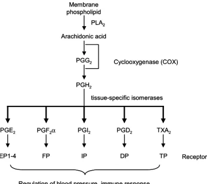
각종 프로스타글란딘들은 세포막의 인지질 분 해 산물인 아라키돈산으로부터 만들어진다. 아라 키돈산은 COX에 의해 일차적으로 각종 prostano- ids 합성 중간체인 prostaglandin G2 (PGG2)로 전환 되며, 이는 peroxidase에 의해 prostaglandin H2
(PGH2)로 전환되어 PGE2, PGD2, PGI2, PGF2α와 thromboxane A2를 생성한다. 합성된 프로스타글란
Fig. 1. General pathway for synthesis of PGs. Arachidonic acid is formed by the action of phospholipase A
2(PLA
2) on membrane phospholipids, and is converted by COX to PGH
2via PGG
2. PGH
2is further metabolized to various PGs by specific isomerases. Each PG has its own coupled receptor on the cell surface.
Arachidonic acid
PGG
2PGH
2PGE
2PGF
2α PGI
2PGD
2TXA
2Cyclooxygenase (COX)
tissue-specific isomerases Membrane
phospholipid
EP1-4 IP TP
Regulation of blood pressure, immune response, water balance, cell division, etc.
PLA
2DP
FP Receptors
Arachidonic acid
PGG
2PGH
2PGE
2PGF
2α PGI
2PGD
2TXA
2Cyclooxygenase (COX)
tissue-specific isomerases Membrane
phospholipid
EP1-4 IP TP
Regulation of blood pressure, immune response, water balance, cell division, etc.
PLA
2DP
FP Receptors
딘들은 세포로부터 분비되어 세포막에 위치하는 수용체, 즉 PGE2는 EP, PGF2α는 FP, PGD2는 DP, TXA2는 TP, PGI2는 IP에 특이적으로 결합하여 세 포 종류에 따라 다양한 생리적인 기능을 수행한 다(Fig. 1).6) PGE2의 수용체인 EP는 EP1, EP2, EP3
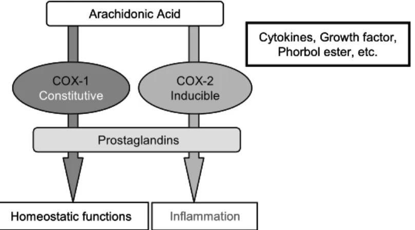
와 EP4 4종류 subtype이 존재한다.7) 한편 프로스타 글란딘 생합성에 관여하는 COX는 COX-1와 COX-2 두 가지 동종효소로 존재한다. 대부분의 조직에서 일정수준으로 발현되는 house-keeping enzyme인 COX-1은 위장관 보호, 신장의 혈류조 절 및 혈소판 응집 등 우리 몸의 정상적인 기능유 지에 중요한 역할을 하는 반면, COX-2는 외부자 극에 의해 발현되는 유도성 효소로서 세포의 성 장, 분화는 물론 염증발현 및 암을 비롯한 각종 퇴행성 질환의 병리학적 현상의 발현 및 진행과 정에 있어서 중요한 역할을 한다(Fig. 2).8∼10) 2) 발암과정에 있어서 PGE2와 COX-2 작용
기전
프로스타글란딘은 면역반응, 호르몬 배출, 혈관 확장, 신경세포 기능 유지를 비롯한 혈액속도, 수 분 및 수면조절 등 다양한 생리적 기능을 수행한 다. 그러나 과량 분비 시 세포의 분열이나 증식에 영향을 줌으로써 유방암, 대장암, 위암, 폐암 등 각종 인체 암 발생 및 진행에 중요한 역할을 한다 는 증거들이 축적되고 있다.1) 특정 프로스타글란 딘 중 특히 PGE2가 각종 인체 암 조직을 비롯한 발암모델에서 비정상적으로 증가됨이 종종 보고
되고 있다. 마우스 피부에 발암물질인 7,12- dimethylbenz [a]anthracence을 국소적으로 처리하 여 암 개시화(initiation)를 유도한 후 TPA 처리로 암화과정을 촉진하여 유발된 유두종에서 대량의 PGE2와 PGF2α가 관찰되었다.11) 비정상적으로 증 가된 PGE2는 COX- 1 보다는 주로 COX-2에 의해 생성되며, PGE2의 세포증식효과는 EP 수용체 활 성화를 통해 이루어짐이 제시되었다. 즉 COX-2의 활성화로 인해 축적된 PGE2는 EP2 수용체에 결합 하여 세포내의 adenylate cyclase를 활성화를 유도 하며 이로 인해 세포 내에 증가된 cAMP는 혈관 신생인자인 vascular endothelial growth factor (VEGF)의 생성을 증가시켜 상피세포의 침윤성 (invasivenesis)을 유도한다.12) 또 다른 보고로는
“가족성 용종증(familial adenomatous polyposis)”의 동물 모델인 APCΔ716 mouse에서 EP2 유전자 제거 시 PGE2에 의해 증가된 용종의 수와 크기가 감소 된 반면, EP1과 EP3를 knockout 시킨 쥐에서는 용 종의 수가 줄어들지 않았다.13) 따라서 PGE2의 세 포증식 효과는 주로 EP2의 수용체 활성화를 통해 유도되며, EP2 수용체를 통한 positive feedback loop에 의해 COX-2의 발현 또한 조절될 것으로 본다. PGE2의 생성 증가 및 COX-2의 발현 증가 외에 hepatocyte growth factor receptor (c-Met-R), epidermal growth factor receptor (EGFR), 및 β- catenin의 발현 증가 또한 대장암 세포의 증식과 침윤성에 연루되어 있다.14) E-cadherin과 결합된 β-catenin과 α-catenin은 세포의 adhesion을 조절
Fig. 2. Differential functions of COX-1 and COX-2.
Arachidonic Acid
Homeostatic functions COX-1 Constitutive
Inflammation
Cytokines, Growth factor, Phorbol ester, etc.
Prostaglandins COX-2 Inducible Arachidonic Acid
Homeostatic functions COX-1 Constitutive
Inflammation
Cytokines, Growth factor, Phorbol ester, etc.
Prostaglandins
COX-2
Inducible
함으로써 상피세포의 표현형을 결정하는데 관여 한다. 세포질에 존재하는 유리형 β-catenin은 TCF/Lef1 DNA 전사인자 활성화를 통해 정상세포 와 암세포의 세포성장에 관여하는 유전자 발현을 조절하는데, 표적 유전자 중 하나로는 암세포의 침윤성과 상당히 관련이 있는 urokinase-type plasminogen activator receptor (uPAR)를 들 수 있 다. Serine proteinase인 urokinase-type plasminogen activator (uPA)가 uPAR에 결합됨으로써 불활성형 의 zymogen plasminogen이 plasmin으로 활성화되 어 extracellular matrix와 세포막 구성성분을 분해 하여 세포의 이동과 침윤을 유도한다. PGE2를 대 장암 세포에 처리시 EGFR의 활성화 유도로 c-Met-R의 인산화가 촉진되어 β-catenin이 인산화 되며, 이로인해 E-cadherin과의 결합에 손상을 초 래하여 β-catenin이 분리됨이 관찰되었다. 유리된 β-catenin이 핵 내로 이행하여 uPAR mRNA의 발 현을 유도함으로써 대장암 세포의 이동과 침윤성 을 유도하였다.15) 또한 PGE2를 랫드의 복강 내로 주입 시 azoxymethane (AOM)으로 유도된 대장암 실험모델에서 대장암 발생률이 증가되고 세포사 멸이 억제되었다.16) PGE2에 의한 침윤성 증가는 COX-2의 억제제나 EP1/2의 수용체 길항제 (antagonist)인 AH6809에 의해 감소되었다.13) 위의
보고를 종합해 볼 때 COX-2의 발현 유도로 인해 증가된 PGE2는 paracrine, autocrine 방법을 통해 EP 수용체를 활성화하여 세포의 침윤성에 관련된 여러 유전자의 발현 및 활성화를 유도하여 세포 증식 및 침윤과정에 관여함을 알 수 있다(Fig. 3).
아울러 PGE2의 생성에 있어서 중요한 역할을 하는 COX-2가 발암과정에 연루되어 있음은 여러 실험적, 임상적 증거를 통해 지지되고 있다. 대장 암, 폐암, 위암, 간암 등 각종 암 조직에서 COX-2 의 발현이 주변의 정상조직에 비해 월등히 높은 것이 관찰되며, 비정상적인 COX-2의 발현 증가가 악성 또는 형질 전환된 세포의 생존을 지연시키 고, 전이능과 관련된 표현형 변화에 관련됨이 보 고 되고 있다.3∼5) 이는 COX-2의 증가가 세포성 장, 전이 및 침윤에 연루되어 있는 신호전달 관련 단백질의 활성화를 통해 암화과정에 관여함을 알 수 있다. 예를 들어 COX-2의 과발현은 Bcl-2 단백 질 발현 증가를 유도하며 transforming growth factor β-2 (TGFβ-2) 수용체, E-cadherin의 감소를 수반한다.17) 결장암 및 유방암 세포에 COX-2를 과 발현 시켰을 때 암전이 및 침윤과 관련된 효소 인 matrix metalloproteinase-2 (MMP-2)의 발현과 활성이 증가됨이 관찰되었고, 이러한 현상은 COX 의 저해제인 sulindac sulfide를 처리함으로서 역전
Fig. 3. PGE
2-mediated intracellular signaling cascade involved in promotion of invasion and growth of cancer cells.
PGE
2generated by COX-2 can increase the cAMP level via EP2/4 in a paracrine and/or autocrine manner, which will lead to induced expression of various signal molecules mediating invasion and cell growth.
AA PGH
2PGE
2COX-2
Metalloproteinase-2, VEGF, HGF receptor,
EGFR, β-catenin
Promotion of Invasion and Growth
PGE
2Signaling Paracrine
Autocrine
Neighboring cells
cAMP
EP2 /4 EP2/4
AA PGH
2PGE
2COX-2
Metalloproteinase-2, VEGF, HGF receptor,
EGFR, β-catenin
Promotion of Invasion and Growth
PGE
2Signaling Paracrine
Autocrine
Neighboring cells
cAMP
EP2 /4 EP2/4
되었다.18) COX-2가 발암과정에 직접 연관되어 있 다는 또 다른 증거로는 ApcΔ716 knockout mouse에 COX-2의 유전자를 돌연변이 시키거나 COX-2의 선택적 저해제를 처리한 경우 소장의 용종(pol- yps) 수와 크기가 상당히 감소한 것을 들 수 있 다.19) 이러한 발견들은 모두 암의 병태생리에 COX-2를 비롯한 PGE2가 관련되어 있음을 시사하 고, COX-2의 부적절한 발현유도를 억제하거나 PGE2의 작용기전을 억제하는 것이 암예방에 있어 효과적이고 전망있는 방법 중의 하나로 제시하고 있다. 이러한 관점에서 최근에 개발된 COX-2의 선택적 억제제인 celecoxib가 실험적으로 유도된 실험동물에서 종양 형성을 억제하고, “가족성 용 종증” 환자들에 있어서 용종의 수를 감소시킨다 는 것은 주목할 만 하다.20)
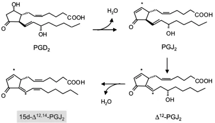
3) 15d-PGJ2의 생성 및 생리적인 역할 COX-2의 활성화로 인해 생성된 프로스타글란 딘 중 PGD2는 탈수과정을 거쳐 PGJ2로 전환되며, PGJ2는 Δ12PGJ2로 전환 후 또 한번의 탈수과정을 거쳐 15d-Δ12,14-PGJ2로 전환된다(Fig. 4).21) 다른 프로스타글란딘과는 달리 15d-PGJ2는 nuclear re- ceptor family 중 하나인 PPAR의 리간드로서 PPAR 활성화를 통해 다양한 생리적인 역할을 수 행한다. PPAR는 서로 다른 유전자에 의해 판독되 는 α, β, γ 세 가지 이성질체로 존재하며, 주로
지방산 및 포도당 대사를 조절하는데 중추적인 역할을 한다.22) 이중 PPARγ는 다양한 암세포에 서 관찰되며, 15d-PGJ2는 PPARγ의 내인성 리간 드로서 염증반응에 관련된 유전자의 발현 억제 및 암 세포의 세포성장 억제 및 세포사멸을 유도 한다(Fig. 5). 또한 15d-PGJ2는 heme oxygenase, HSP40, HSP70, HSP28과 같은 heat shock response 에 관련된 유전자들과 γ-glutamylcysteine synth- ase, thioredoxin reductase, 그리고 glutathione pero- xidase와 같은 산화환원 조절 및 해독화 과정에 관련된 효소 합성을 통해 활성산소종 으로부터 세포를 보호하는 역할을 한다(Fig. 5).23) PPARγ의 리간드로는 프로스타글란딘, 당뇨병 치료제, 지방 산 유도물질들을 비롯한 다양한 NSAIDs들이 알 려져 있다.
(1) 15d-PGJ2의 세포성장 억제 기전: 최근 보고 에 따르면 15d-PGJ2를 비롯한 다양한 PPARγ 리 간드들이 유방암, 대장암, 폐암, 전립선 암, 위암, 췌장암과 같은 다양한 암세포에서 세포사멸을 유
도하며,24∼26) 유방암 세포나 대장암 세포 이식으
로 형성된 마우스의 종양 생성을 억제하는 등 이 들의 항암효과에 대한 많은 연구결과가 보고되고
있다.26∼28) 15d-PGJ2를 비롯한 PPARγ 리간드에
의해 유도되는 세포사멸은 이들 수용체인 PPAR γ의 활성정도와 상관관계가 있음이 보고되었
다.24,29,30) PPARγ의 합성 리간드인 GW7845를 두
Fig. 4. Formation of cyPGs from the cyclopentane PGD
2via dehydration. Asterisks indicate the positions of chemically reactive electrophilic carbon atoms.
OH
O
COOH
OH
O
COOH
OH
*
O
COOH
OH
*
*
*
O
COOH
*
PGD2 PGJ2
∆12-PGJ2 15d-∆12,14-PGJ2
H
2O
H
2O OH
O
COOH
OH
O
COOH
OH
*
O
COOH
OH
*
*
*
O
COOH
*
PGD2 PGJ2
∆12-PGJ2 15d-∆12,14-PGJ2
H
2O
H
2O
달 동안 투여시 nitrosomethylurea에 의해 유도되는 유방암의 발생빈도, 중량 감소가 관찰되었다.31) 최근에는 PPARγ와 무관하게 15d-PGJ2에 의하 여 세포사멸 및 세포성장 억제가 유도됨이 관찰 되었다. 15d-PGJ2를 hepatic myofibroblasts 세포 및 human neuroblastoma cell에 처리시 활성산소종이 생성되며, N-acetyl-L-cysteine (NAC)와 같은 항산 화제 처리로 15d-PGJ2에 의한 세포사멸이 억제됨 이 확인되었다.32,33) 또한 활성산소종에 의해 활성 화되는 mitogen-activated protein kinase (MAPK)와 apoptosis signal-regulating kinase-1 (ASK1)들이 15d-PGJ2에 의해 유도되는 세포사멸 시 활성화됨 이 관찰되었다.34,35)
15d-PGJ2에 의한 세포사멸 및 세포성장 억제에 대한 또 다른 기전으로는 cell cycle 조절 및 세포 사멸에 관련된 단백질의 생합성을 들 수 있다.
Oligonucleotide microarrays 분석을 통해 15d-PGJ2
에 의해 유도되는 neuroblastoma cell의 세포사멸 시 p53의 발현, 인산화 및 DNA binding activity가 증가되며, p53 antisense oligonucleotide 처리로 15d-PGJ2에 의한 세포사멸이 억제됨이 보고되었 다.23) 15d-PGJ2는 cyclin-dependent kinase inhibitor 인 p21waf/cip1, p27과 p18INK4c의 발현 증가를 통해 유방암, 췌장암, 간암세포 등의 성장을 억제하였 다.36∼38)
PPAR 활성화로 인한 유전자의 발현 조절에 대 한 분자적 기전에 대해 명확히 알려진 바는 없지 만, PPARs는 retinoid X receptor (RXR)와 hetero-
dimer를 형성하며 존재하여, 리간드가 없는 상태 에서는 N-CoR 또는 SMART와 같은 nuclear recep- tor corepressor가 결합되어 표적 유전자의 프로모 터 부위와의 결합 억제로 유전자의 발현이 저해 된다. 반면에 PPAR 리간드가 이들 수용체에 결합 하게 되면 수용체의 구조적인 변화로 corepressor 가 떨어져 나가고 CREB binding protein (CBP), CBP homologue p300, SRC-1와 같은 coactivator가 결합함으로서 이들 complex가 적절한 표적 유전 자의 프로모터 부분에 존재하는 peroxisome proli- ferator response element (PPRE)에 결합함으로서 표적 유전자의 전사를 조절할 것으로 보고 있 다.22,39)
또 다른 보고에 따르면 PPARγ 리간드가 tumor suppressor 유전자인 PTEN의 mRNA의 발현을 유 도함으로서, phosphatidylinositol 3-kinase 활성을 억제하고 이로 인해 세포의 생존에 있어서 중요 한 역할을 하는 Akt의 활성 억제를 수반하여 세 포사멸을 유도할 것으로 본다.40,41) 아울러 15d- PGJ2의 cyclopentenone 링에 위치하는 α, β-unsa- turated carbonyl group으로 인해 형성되는 친전자 성 탄소가 단백질과 같은 거대분자의 활성 부위 에 존재하는 잔기와 공유결합물을 형성함으로서 세포의 성장에 관련된 단백질의 기능 손실을 초 래하여 세포 성장을 억제할 것이라는 가능성 또 한 배제할 수 없다.
최근 15d-PGJ2 외에 troglitazone, rosiglitazone, ciglitazone 등과 같은 당뇨병 치료제가 PPARγ 리
Fig. 5. Various biological functions of 15d-PGJ
2.
15d-∆12,14-PGJ2
Apoptosis
Growth of cancer cells Inflammation
Detoxification Inhibition of NF-kB
& AP-1 DNA binding activity
Induction of ROS, PTEN expression, etc
Induction of antioxidative- detoxification enzyme
Induction of cell cycle related proteins expression
15d-∆12,14-PGJ2
Apoptosis
Growth of cancer cells Inflammation
Detoxification Inhibition of NF-kB
& AP-1 DNA binding activity
Induction of ROS, PTEN expression, etc
Induction of antioxidative- detoxification enzyme
Induction of cell cycle related
proteins expression
간드로 작용하여 암세포의 성장억제, 세포사멸 및 분화를 유도한다는 연구 결과들도 있다.27,30) 이런 측면에서 세포의 증식과 종양형성 억제에 대한 PPARγ 리간드의 작용기전에 대해 많은 관심이 집중되고 있다.
(2) PPARγ 리간드에 의한 세포사멸과 COX-2 의 발현: COX-2는 다양한 사이토카인이나 발암물 질에 의해 유도되는 유도성 효소로 염증반응은 물론 세포의 암화과정에 관련되어 있다. COX-2의 과도한 발현이 암조직의 혈관신생 및 전이능을 높이고 세포사멸을 억제하는 것으로 알려져 있다.
PPARγ 리간드에 의한 COX-2의 발현 조절에는 서로 상반된 결과들이 보고되고 있다. PPARγ의 내인성 리간드인 15d-PGJ2이 NF-κB 활성 억제를 통해 lipopolysaccharide와 interleukin-1β (IL-1β)에 의한 COX-2의 발현을 억제하였다.43,44) 이는 15d- PGJ2가 NF-κB의 활성에 있어서 중요한 역할을 하는 IκB kinase의 cysteine 잔기와 공유결합을 형 성함으로서 IκB의 분해를 막아 핵내로의 NF-κB 의 이행을 막거나, NF-κB와 표적 DNA와의 결합 을 방해함으로서 NF-κB에 의해 조절되는 유전자 의 발현을 억제함으로써 일어나는 것으로 짐작된 다.45) COX-2 발현은 NF-κB 외의 다른 전사인자 들에 의해 조절되는데 Subbaramaiah 등46)에 의하 면 15d-PGJ2는 AP-1의 구성성분 중 하나인 c-jun 발현 억제를 통해 COX-2의 promoter 부위에 존재 하는 CRE site와 AP-1과의 complex 형성을 방해 함으로써 phorbol ester에 의한 COX-2의 발현을 저해한다고 제시하였다. 또한 15d-PGJ2에 의한 PPARγ 활성화 시 coactivator인 CBP/p300과 결합 함으로서 AP-1과 같은 다른 전사인자의 활성화에 필요한 coactivator가 상대적으로 부족하여 COX-2 의 발현이 억제될 수 있다.
이와는 반대로 다양한 PPAR 리간드에 의해 COX-2의 발현이 증가되었다는 연구 결과 또한 제시되고 있다. Meade 등47)은 COX 억제제, 기질, 산물들인 PGD2, 15d-PGJ2, PGF2α를 epithelial cells 에 처리시 COX-2의 발현이 증가됨을 보고하였다.
Sulindac에 의해 유도된 oral squamous cells의 세포 사멸 시 COX-2의 발현이 유도되었으며,48) COX-2 의 선택적 억제제인 NS-398에 의한 대장암 세포 의 세포사멸시 COX-2의 발현이 유도되었다.49) 본
실험실의 연구 결과에서도 항암 효과가 있는 alkylisophospholipid 계열인 ET-18-O-CH3이
ras
oncogene으로 변형된 인체유방세포(MCF10A- ras) 의 세포사멸을 유도하는 조건에서 COX- 2 발현 을 유도함을 관찰하였다.50) 또한 15d-PGJ2에 의한 MCF10A-ras cells의 세포사멸 유도 시 COX-2 발 현이 유도되었으며 이러한 현상은 PPARγ anta- gonist인 GW9662에 의해 부분적으로 저해됨을 확 인하였다(H.-K. Na and Y.-J. Surh, unpublished data). 최근 들어 PPARγ 리간드에 의한 세포사멸 유도 시 COX-2의 발현이 증가된다는 보고들이 제시되고 있지만 이에 대한 분자적인 기전이 밝 혀지진 않았다. 그러나 최근 Pang 등51)에 의해 COX-2 promoter에 PPAR 리간드 복합체가 결합하 는 PPRE가 존재함을 확인하였다. NSAIDs를 포함 한 PPARγ 리간드에 의한 세포사멸시 유도되는 COX-2의 발현은 리간드에 의해 활성화된 PPAR γ complex가 COX-2 promoter에 존재하는 PPRE에 결합함으로써 유도되는 것으로 사료된다. 또한 COX-2 발현으로 인해 생성된 다양한 프로스타글 란딘의 상대적 농도, 특히 PGE2와 15d-PGJ2의 생 성 비율이 세포성장과 사멸을 결정하는 중요한 인자로 작용할 것으로 사료된다.결 론
COX-2는 다양한 사이토카인이나 암 촉진제에 의해 유도되는 유도성 효소로서 비정상적인 COX-2의 증가는 암세포의 세포사멸에 저항성을 주고, 세포성장을 촉진하며, 암세포의 침윤성을 증가시킴으로서 암화과정에 있어 중요한 역할을 하는 것으로 알려져 있다. 아울러 COX-2의 주요 산물 중 하나인 PGE2도 EP 수용체 활성화를 통해 암세포의 침윤성에 관련된 유전자의 발현을 조절 함으로서 암화과정에 관여한다는 실험적 증거가 제시되고 있다. 따라서 COX-2와 이의 주요산물인 PGE2의 작용기전에 대한 분자 수준에서의 규명은 암을 치료하거나 예방하는데 좋은 전략이 될 것 으로 기대하고 있다. 실제로 celecoxib를 비롯한 COX-2의 선택적 억제제들의 암세포 증식 억제 효과 및 세포사멸 효과들이 속속들이 보고되고 있다.
그러나 최근 COX-2의 또 다른 최종산물인 15d-PGJ2를 처리시 세포사멸이 유도됨이 보고되 고 있으며, 15d-PGJ2에 의한 세포사멸시 COX-2의 증가가 관찰되기도 한다. 최근 COX-2 promoter 부 위에 PPRE site가 존재함이 확인됨으로서 PPARγ 는 COX-2의 발현을 조절하는 새로운 전사인자로 서 관심을 받고 있다. 위의 여러 보고를 종합해 볼 때 암세포의 성장을 조절하는 측면에서 COX- 2를 비롯한 COX-2 산물들이 갖는 역할은 세포의 종류 및 사용한 약물에 의존적이라 할 수 있으며, 아울러 COX-2에 의해 최종적으로 형성된 프로스 타글란딘 종류의 비율에 따라 그 세포의 증식 및 사멸의 방향이 결정될 것으로 사료된다.
참 고 문 헌