• 검색 결과가 없습니다.

Effects of Bridge Exercise with Pelvic Compression Belt on Electromyographic Activities of Selected Lumbopelvic Muscles in Young Adults with Lumbar Instability

N/A
N/A
Protected

Academic year: 2021

Share "Effects of Bridge Exercise with Pelvic Compression Belt on Electromyographic Activities of Selected Lumbopelvic Muscles in Young Adults with Lumbar Instability"

Copied!
10
0
0

로드 중.... (전체 텍스트 보기)

전체 글

(1)

Research Article Open Access

Effects of Bridge Exercise with Pelvic Compression Belt on Electromyographic Activities of Selected Lumbopelvic Muscles in Young Adults with Lumbar Instability

Hyun-Gyu Cha⋅Yu-Won Choe

1

⋅Yan-Ting Wu

1

⋅Myoung-Kwon Kim

2†

Dept. of Physical Therapy, Joongbu University

1

Dept. of Rehabilitation Sciences, Graduate School, Daegu University

2

Dept. of Physical Therapy, College of Rehabilitation Sciences, Daegu University

Received: April 5, 2017 / Revised: April 6, 2017 / Accepted: May 22, 2017

ⓒ 2017 J Korean Soc Phys Med

| Abstract |

1)

PURPOSE: This study assessed the effect of the pelvic compression belt on the electromyographic activity of erector spinae (ES), internal oblique (IO), rectus femoris (RF), and biceps femoris (BF) after bridge exercise with pelvic belt compression in subjects with lumbar instability.

METHODS: Forty subjects with lumbar instability volunteered for this study. We asked them to perform the bridge exercise while wearing a pelvic belt compression for 30 minutes five times weekly over a six week period. The pelvic compression belt was adjusted just below the anterior superior iliac spines with stabilizing pressure using elastic compression bands during bridge position. Surface electromyographic data were collected from the erector spinae (ES), internal oblique (IO), rectus femoris (RF) and biceps femoris (BF).

RESULTS: After the six week intervention, the ex- perimental group improved significantly. Muscle activation

† Corresponding Author : [email protected]

This is an Open Access article distributed under the terms of the Creative Commons Attribution Non-Commercial License (http://creativecommons.org/licenses/by-nc/3.0) which permits unrestricted non-commercial use, distribution, and reproduction in any medium, provided the original work is properly cited.

significantly decreased in the erector spinae, rectus femoris, and biceps femoris, and increased in the internal oblique mus- cle in bridge position while wearing a PCB (P <.01).

CONCLUSION: Our findings suggest that the bridge exercise with pelvic belt compression is helpful to reduce activation in superficial muscles and lower extremity muscles such as erector spinae (ES), rectus femoris (RF), biceps femoris (BF) and increase activation in deep trunk muscle such as the internal oblique (IO).

Key Words: Lumbar instability, Muscle activation, Pelvic compression belt

Ⅰ. Introduction

The pelvic compression belt (PCB) is effective for stability of the pelvic and lumbar, allowing exercises addressing coordination and stabilization (Vleeming et al., 1992). The PCB facilitates neuromuscular performance and decreases pain in patients with lumbopelvic problems (Lee, 2004).

Spinal instability is commonly considered a part of

chronic LBP (Fritz et al., 1998; Silfies et al., 2009) and

can have a negative effect on trunk alignment (Jeong and

(2)

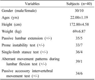
Variables Subjects (n=40)

Gender (male/female) 30/10

Ages (yrs) 22.00±1.19

Height (cm) 172.80±4.58

Weight (kg) 69±6.87

Passive lumbar extension (+/-) 35/5 Prone instability test (+/-) 33/7 Single-limb stance test (+/-) 36/4 Aberrant movement patterns during

lumbar flexion test (+/-) 39/1 Passive accessory intervertebral

movement test (+/-) 34/6

+: positive, -: negative

Table 1. Subject characteristics Kim, 2016). Lumbar segmental instability in the lack of

defects of stability of the lumbar spine has been cited as a main cause of chronic LBP. Abnormal inter-segmental motion and increased movement is frequent in subjects with chronic LBP, often in the absence of other radiological findings (O'Sullivan, 2000). Chronic LBP often causes dysfunction and de-conditioning as well as changes to the neural control system, which influence timing of co-contraction, balance, reflex and righting responses (O'Sullivan et al., 1997).

Proper activation of abdominal muscles is needed in patients with LBP (Bang, 2015; Cho, 2013; Cho et al., 2013; Hwang and Kim, 2011; Kim et al., 2016; van Dieen et al., 2013). Deep trunk muscles are important for trunk stabilization (Gong, 2013; Lee et al., 2016). Deep trunk muscles such as the internal oblique (IO), transverse abdominis (TrA), and lumbar multifidus (LM), important in lumbar stability, activate insufficiently in patients with low back pain (Butcher et al., 2007; Danneels et al., 2002;

Kim et al., 2016). Poor function of trunk deep muscles is associated with LBP (Abdelraouf and Abdel-aziem, 2016). Moreover, activation of deep abdominal muscles such as TrA and IO is delayed and co-activation is absent in patients with low back pain (Massé-Alarie et al., 2012).

Increased activation of deep local muscles could improve spinal joint stability (Vleeming et al., 2014; Unsgaard- Tøndel et al., 2016).

Hu et al. (2010) reported that a PCB during active straight leg raise test (ASLR) improves ASLR performance and reduces abdominal muscle activation. Lumbar stabilization can be achieved by either isometric action of the abdominal muscles or external pelvic compression. Park et al. (2010) and Takasaki et al. (2009) found that a PCB can improve neuromuscular control and prevent unwanted substitution movement during hip joint movement.

Therefore, use of a PCB might improve compensatory movement in a neutral position and provide proper load transfer to the lumbar in females with chronic LBP during

prone hip extension. Furthermore, application of PCB is a conservative method in sacroiliac joint pain therapy (Arumugam et al., 2012a). This belt is inexpensive and has no side effects. There are many studies investigating the muscle activity and mobilization pattern of trunk muscles after wearing a PCB.

Most studies observed immediate change in trunk muscle activity during straight leg raise and bridge exercise while wearing a PCB. However, research on whether any changes in muscle activity of the trunk and lower extremity in bridge position will occur in adults with spinal instability after wearing the PCB is lacking.

Therefore, the purpose of this study was to evaluate the effect of PCB on muscle activity of the trunk and lower extremity muscles in adults with lumbar instability after six week intervention.

Ⅱ. Methods

1. Subjects

Forty subjects with lumbar instability volunteered for

this study (Table 1). With the lumbar apprehension test,

we measured the muscle activity of pelvic limb and trunk

(3)

muscles for 40 adults with potentially unsafe vertebrae while wearing the pelvic compression belt (Kasai et al., 2006; Wadsworth et al., 1988; Tidstrand and Horneij, 2009). Our study is based on the inspection method used in previous research to select subjects with lumbar instability (Hidalgo et al., 2014).

Subjects were included if they scored positive on each of the following tests: passive lumbar extension, prone instability test, single-limb stance test, aberrant movement patterns during lumbar flexion test, and passive accessory intervertebral movement test.

Subjects were excluded if they have had orthopedic operations, neurological disorders, and difficulties performing the tasks required because of limitations in range of motion.

Information on the study and written informed consent according to the ethical standards of the Declaration of Helsinki were provided to all subjects prior to their participation, and all agreed to participate in the experiment.

2. Application of the Pelvic Compression Belt (PCB)

The pelvic compression belt (COMPRESSOR, OPTP, USA) consists of a main belt (body belt) and two compression bands. The main belt is attached to pass the anterior superior iliac spine (ASIS) (Damen et al., 2002), and the two compression bands are attached to the main belt to provide additional pressure. One compression band is attached toward the navel in ASIS to prompt transverse abdominals activity, and the other band to the mean physiognomy from anterior superior iliac spine (ASIS) to prompt multifidus muscles activity (Lee, 2004).

In passive lumbar extension test, the subjects are in prone position. An examiner passively elevates legs with knees extended to 30cm and gently pulls the legs. It is considered positive if pain is present during this test (Kasai et al., 2006). In prone instability test, the patient lies prone on the examining table with legs over the edge and feet resting on the floor. While the patient rests in this position with the trunk muscles relaxed, the examiner applies posterior

to anterior pressure to spinous process of the lumbar spine.

Any provocation of pain is reported. Then the subjects lift the legs off the floor and posterior to anterior compression is applied again to the lumbar spine while the trunk muscle is contracted. The test is considered positive if pain is present in the resting position but subsides in the second position, suggesting lumbopelvic instability. The reliability for this inspection is r=.089 (Wadsworth, 1988). The single leg stance test is considered positive if there is excessive pelvic displacement and there is compensatory movement of the upper and lower extremity while maintaining a one leg standing posture for 20 seconds. The reliability for this inspection is r=.88~1.00 (Tidstrand and Horneij, 2009).

For aberrant movement patterns during lumbar flexion test, we labeled the patient as positive when the patient felt pain at bending from a standing posture, and extending from bending, or reversal phenomenon of lumbopelvic rhythm, grower's sign or thigh climbing, or instability catch (Ahn et al., 2003).

The purpose of passive accessory intervertebral movements is to assess the amount and quality of movement at various intervertebral levels, and to treat pain and stiffness of the cervical and lumbar spine. A posterior to anterior force of varying strength is applied by the examiner either centrally onto the spinous process, or unilaterally on either the left or right articular pillar. The test is considered positive if there was excessive motion of the vertebral body and pain (Hidalgo et al., 2014).

3. EMG Recording and Data Processing

We collected the data using an electromyogram (EMG)

MP150 system (BIOPAC System Inc., CA, USA) to meas-

ure trunk muscle activity. With the subjects in the bridge

position, we obtained four analogue electromyography

signals using the MP 150, then digitized and filtered sig-

nals using Acknowledge 3.7.3 (BIOPAC System Inc.,

Santa Barbara, USA). We established a sampling rate of

(4)

electromyogram signals to 1,000 Hz, and filtered the am- plified waveform using 60~500 Hz band pass filter and 60 Hz notch filter. To quantify the signals, we used the average root-mean-square (%RVC) value, and normal- ized the signals collected from each muscle to reference voluntary contraction (%RVC) (Beimborn and Morrissey, 1988).

To reduce errors due to repeated measurements, we designated electrode sites of 1 cm in diameter and kept each electrode attachment distanced equally. To reduce electrode to skin contact errors, electrodes were attached after removing skin hair at measurement sites and cleaning skin with medical alcohol. The surface electrodes used for collecting electromyogram values were of the disposable Ag/Agcl type of diameter 11.4 mm. For surface EMG, 2-pole electrode shield cable, consisting of ground, active, and reference electrode, was connected to each electrode (Kasman et al., 1998).

We measured EMG electrode finding muscle positions by pressing the muscle to trace the grain direction of muscle fibers (Hungerford et al., 2003). The attachment sites of surface electrodes are as follows (Kasman et al., 1998).

1) for the elector spinae, 2 cm lateral to the spinous process at the L4-5 interspace; 2) for the oblique internal oblique (IO), in the center of the triangle formed by a horizontal line between the anterior superior iliac spine of the innominate and the umbilicus, midline, and the inguinal ligament; 3) for the rectus femoris, the midpoint between the upper margin of the patella and ASIS; and 4) for the long head of the biceps femoris, the midpoint between the gluteal fold and the knee joint.

To determine the pressure area of the pelvis belt, we conducted pre-pressing pelvis inspection in the supine position. We used the method suggested by Lee (1999).

There are four pressure techniques adapted for pelvis pressure method. For sacroiliac articulation pressure, 1) press both iliac spine of pelvis to rear and inner direction,

2) press both iliac spine of pelvis to front and inner direction, 3) press iliac spine of right pelvis to rear and inner direction and left iliac spine to front and inner, and 4) press iliac spine of right pelvis to front and inner direction, left iliac spine to rear and inner direction. We instructed the subjects to perform an active straight leg raise while pressing sacroiliac articulation in the above four ways. We applied pressure in a random sequence. We selected the appropriate way to fasten pelvis belts by how subjects felt it easiest to do active straight leg raises when pressure was applied randomly.

The bridging position method while wearing the pelvic belt is as follows. At the beginning position, subjects bend their knees to 90 degrees, open their arms to 30 degrees, and have their palms face the ground. The spacing between feet is 20 cm and subjects place their feet in a straight line. Subjects keep their head and neck straight and look at the ceiling while a supervisor instructs the subject to pull the navel toward the spine as if exhaling.

We conducted the bridge exercise to measure muscle activities for reference voluntary contraction (RVC) of pelvic limb and trunk muscles (Choi et al., 2015; Kim et al., 2013). We first obtain data value for five seconds at maximal voluntary isometric contraction, and we use the average of the EMG signal for three seconds, excluding the first and last second as %RVC. This procedure is conducted three times.
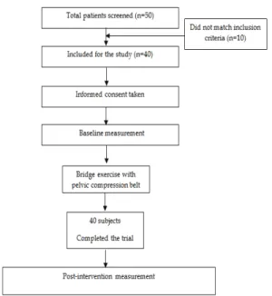
4. Procedures

We asked subjects to perform the bridge exercise

with PCB for 30 minutes five times weekly over a six

week period (Fig. 1). All procedures for EMG measure-

ments were performed with the subject in the bridge po-

sition on the table. The EMG activities were measured

in the erector spinae (ES), internal oblique (IO), rectus

femoris (RF) and biceps femoris (BF) while performing

the bridge position.

(5)

Pre test Post test Change value Effect size t

Erector spinae 112.16±16.24

a

85.16±13.80 27.00±13.42 2.84 9.00**

Internal oblique 81.16±12.52 96.30±12.19 15.13±15.24 1.40 4.44**

Rectus femoris 104.08±14.22 82.48±7.52 21.60±12.28 2.48 7.87**

Biceps femoris

(lateral) 106.98±16.35 88.48±10.17 18.50±16.03 1.63 5.16**

** Significant intergroup difference in the gains achieved ( p <.01)

a

Effect size>.70

Table 2. Pre- and Post-intervention electromyography values of subjects with and without pelvic belt (values are presented as means (standard deviations)) (unit: %RVC) (n=40)

Fig. 1. Study flowchart. PCB; Pelvic compression belt

5. Statistical Analysis

The data are described as the mean±SD. Outcome variables with and without PCB were compared using paired samples t-tests. The statistical software SPSS (IBM Corp. Released 2011. IBM SPSS Statistics for Windows, Version 20.0. Armonk, NY: IBM Corp.) was used for statistical analysis. Null hypotheses of no difference were rejected if p-values were less than .05. The sample size for this study was calculated using the G* power program 3.1.0 (G power program Version 3.1, Heinrich-Heine- University Dusseldorf, Germany). Based on the data from

the pilot study, the estimated sample to obtain a power of minimum 80% at an alpha level of 95% was 40 participants.

Intergroup effect sizes were calculated using the Cohen d coefficient. An effect size < .2 reflects a negligible mean difference; .2~.5, a small difference; .5~.8, a moderate mean difference; and > .8, a large difference. Statistical analysis was performed at a 95% confidence level.

Ⅲ. Results

After the six week intervention, the experimental group improved significantly in decreased use of superficial muscles; ES (without the pelvic compression belt, 112.16±16.24% RVC; with the pelvic compression belt, 85.16±13.80% RVC), RF (without the pelvic compression belt, 104.08±14.22% RVC; with the pelvic compression belt, 82.48±7.52% RVC), and BF (without the pelvic compression belt, 106.98±16.35% RVC; with the pelvic compression belt, 88.48±10.17% RVC), and deep trunk muscle activation significantly increased; IO (without the pelvic compression belt, 81.16±12.52% RVC; with the pelvic compression belt, 96.30±12.19% RVC) in bridge position while wearing a PCB (P <.01) (Table 2).

The effect size for gains when using the PCB was small

in erector spinae (Large), internal oblique (Large), rectus

femoris (Large), and biceps femoris (lateral) (Large).

(6)

Ⅳ. Discussion

We evaluated the muscle activities of pelvic, trunk, and lower extremity muscles after bridge exercise with PCB for adults who have potentially lumbar instability. While wearing the PCB, muscle activities are reduced in the ES, RF, BF muscles, while muscle activity increased in IO muscle. The reason for choosing the RF and BF muscle is that the reduction of the ES of the trunk and increase of the IO due to wearing the pelvic belt may increase the stability of the lumbar and reduce unnecessary hyperactivity of the RF and BF in the lower extremity (Jung et al., 2013, Jung et al., 2012).

Bridge position is widely used for lumbo-pelvic stabilization in patients with low back pain, and it activates abdominal muscles and low extremity muscles (Kong et al., 2013; Yoo et al., 2016). Many studies used bridge position to measure muscle activation of abdominal muscles and lower extremities (Kong et al., 2013; Park et al., 2014;

Stevens et al., 2006). A previous study used bridge position to measure muscle activations of ES, IO, RF, BF while the patients were wearing a PCB (Cha et al., 2016). Thus our study selected this position to identify muscle activations of abdominal muscles (ES, IO) and lower extremity muscles (RF, BF).

The main finding of our study is that activation of the core muscles such as IO muscle was greatly increased with the PCB, while global muscles including ES and RF declined. Poor motor control is related to lumbar instability, and weakness of deep trunk muscles caused trunk instability, a risk factor for chronic low back pain (Soundararajan and Thankappan, 2016; Hides et al., 1996;

Richardson et al., 2004). Internal oblique (IO) muscles are important for spine stabilization (Cho, 2013). In our study, we find superficial muscle activity is reduced and deep muscle increased after mediation. When a normal person's coxa is bent or extended, contraction of their deep trunk muscles before the agonist movement is generated, and this effect produces lumbosacral site stabilities (Richardson

et al., 2002). Deep muscles in the trunk can adjust the movement of lumbosacral site during functional movement by increasing stability of the spinal segment (Pool- Goudzwaard et al., 1998). This result is broadly similar to studies that investigated the effects of pelvic belts on patients with back instability.

According to Jung et al. (2013), use of the PCB during one-leg standing helped activate the gluteus maximus while inhibiting activation of the biceps femoris in both the sacroiliac joint pain patients and the asymptomatic patients.

Use of the PCB increased gluteus maximus activity and decreased biceps femoris activity during walking (Hu et al., 2010). During walking, the initial contact and loading response events require hip extensor muscular effort to maintain sacroiliac joint stability, and to transmit weight load and ground reaction force efficiently (Hossain and Nokes, 2008). Therefore, during the loading response, lumbopelvic stability may be important for balanced muscle control between the gluteus maximus and the biceps femoris (Hossain and Nokes, 2005). Use of a PCB reduces the gluteus maximus (GM) and elector spinae (ES) activity, which could be due to decreased unwanted movement, such as anterior pelvic tilt or excessive lumbar extension during prone hip extension test (PHE) in females with chronic low back pain (CLBP) (Oh, 2014).

External pelvic compression applied with a pelvic belt can augment the stability of the pelvic joints, and alter neuromotor control of the lumbopelvic and thigh muscles (Arumugam et al., 2012b). The mechanisms of pelvic compression belts are unclear. However, one theory suggests that sacroiliac joint stiffness can be improved when a pelvic compression belt is worn (Damen et al., 2002).

Brizzolara et al. (2015) hypothesize that the behavior of

the deep abdominal muscles could be influenced by this

enhanced stiffness and therefore, the proportionate change

of muscles thickness during the ASLR would be different

while wearing the PCB. However, this was not demon-

strated in the results. It appears that although PCB can

be used to address the passive systems of the pelvis, specific

(7)

localized exercises are necessary to address the ‘active’

deep abdominal muscles to improve change during the ASLR.

Patients with low back pain have a lack of proprioception that can cause impairment of position sense and trunk instability (Kong et al., 2015). A pelvic compression belt could provide proprioceptive input (Arumugam et al., 2012a, Arumugam et al., 2015). Kinesiology taping that supports lumbar muscles could normalize muscle function by activating mechanical receptors (Fong et al., 2015;

Paoloni et al., 2011). Our intervention method, the pelvic compression belt, might change trunk muscle activation by affecting sensory receptors on the trunk.

Most patients with chronic LBP lack motor control of trunk muscles (Halliday et al., 2016). Patients with chronic LBP showed changed activation of trunk muscles such as increased activation of superficial muscles and decreased activation of deep muscles (Smith and Kulig, 2016;

Danneels et al., 2016). Using a PCB could change muscles recruitment through neurophysiological effects (Arumugam et al., 2012b, Arumugam et al., 2015). Likewise, our intervention might have an effect on the activation of trunk muscles by changing muscle recruitment.

The present study has some limitations. First, the small sample size may have influenced variability of results.

Second, the absence of observation after the end of the pelvic belt intervention does not allow for determination of the durability of the effect of this therapy.

Ⅴ. Conclusion

In our study, activation of ES, RF, and BF significantly decreased and deep trunk muscle activation (IO) significantly increased after bridge exercise with PCB. For this reason, we suggest that the bridge exercise with PCB helps reduce muscle activation in unnecessary superficial muscles and lower extremity and increase activation of deep trunk muscle. It could have a positive effect on the

patients with low back pain and dysfunction of lumbar structures due to the lumbar instability.

References

Abdelraouf OR, Abdel-aziem AA. The relationship between core endurance and back dysfunction in collegiate male athletes with and without nonspecific low back pain. Int J Sports Phys Ther. 2016;11(3):337.

Ahn NU, Hammerberg KW, DeWald CJ, et al.

Spondylolisthesis: classification, diagnosis, and natural history. In Seminars In Spine Surgery.

2003;15(2):112-24.

Arumugam A, Milosavljevic S, Woodley S, et al. Can applica- tion of a pelvic belt change injured hamstring muscle activity?. Medical Hypotheses. 2012a;78(2):277-82.

Arumugam A, Milosavljevic S, Woodley S, et al. Effects of external pelvic compression on form closure, force closure, and neuromotor control of the lumbopelvic spine–A systematic review. Man Ther. 2012b;17(4):

275-84.

Arumugam A, Milosavljevic S, Woodley S, et al. The effect of a pelvic compression belt on functional hamstring muscle activity in sportsmen with and without previous hamstring injury. Int J Sports Phys Ther.

2015;10(3):291.

Bang HS. The Effects of Lumbar Stabilization Exercise on Muscle Activity and Isokinetic Muscle Strength of Female Patients with Chronic Low Back Pain. J Korean Soc Phys Med. 2015;10(2):63-71.

Beimborn DS, Morrissey MC. A review of the literature related to trunk muscle performance. Spine. 1988;

13(6):655-60.

Brizzolara K., Wang-Price S, Roddey T. The effect of pelvic

compression on deep abdominal muscle thickness

during the active straight leg raise test. Int J Ther

Rehabil. 2015;22(2):60-7.

(8)

Butcher SJ, Craven BR, Chilibeck PD, et al. The effect of trunk stability training on vertical takeoff velocity.

J Orthop Sports Phys Ther. 2007;37(5):223-31.

Cha HG, Wu YT, Kim MK. The effects of a pelvic belt on trunk and lower extremity muscles in the bridge position. J Phys Ther Sci. 2016;28(12):3293-5.

Choi SA, Cynn HS, Yi CH, et al. Isometric hip abduction using a Thera-Band alters gluteus maximus muscle activity and the anterior pelvic tilt angle during bridging exercise. J Electromyogr Kinesiol. 2015;

25(2):310-5.

Cho MS. The effects of modified wall squat exercises on average adults’ deep abdominal muscle thickness and lumbar stability. J Phys Ther Sci. 2013;25(6):689-92.

Cho SH, Kim JH, Choi MH. The Effect of Short-term Lumbar Stabilization Exercise for Lumbar Muscle Strength and Postural Balance on Chronic LBP. J Korean Soc Phys Med. 2013;8(3):295-302.

Damen L, Spoor CW, Snijders CJ, et al. Does a pelvic belt influence sacroiliac joint laxity?. Clinical Biomechanics. 2002;17(7):495-8.

Danneels L, Cagnie B, D'hooge R, etal. The effect of experimental low back pain on lumbar muscle activity in people with a history of clinical low back pain:

a muscle functional MRI study. J Neurophysiol.

2016;115(2):851-7.

Danneels L, Coorevits P, Cools A, et al. Differences in electromyographic activity in the multifidus muscle and the iliocostalis lumborum between healthy subjects and patients with sub-acute and chronic low back pain. Eur Spine J. 2002;11(1):13-9.

Fong SS, Tam YT, Macfarlane DJ, et al. Core muscle activity during TRX suspension exercises with and without kinesiology taping in adults with chronic low back pain: implications for rehabilitation. Evid Based Complement Alternat Med. 2015.

Fritz JM, Erhard RE, Hagen BF. Segmental instability of the lumbar spine. Phys Ther. 1998;78(8):889-96.

Gong WT. Correlations between transversus abdominis thickness, lumbar stability, and balance of female university students. J Phys Ther Sci. 2013;25(6):

681-3.

Halliday MH, Pappas E, Hancock MJ, et al. A randomized controlled trial comparing the McKenzie method to motor control exercises in people with chronic low back pain and a directional preference. J Orthop Sports Phys Ther. 2016;46(7):514-22.

Hidalgo B, Hall T, Nielens H, et al. Intertester agreement and validity of identifying lumbar pain provocative movement patterns using active and passive ac- cessory movement tests. J Manipulative Physiol Ther. 2014;37(2):105-15.

Hides JA, Richardson CA, Jull GA. Multifidus Muscle Re- covery Is Not Automatic After Resolution of Acute, First ‐Episode Low Back Pain. Spine. 1996;21(23):

2763-9.

Hossain M, Nokes LD. A biomechanical model of sacro-iliac joint dysfunction as a cause of low back pain. J Bone Joint Sur. 2008;90:222-3.

Hossain M, Nokes LD. A model of dynamic sacro –iliac joint instability from malrecruitment of gluteus maximus and biceps femoris muscles resulting in low back pain. Medical Hypotheses. 2005;65(2):278-81.

Hu H, Meijer OG, Van Dieen JH, et al. Muscle activityduring the active straight leg raise (ASLR), and the effects of a pelvic belt on the ASLR and on treadmill walking. J Biomech. 2010;43(3):532-9.

Hungerford B, Gilleard W, Hodges P. Evidence of altered lumbopelvic muscle recruitment in the presence of sacroiliac joint pain. Spine. 2003;28(14):1593-600.

Hwang BJ, Kim JW. Effects of Lumbar Stabilization Exercise on Lumbar and Lower Extremity Strength of the Elderly women. J Korean Soc Phys Med. 2011;

8(3):267-75.

Jeong EY, Kim SY. The Effect of Lumbar Stabilization

Exercise and The Neurodynamic Technique on

(9)

Patients with Low Back Pain and Lumbar Instability.

J Korean Soc Phys Med. 2016;11(4):115-25.

Jung HS, Jeon HS, Oh DW, et al. Effect of the pelvic compression belt on the hip extensor activation patterns of sacroiliac joint pain patients during one-leg standing: a pilot study. Man Ther. 2013;18(2):143-8.

Jung HS, Jeon HJ, Yi CH, et al. Effect of Applying the pelvic compression belt on the trunk and hip extensor electromyography pattern in female patients with sacroiliac joint pain during the one leg standing. Phys Ther Kor. 2012;19(2):1-11.

Kasai Y, Morishita K, Kawakita E, et al. A new evaluation method for lumbar spinal instability: passive lumbar extension test. Phys Ther. 2006;86(12):1661-7.

Kasman GS, Cram JR, Holtz J. Introduction to Surface Electromyography. Canada. Aspen Publeshers. 1998.

Kim HK, Kim CY, Kang JH, et al. Influence of the Supplementary Ki-hap Technique and Verbal Encouragement on Abdominal Muscle Activation during Crunch Exercise in Healthy Subjects: A Pilot Randomized Controlled Trial. J Korean Soc Phys Med, 2016;11(2):53-62

Kim MJ, Oh DW, Park HJ. Integrating arm movement into bridge exercise: Effect on EMG activity of selected trunk muscles. J Electromyogr Kinesiol. 2013;23(5):

1119-23.

Kim SY, Kim JH, Jung GS, et al. The effects of transcutaneous neuromuscular electrical stimulation on the activation of deep lumbar stabilizing muscles of patients with lumbar degenerative kyphosis. J Phys Ther Sci.

2016;28(2):399-406.

Kong YS, Cho YH, Park JW. Changes in the Activities of the Trunk Muscles in Different Kinds of Bridging Exercises. J Phys Ther Sci. 2013:25(12):1609–12.

Kong YS, Lee WJ, Park S, et al. The effects of prone bridge exercise on trunk muscle thickness in chronic low back pain patients. J Phys Ther Sci. 2015;27(7):

2073-6.

Lee DG. The pelvic girdle. An approach to the examinationand treatment of the lumbo-pelvic-hip region (2nd ed).

Edinburgh. Churchill Livingstone. 1999.

Lee DG. The Pelvic Girdle: An approach to the examination and treatment of the lumbo-pelvic-hip region.

Churchill Livingstone. 2004.

Lee DG. The pelvic girdle: an integration of clinical expertise and research (3rd ed). China. Elsevier Health Sciences.

2004.

Lee TG, Park CH, Son HH. The Effects of a Bridging Exercise Applying Changes in the Base of Support for the Shoulders on Trunk Muscle Activation. J Korean Soc Phys Med. 2016;11(3):97-104

Massé-Alarie H, Flamand VH, Moffet H, et al. Corticomotor control of deep abdominal muscles in chronic low back pain and anticipatory postural adjustments.

Experimental brain research. 2012;218(1):99-109.

Oh JS. Effects of pelvic belt on hip extensor muscle EMG activity during prone hip extension in females with chronic low back pain. J Phys Ther Sci. 2014;

26(7):1023-4.

O'sullivan PB. Masterclass. Lumbar segmental ‘instability’:

clinical presentation and specific stabilizing exercise management. Man Ther. 2000;5(1):2-12.

O'Sullivan PB, Twomey L, Allison GT. Dysfunction of the neuro-muscular system in the presence of low back pain—implications for physical therapy management.

J Man Manip Ther. 1997;5(1):20-6.

Paoloni M, Bernetti A, Fratocchi G, et al. Kinesio Taping applied to lumbar muscles influences clinical and electromyographic characteristics in chronic low back pain patients. Eur J Phys Rehabil Med. 2011;

47(2):237-44.

Park HJ, Oh DW, Kim SY. Effects of integrating hip move- ments into bridge exercises on electromyographic activities of selected trunk muscles in healthy individuals. Man Ther. 2014;19(3):246-51.

Park KM, Kim SY, Oh DW. Effects of the pelvic compression

(10)

belt on gluteus medius, quadratus lumborum, and lumbar multifidus activities during side-lying hip abduction. J Electromyogr Kinesiol. 2010;20(6):

1141-5.

Pool-Goudzwaard AL, Vleeming A, Stoeckart R, et al.

Insufficient lumbopelvic stability: a clinical, anatomical and biomechanical approach to ‘a-specific’

low back pain. Man Ther. 1998;3(1):12-20.

Richardson C, Hodges PW, Hides J, et al. Manipulation Association of Chartered Physiotherapists. Therapeutic Exercise for Lumbopelvic Stabilization: A Motor Control Approach for the Treatment and Prevention of Low Back Pain. New York. Churchill Livingstone.

2004.

Richardson CA, Snijders CJ, Hides JA, et al. The relation between the transversus abdominis muscles, sacroiliac joint mechanics, and low back pain. Spine. 2002;

27(4):399-405.

Silfies SP, Mehta R, Smith SS, et al. Differences in feedforward trunk muscle activity in subgroups of patients with mechanical low back pain. Arch Phys Med Rehabil.

2009;90(7):1159-69.

Smith JA, Kulig K. Altered multifidus recruitment during walking in young asymptomatic individuals with a history of low back pain. J Orthop Sports Phys Ther.

2016;46(5): 365-74.

Soundararajan LRA, Thankappan SM. Efficacy of the Multifidus Retraining Program in Computer Professionals with Chronic Low Back Pain. Asian Spine J. 2016;

10(3):450-6.

Stevens VK, Bouche KG, Mahieu NN, et al. Trunk muscle activity in healthy subjects during bridging

stabilization exercises. BMC Musculoskelet Disord.

2006;7(1):75.

Takasaki H, Iizawa T, Hall T, et al. The influence of increasing sacroiliac joint force closure on the hip and lumbar spine extensor muscle firing pattern. Man Ther.

2009;14(5):484-9.

Tidstrand J, Horneij E. Inter-rater reliability of three standardized functional tests in patients with low back pain. BMC Musculoskelet Disord. 2009;10(1):58.

Unsgaard-Tøndel M, Vasseljen O, Woodhouse A, et al.

Exercises for Women with Persistent Pelvic and Low Back Pain after Pregnancy. Glob J Health Sci.

2016;8(9):107.

van Dieen JH, Selen LP, Cholewicki J. Trunk muscle activation in low-back pain patients, an analysis of the literature.

J Electromyogr Kinesiol. 2013;13:333–51.

Vleeming A, Buyruk HM, Stoeckart R, et al. An integrated therapy for peripartum pelvic instability: a study of the biomechanical effects of pelvic belts. Am J Obstet Gynecol. 1992;166(4):1243-7.

Vleeming A, Schuenke MD, Danneels L, et al. The functional coupling of the deep abdominal and paraspinal muscles: the effects of simulated paraspinal muscle contraction on force transfer to the middle and posterior layer of the thoracolumbar fascia. J Anat. 2014;

225(4):447-62.

Wadsworth CT. Manual examination and treatment of the spine and extremities. Baltimore. Williams &

Wilkins. 1988.

Yoo WG. Effects of bridging plus exercises with heel lift on lower extremity muscles. J Phys Ther Sci. 2016;

28(5):1582-3.

수치

Table 1. Subject characteristicsKim, 2016). Lumbar segmental instability in the lack of
Table 2. Pre- and Post-intervention electromyography values of subjects with and without pelvic belt  (values are presented as means (standard deviations)) (unit: %RVC) (n=40)

참조

관련 문서

This study aims to examine the effects of fun factors on exercise commitment and schol happiness in school sports club activity.. The population was

This study investigates the effect of complex exercise program activities using Motion-based games on high school students with developmental disabilities on

In conclusion, applications of combined exercise, which can be easily carried out in one’s daily life for elderly women, would be helpful for their

With the opening of the Songdo Bridge 4, IFEZ is looking forward to less congestion in traffic that connects from Songdo International City locations, including Incheon New

The purpose of this study was to investigate how the BOSU Ball exercise, which can develop core muscles in middle-aged women, affects body composition and

In conclusion, Pilates chair exercise is a positive factor in improving skeletal muscle mass among menopausal women's body composition and lower muscles EMG

The Relationship between Physical Activities and Health-related Quality of Life in Korean Adults with Diabetes

Eucommia ulmoides is a Traditional Chinese Medicine that has been used for longevity, being strengthen the muscles and joints. While traditional application of Eucommia was