*동아대학교 의과대학 동아대학교병원 흉부외과학교실
Department of Thoracic and Cardiovascular Surgery, Dong-A University Hospital, College of Medicine, Dong-A University
**동아대학교 의과대학 동아대학교병원 병리학교실
Department of Pathology, Dong-A University Hospital, College of Medicine, Dong-A University
†본 논문은 2006학년도 동아대학교 학술연구조성비에 의하여 연구되었음.
논문접수일:2007년 9월 12일, 심사통과일:2007년 10월 15일
책임저자:노미숙 (602-715) 부산시 서구 동대신동 3가 1번지, 동아대학교병원 해부병리과 (Tel) 051-240-2833, (Fax) 051-247-8753, E-mail: msroh@dau.ac.kr
본 논문의 저작권 및 전자매체의 지적소유권은 대한흉부외과학회에 있다.
식도암에 있어서 Fascin의 발현과 예후와의 상관관계에 대한 연구
최필조*ㆍ정상석*ㆍ방정희*ㆍ조광조*ㆍ우종수*ㆍ노미숙**
Independent Prognostic Value of the Fascin Expression in Patients with Esophageal Cancer
Pill Jo Choi, M.D.*, Sang Seok Jeong, M.D.*, Jung Heui Bang, M.D.*, Kwang Jo Cho, M.D.*, Jong Soo Woo, M.D.*, Mee Sook Roh, M.D.**
Backgrond: Fascin is an actin-bundling protein that induces membrane protrusions and it increases cell motility in various transformed cells. Esophageal cancer is one of the most lethal malignancies, and it exhibits extensive local invasion or frequent regional lymph node metastasis even after curative surgery. We investigate the expression of fascin by performing immunohistochemistry to evaluate the clinical characteristics and prognostic significance of its expression in esophageal cancer patients. Material and Method: Immunochemistry for fascin was performed on 76 tumor samples from 76 patients who underwent esophageal cancer operations. The expression levels of fascin in the 76 esophageal cancer tissues were compared with those in the corresponding normal esophageal epithelium.
The fascin-positive samples were defined as those showing more than 75% of fascin-positive cells. Result: Overall, a fascin positive expression was detected in 39 (51.3%) out of the total 76 cases. The tumors with positive fascin expression tended to more frequently show a higher stage (p=0.030), and a higher T-factor (p=0.031). The prog- nosis of the fascin negative group was significantly better than that of the fascin positive group (p=0.004).
Multivariate analysis revealed that lymphovascular invasion and the fascin expression were independent prognostic factors. Conclusion: Fascin was expressed in 51.3% of the esophageal cancer tissues, and a positive expression of fascin was associated with more advanced tumor progression and recurrence. Our study suggests that the fas- cin expression may be an independent prognostic factor for an unfavorable clinical course for those patients suffer- ing with esophageal cancer.
(Korean J Thorac Cardiovasc Surg 2008;41:74-81) Key words: 1. Esophageal neoplasms
2. Immunohistochemistry 3. Neoplasm proteins 4. Neoplasm marker
서 론
식도암은 근치적 수술 후에도 5년 생존율이 20∼30%를
보일 정도로 굉장히 치명적인 암의 하나이다. 이처럼 좋 지 못한 예후를 보이는 이유는 식도암이 조기에 광범위한 국소침윤이나 국소 임파절 전이를 자주 일으키는데 기인
한다고 할 수 있다.
국소침윤과 원격전이 발생에 있어서 암세포, 세포외바 탕질(extracellular matrix) 단백, 그리고 내피세포 사이의 상 호 작용이 기본적인 중요한 역할을 한다. 정상 상피 조직 에서는 세포-바탕질(cell-matrix)과 세포-세포사이의 유착성 상호작용이 세포층의 정상적인 구성과 안정화에 중요한 역할을 한다. 그러나 종양의 침습과 전이의 경우에는 종 종 세포와 세포사이의 유착(adhesion)과 연접부 교통 (junctional communication)의 소실 분만 아니라 세포막 돌 출이 나타면서 세포 모양이 바뀌는 등의 특징을 가진다 [1-3]. 이런 변화의 많은 부분이 몇몇 actin cross-linking protein의 작용에 의한 세포골격잔섬유(cytoskeletal micro- filament)의 재조합에 기인한다고 한다[4-6].
Fascin은 55 kDa의 actin-bundling protein으로 filopodia, spikes, lamelipodial rib, 수상돌기(dendrite) 그리고 미세융모 (microvilli) 같은 actin-based structure의 기질화에 중요한 역 할을 한다[7]. Fascin의 과발현은 세포와 세포사이의 유착 을 감소시키고 세포의 선도 가장자리(leading edge)에서 세 포막 돌출을 야기하고 상피 세포의 운동성을 상당히 증가 시킨다고 알려져 있다[7]. 정상 상피 세포에서는 fascin 발 현이 없거나 그 정도가 보통 매우 낮으나 몇몇 상피성 종 양의 경우 fascin 발현이 종종 높은 경우가 보고되며 이 들 종양 세포에서 fascin의 높은 발현은 병이 진행성이고 전이성 경향이 높은 종양과 연관성이 있다고 보고된다 [8-13].
현재의 예후 인자들은 개별적 위험 인자에 대한 정확하 고 충분한 정보를 제공하지 못하는 것이 현실적인 상황이 며 식도암에 대한 새로운 예후 결정 지침과 치료 지침이 필요한 현실이다. 본 연구의 목적은 식도암에 있어서 fas- cin에 대한 연구가 국내에서는 거의 없는 상황에서 면역 조직화학 염색 방법을 이용하여 fascin의 발현 양상을 알 아보고 발현 여부에 따른 임상 및 병리학적 특성과 예후 인자로서의 의의 등을 알아보고자 하였다.
대상 및 방법
1) 연구 대상
1995년 1월부터 2005년 12월까지 식도의 편평상피암으 로 진단되어 근치적 식도암 절제 수술을 받았으며 파라핀 포매괴의 보존상태가 양호한 76명의 환자를 대상으로 하 였다. 수술 전 화학요법 또는 방사선 치료를 시행한 환자, 수술 중에 원격 전이가 발견되었거나 절제연에 육안적,
현미경적 잔존암이 남은 환자와 수술 후 합병증으로 사망 한 환자는 대상에서 제외하였다. 이들 76명을 대상으로 하여 임상적 자료를 후향적인 방법으로 조사하였다.
2) 연구방법
(1) 면역조직화학 염색 및 판독: 대상 환자 76명으로부 터 수술 시 절제된 후 보관된 조직을10% 중성 포르말린 에 고정하여 파라핀에 포매한 조직을 4μm 두께로 박절하 여 probe-on 슬라이드에 부착하여 자일렌으로 파라핀을 제 거한 후 100%, 90%, 80% 및 70% 알코올에 순서대로 1분 씩 두어 함수시켰다. 조직을 포르말린에 고정하고 파라핀 으로 포매하는 과정에서 감추어졌던 항원을 노출시키고 복구하여 예민도를 높이기 위하여 citrate 완충액(pH 6.0)에 조직 슬라이드를 담근 후 700 W 마이크로파로 5분간 2회 처리하였다. PBS 완충액으로 씻은 후 3% 과산화수소수로 5분간 처리하여 세포내의 내인성 과산화효소 활성을 저지 시킨 다음 면양 혈청에 5분간 접촉시켜 비특이적 항체 결 합을 억제시켰다. 단클론 항체인 mouse anti-fascin (Dako- Cytomation, Carpinteria, Ca, USA, A;50)을 떨어뜨려 습윤 상태의 실온에서 1시간 반응시킨 후 PBS 완충액으로 5분 간 2번 씻었다. Anti-mouse IgG 2차 항체(LSAB+ Kit, DakoCytomation, Denmark)로 30분간 반응시켜 PBS 완충액 으로 씻은 후 streptavidin peroxidase로 30분간 염색하였다.
다시 PBS 완충액으로 씻고 발색시약 3-amino-9-ethyl- carbazole (AEC)로 10분간 염색한 후 물로 세척하고 hema- toxylin으로 3분간 대조 염색을 한 후 다시 물로 씻고 crys- tal mount로 봉입하여 광학 현미경으로 검경 하였다.
염색된 식도암 조직을 현미경으로 관찰하여 염색 강도가 internal control인 내피세포(endothelial cell)와 같거나 더 진 하고 염색된 세포의 수가 전체 암 조직의 75% 이상일 때 fascin 양성으로 판독하였다.
(2) 분석 및 통계학적 처리: 환자의 임상 기록지와 병 리 조직 판독지로부터 임상 자료를 후향적으로 분석하였 고 면역조직화학염색은 대상환자의 생존 유무를 포함한 임상자료가 없는 병리의에 의해 실시되었다. 통계분석은 SPSS 12.0 version (SPSS Inc., Chicago, IL, USA)을 사용하 였다. Fascin의 발현 여부와 임상병리학적 요소들과의 관 계는 카이 제곱 검정을 이용하여 분석하였다. 환자들의 생존율과 무병생존율은 Kaplan-Meier 방법을 이용하였고 Log rank test로 생존율을 비교하였다. 식도암의 예후와 관 련된 인자를 알아보기 위해 Cox's proportional hazard mod- el을 이용하여 다변량 분석을 하였다. 통계적 유의성은 p
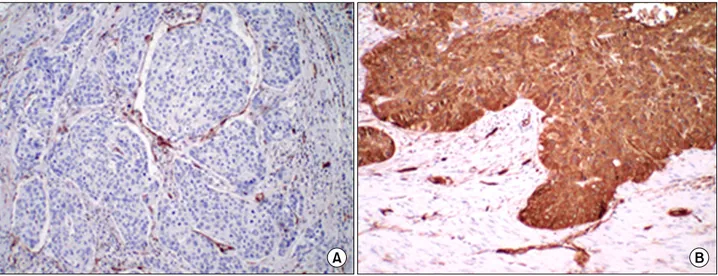
Fig. 1. Typical results of fascin immunohistochemical staining. (A) fascin negative: In normal esophageal epithelium, fascin in invariably ex- pressed in endothelial cells of microvessels as well as the stromal cells of the interstitium (×100). (B) fascin positve: ESCC demostrating positive immunoreacitivity for fascin (×100).
<0.05로 하였다.
결 과 1) Clinicopathological chracteristics
환자의 연령은 29세에서 78세로 평균 59.3세였다. 남녀 비는 69:7로 남자가 월등히 많았다. 수술 후 병기는 I기 17 명, II기 34명, III기 25명이었다. 조직학적 분화정도는 well differentiated 27명(35.5%), moderately differentiated 35명 (46.1%), poorly differentiated 14명(18.4%)이었다. 종양의 침 습이 점막이나 점막하부까지인 경우(T1)가 21명(27.6%), 근육층까지(T2)가 13명(17.1%), 그 이상인 T3가 40명(52.6%), T4가 2(2.6%)명이었다. 국소 임파절 전이가 없었던 경우 (N0)가 40명(52.6%), 있었던 경우(N1)가 36명이었다. 림프 관ㆍ혈관 침범(lymphovascular invasion)이 있었던 경우가 50명이었으며 신경주위침범(perineural invasion)을 보였던 경우는 61명이었다.
2) The relationship between clincopathological char- acteristics and the expression of fascin
전체 76명의 식도암 환자 조직에서 면역조직화학적 염 색 결과에 따른 fascin의 발현 양성은 39명(51.3%), 발현 음성은 37명(48.7%)이었다(Fig. 1). Fascin 발현과 각 임상 및 병리학적 요인과의 연관성을 조사하였다. T 인자(p=0.031) 와 병기(p=0.030)가 fascin의 양성발현과의 통계적 유의성
이 있음을 보였다. 그러나 환자의 연령, 성별, 조직학적 분 화도, 임파절의 전이유무, 림프관ㆍ혈관 침범 여부, 신경 주위 침범 여부 등과는 통계적 유의성을 보이지 못하였다 (Table 1).
3) Influence of fascin expression on recurrence and survival
중앙 추적관찰기간은 38개월(범위, 3∼95개월)이었다.
29명은 추적기간 중 사망하였다. 사망 원인은 17명은 식 도암의 진행으로 인하여 사망하였고 나머지는 식도암 이 외의 원인으로 사망하였다. 재발은 33명에서 발생하였는데 국소재발이 15명, 원격전이가 18명에서 발생하였다. Fascin 의 발현 양상에 따른 종양의 재발과 환자의 사망과의 상 관성을 조사에서 fascin의 양성 발현군이 음성군에 비해서 종양의 재발(p=0.005)과 환자의 사망(p=0.000)에 있어서 통 계적 유의성이 있게 높음을 알 수 있었다(Table 2).
각 임상병리학적 인자들의 예후인자로서 단변량 분석 결과 임파절의 전이유무, 림프관ㆍ혈관 침범 여부, 신경주 위침범 여부, 병기, fascin 발현 여부 등이 통계적으로 유 의한 인자들로 나타났다(Table 2). Fascin 발현이 양성인 경우 5년 생존율이 32% 이었고, 발현이 음성인 경우 58%
로 양성 발현군의 예후가 유의하게 불량하였다(Fig. 2).
단변량 분석에서 통계적 의의가 있었던 임파절의 전이 유무, 림프관ㆍ혈관 침범 여부, 신경주위침범 여부, 병기, fascin 발현 여부 등을 예후인자로 조사한 다변량 분석에
Table 1. The relationship between clincopathological characteristics and the expression of fascin Expression of fascin Clinicopathological
p-value
characteristics Negative (n=37) Positive (n=39)
n (%) n (%)
Age (years) 0.642
≤59 (n=37) 17 (45.9) 20 (51.3)
>59 (n=39) 20 (54.1) 19 (48.7)
Gender 0.638
Male 33 (89.2) 36 (92.3)
Female 4 (10.8) 3 (7.7)
Pathologic stage 0.030
1 13 (35.1) 4 (10.3)
2 16 (43.2) 18 (46.2)
3 8 (21.6) 17 (43.6)
T factor 0.031
1 16 (43.2) 5 (12.8)
2 5 (13.5) 8 (20.5)
3 16 (43.2) 26 (66.7)
4 1 (2.7) 1 (2.6)
Histologic grade 0.431
Well 12 (32.4) 15 (38.5)
Moderate 16 (43.2) 19
Poorly 9 5
Lymphovascular invasion 0.423
Negative 26 24
Positive 11 15
Perineural invasion 0.453
Negative 31 30
Positive 6 9
Lymph node metastasis 0.105
Negative 23 17
Positive 14 22
Table 2. The relationship between clincopathological characteristics and the expression of fascin Expression of fascin Clinicopathological
p-value
characteristics Negative (n=37) Positive (n=39)
n (%) n (%)
Recurrence 0.005
Negative 27 (73) 16 (41)
Positive 10 (27) 23 (59)
Death 0.000
Negative 31 (83.8) 16 (41)
Positive 6 (16.2) 23 (59)
Fig. 2. Survival curves of 76 patients with esophageal cancer ac- cording to fascin expression. The prognosis of fascin () patients was significantly better than that of fascin (+) patients (p=0.004).
Table 3. Univariate prognositc value of tumor variables for survival
Variable No of patients d.f p value
Age (years) 1 0.333
≤59 37
>59 39
Gender 1 0.515
Male 69
Female 7
Pathologic stage 2 0.005
1 17
2 34
3 25
T factor 3 0.276
1 21
2 13
3 42
4 2
Histologic grade 2 0.612
Well 27
Moderate 35
Poorly 14
Lymphovascular invasion 1 0.031
Negative 50
Positive 26
Perineural invasion 1 0.000
Negative 61
Positive 15
Lymph node metastasis 1 0.05
Negative 40
Positive 26
Fascin positivity 1 0.004
Negative 37
Positive 39
d.f=Degree of freedom.
서는 림프관ㆍ혈관 침범 여부, fascin 발현 여부가 유의한 독립적 예후 인자였으나 임파절의 전이유무, 신경주위침 범 여부, 병기는 통계적 유의성이 없었다(Table 3, 4).
고 찰
세포 바탕질(cell-matrix)과 세포간 유착은 상피 조직에 서 정상적인 기질화와 안정화에 중요한 역할을 한다. 정 상 상피 세포가 악성화로 전환되는 것은 종양의 침습과 원격 전이를 가능하게 하는 이런 유착 체계의 발현과 기 능에 있어서의 변화와 관련이 있다. 종양의 침습과 원격 전이는 몇 개의 연속적인 단계의 결과인데 그들은, 부분 적으로는 세포와 세포 사이의 유착(adhesion)과 세포와 바 탕질 사이의 유착을 극복할 수 있고 그리하여 주변 조직 으로 침범해 들어갈 수 있는 종양세포의 운동성에 기인한 다고 할 수 있다[3]. 최근의 연구들은 세포운동성(cellular motility)의 중요한 조절 단백질인 fascin이 폐암, 식도암, 위암, 대장암, 유방암, 췌장암과 같은 몇몇 흔한 암종에서 병의 진행과 원격전이에 연관성이 있음을 보여주었다 [8-13]. Pelosi 등은 제1병기 편평상피폐암 116예의 중 98%, 제1병기 선암 96예의 78%에서 fascin 발현을 보였다 고 한다. 그의 연구에서 fascin의 면역반응성(immunoreac- tivity)은 종양의 악성도와 연관성이 있다고 하였으며 fas- cin에 대해 광범위하고 강력한 면역반응을 보이는 경우 (diffuse, >60% immunoreactive neoplastic cells)가 장기생존 에 좋지 못한 영향을 미치는 것을 보고하였다. 특히 강력 한 면역반응을 보였던 편평상피 폐암의 경우 예후가 좋지
못하였고 fascin intensity는 독립적인 예후인자임을 알 수 있었다고 하였다[12]. 본 연구에서 fascin 양성 발현이 전 체 76예 중 39예(51%)에서 보였으며 fascin의 양성 발현군 이 음성군에 비해서 종양의 재발(p=0.005)과 환자의 사망 (p=0.000)에 있어서 통계적 유의성이 있게 높음을 알 수 있었다.
식도 편평상피암에 대한 Hashimoto의 보고에서도 fascin 의 발현율이 종양의 범위와 임파절 전이와 밀접한 관계가 있다고 하였다[14]. 본 연구에서 T 인자(p=0.031)와 병기 (p=0.030)가 fascin의 양성발현과의 통계적 유의성이 있음
Table 4. Multivariate analysis of prognostic factors by Cox propor- tional hazard model
Variable RR 95% CI p-value
Age (>59 y) 1.52 0.42∼5.42 0.531
Sex (male) 1.25 0.00∼8.94 0.780
Histologic grading 0.03 0.001∼0.68 0.028 Lymph node metastasis 0.01 0.00∼4.22 0.733 Lymphovascular invasion 0.14 0.03∼0.582 0.007 Perineural invasion 1.50 0.39∼5.77 0.558
Stage 1.54 0.00∼2.35 0.777
T factor 0.28 0.02∼3.86 0.344
Fascin positivity 1.79 1.15∼2.77 0.0094 RR=Relative risk; CI=Confidence interval.
을 보았다. 이는 식도 편평상피암에 있어서 병의 진행 (progression) 과정에 있어서 이 단백질의 역할을 짐작할 수 있다고 여겨지며 이런 소견들이 식도 편평 상피암의 공격성(aggressiveness)을 조기에 짐작할 수 있는 생물학적 지표(biomarker)의 역할을 할 수 있는 가능성을 보인다고 하겠다. 다른 보고에서도 폐신경내분비종양(pumonary neu- roendocrine tumor)의 경우 fascin 발현 정도가 임파절 전이 의 발생과 전이 임파절의 개수와 밀접한 관계가 있음을 보고하였다[15]. 위암의 경우, fascin의 발현이 임파절 전이 와 연관성이 있으며 임파절의 전이성 종양에서 fascin의 발현이 특별히 우세함을 보인다고 하였다[16]. 그러나 원 발성 종양에 있어서 임파절 전이의 진행 과정에 대한 fas- cin의 정확한 역할에 대해서는 아직 불분명하다. 식도 편 평상피암은 전형적으로 식도염(esophagitis), 형성이상(dys- plasia), 상피내암종(carcinoma in situ), 그리고 결국에는 침 습성암(invasive cancer)이라는 일련의 조직병리학적인 과 정을 겪는다. 최근 fascin에 대한 연구들은 보통 정상조직 과 암성조직에서 fascin의 발현의 정도에 초점을 맞추었 다. 그러나 fascin의 up regulation이 종양의 변형과 발생의 과정동안 언제 일어나는지를 아는 것이 필요하다고 본다.
Maitra 등은 췌장의 낮은 악성도의 상피내종양(low-grade intraepithelial neoplasia)의 89%가 fascin 발현이 없었던 반 면 높은 악성도의 상피내 종양의 40%에서만 fascin 발현 을 보였기 때문에 fascin의 up regulation이 췌장의 다단계 발병기전 중에서 후기에 일어나는 현상으로 보인다고 하 였다[10]. 그러나 Zhang 등은 fascin 발현 정도와 과발현의 빈도가 정상 상피조직에서 단순 과다형성(hyperplasia), 형 성이상, 상피내암종, 침습성식도암에 이르는 진행 과정에
서 점차 증가하며 식도암의 전암성병변을 형성하는데 fas- cin의 up regulation이 역할을 한다고 보고하였다. 이는 종 전의 fascin의 발현은 췌장이나 폐의 전암성 병변이나 상 피내암종에서는 거의 발현이 되지 않는다는 보고에 상반 되는 것이다[17].
전이 기전은 강한 fascin발현을 보이는 적극적으로 옮겨 다니는 종양세포에 의한 직접적인 혈관 침투 과정이거나 강한 fascin 면역 반응성을 보이는 새로 형성된 신생 혈관 의 형성 과정을 포함한다. 향후 fascin의 전이 잠재성을 예 측하는 종양 표식자로서의 가능성에 대한 더 많은 연구가 진행될 필요가 있다고 본다.
본 연구에서 단변량 분석에서 의미가 있던 병기와 림프 절 전이가 다변량 분석에서는 의미가 없어진 것은, Fascin 이 병기와 림프절 전이와 연관이 있으면서, 예후 인자로 서의 영향이 병기나 림프절 전이에 비하여 더욱 크기 때 문으로 생각되며 또한 censoring의 숫자가 많음에 기인된 것으로도 해석이 되기 때문에 좀더 환자수를 보충하고 추 적관찰 기간을 늘려 조사하는 것이 필요하다고 생각된다.
결 론
Fascin은 식도암조직의 51.3%에서 발현되었으며 fascin 의 양성발현이 T 인자(p=0.031)와 병기(p=0.030)가 진행할 수록 높은 발현을 보였으며 fascin의 양성 발현군이 음성 군에 비해서 종양의 재발과 환자의 사망에 있어서 통계적 유의성이 있게 높음을 확인할 수 있었는바 이의 양성 발 현은 보다 진행된 종양 및 재발과 연관성이 있음을 알 수 있었다. 또한 fascin의 음성 발현군이 양성 발현군에 비해 유의하게 예후가 좋았으며 fascin의 발현 여부가 식도암의 예후 인자로서 의의가 있음을 알 수 있었다. 향후 censor- ing을 줄이고 보다 많은 자료를 보충한 연구가 필요할 것 으로 생각된다.
참 고 문 헌
1. Aznavoorian S, Murphy AN, Stetler-Stevenson WG, Liotta LA. Molecular aspects of tumor cell invasion and metastasis.
Cancer 1993;71:1368-83.
2. Partin AW, Schoeniger JS, Mohler JL, Coffey DS. Fourier analysis of cell motility: correlation of motility with meta- static potential. Proc Natl Acad Sci USA 1989;86:1254-8.
3. Liotta LA, Kohn EC. The microenvironment of the tu- mour-host interface. Nature 2001;411:375-9.
4. Otto JJ. Actin-bundling proteins. Curr Opin Cell Biol 1994;
6:105-9.
5. Tilney LG, Connelly PS, Vranich KA, Shaw MK, Guild GM. Why are two different cross-linkers necessary for actin bundle formation in vivo and what does each cross-link con- tribute? J Cell Biol 1998;143:121-33.
6. Matsudaira P. Actin crosslinking proteins at the leading edge. Semin Cell Biol 1994;5:165-74.
7. Kureishy N, Sapountzi V, Prag S, Anilkumar N, Adams JC.
Fascins, and their roles in cell structure and function.
Bioessays 2002;24:350-61.
8. Hu W, McCrea PD, Deavers M, Kavanagh JJ, Kudelka AP, Verschraegen CF. Increases expression of fascin, motility as- sociated protein, in cell cultures derived from ovarian can- cer and in borderline and carcinomatous ovarian tumors.
Clin Exp Metastasis 2000;18:83-8.
9. Grothey A, Hashizume R, Sahin AA, McCrea PD. Fascin, an actin-bundling protein associated with cell motility, is upre- gulated in hormone receptor negative breast cancer. Br J Cancer 2000;83:870-3.
10. Maitra A, Iacobuzio-Donahue C, Rahman A, et al.
Immunohistochemical validation of a novel epithelial and a novel stromal marker of pancreatic ductal adenocarcinoma identified by global expression microarrays: sea urchin fascin homolog and heat shock protein 47. Am J Clin Pathol 2002;
118:52-9.
11. Jawhari AU, Buda A, Jenkins M, et al. Fascin, an ac- tin-bundling protein, modulates colonic epithelial cell in- vasiveness and differentiation in vitro. Am J Pathol 2003;
162:69-80.
12. Pelosi G, Pastorino U, Pasini F, et al. Independent prog- nostic value of fascin immunoreactivity in stage I nonsmall cell lung cancer. Br J Cancer 2003;88:537-47.
13. Goncharuk VN, Ross JS, Carlson JA. Actin-bundling protein fascin expression in skin neoplasia. J Cutan Pathol 2002;
29:430-8.
14. Hashimoto Y, Ito T, Inoue H, et al. Prognostic significance of fascin overexpression in human esophageal squamous cell carcinoma. Clin Cancer Res 2005;11:2597-605.
15. Pelosi G, Pasini F, Fraggetta F, et al. Independent prognostic value of fascin immunoreactivity for predicting lymph node metastases in typical and atypical pulmonary carcinoids.
Lung Cancer 2003;42:203-13.
16. Hashimoto Y, Shimada Y, Kawamura J, Yamasaki S, Imamura M. The prognostic relevance of fascin expression in human gastric carcinoma. Oncology 2004;67:262-70.
17. Zhang H, Xu L, Xiao D, et al. Fascin is a potential bio- marker for early-stage oesophageal squamous cell car- cinoma. J Clin Pathol 2006;59:958-64.
=국문 초록=
배경: Fascin은 actin-bundling protein으로 세포막 돌출(cell membrane protrusion)을 야기하고 상피세 포의 운동성(motility)을 증가시킨다고 알려져 있다. 식도암은 근치적인 수술 후에도 조기에 광범위한 국소침윤이나 국소 임파절의 전이를 자주 일으키는 치명적인 암의 하나이다. 본 연구의 목적은 식도 암에 있어서 fascin에 대한 연구가 국내에서는 거의 없는 상황에서 면역조직화학 염색 방법을 이용하 여 fascin의 발현 양상을 알아보고 발현여부에 따른 임상 및 병리학적 특성과 예후인자로서의 의의 등을 알아보고자 하였다. 대상 및 방법: 식도 편평 상피암으로 진단받고 수술을 시행 받은 76명의 환 자의 암조직에서 fascin의 발현여부를 면역조직화학염색을 통해 조사하였다. Fascin에 대한 면역조직 화학 염색에 대한 판독은 반정량적으로 2 등급으로 나누었다. 발현양상에 따른 임상 및 병리학적 특 성 및 예후분석을 시행하였다. 결과: 전체 76개의 암조직중 39예(51.3%)에서 Fascin의 발현이 양성이 었다. Fascin의 발현은 임상병기가 진행될수록(p=0.030), T 인자가 높을수록(p=0.031) 양성률이 높게 나타났으며 추적기간 중 재발이 있었던 군(p=0.005)과 사망을 한 군(p=0.000)에서 양성 발현율이 통 계적으로 유의하게 높았다. 각 임상병리학적 인자들의 예후인자로서 단변량 분석 결과 임파절의 전 이유무, 림프관ㆍ혈관 침범(lymphovascular invasion)여부, 신경주위침범(perineural invasion)여부, 병 기, fascin 발현 여부가 통계적으로 유의한 인자들로 나타났다. 다변량 분석에서는 림프관ㆍ혈관 침범 (lymphovascular invasion)여부, fascin 발현 여부가 예후인자로 유의하였다. 결론: Fascin은 식도암조 직의 51.3%에서 발현되었으며 이의 양성 발현은 보다 진행된 종양 및 재발과 연관성이 있었다.
Fascin의 음성발현군이 양성발현군에 비해 유의하게 예후가 좋았으며 Fascin의 발현 여부가 식도암의 예후 인자로서 유용할 수 있음을 알 수 있었다.
중심 단어:1. 식도암
2. 면역조직화학 3. 종양단백질 4. 종양표식인자