52 337
This is an Open Access article distributed under the terms of the Creative Commons Attribution Non-Commercial License (http://creativecommons.org/licenses/by-nc/4.0/) which permits unrestricted non-commercial use, distribution, and reproduction in any medium, provided the original work is properly cited.
Copyright © 2019. Anatomy & Cell Biology
Introduction
The human tentorium cerebelli is a crescentic fold of dura mater, situate above the posterior cranial fossa, separating the occipital and temporal cerebral hemisphere from the cerebel- lum and infra-tentorial part of brainstem. The tentorium cerebelli is a differentiating landmark and divides the cranial cavity into the supratentorial and infratentorial spaces [1].
The tentorium cerebelli is more developed in humans than subhuman species, which may reflect its role in supporting the heavier cerebral hemispheres in the humans [2].
The falx cerebelli is a small sickle-shaped fold of dura ma- ter located below the tentorium cerebelli. Occipital sinus is usually placed along its posterior attachment to the internal occipital crest [3]. Variations in the morphology of the falx
cerebelli are very rare. Reported variations include its com- plete absence, duplication, triplication, fenestration, and varia tion in dimensions.
The dura mater and its partitions are noticed as early as the 14th gestational week [4]. The developing nervous system induces the formation of the dura mater from adjacent mes- enchymal cells [5]. As the development of the dural venous sinuses goes hand in hand with the development of the dural folds, any changes in the morphology of the dural folds may possibly be related with variations of the venous sinuses too.
Case Report
During routine dissection classes for medical students, a concurrent variation of tentorium cerebelli and falx cerebelli was noted in an adult male cadaver aged approximately 75 years. While removing the brain, the falx cerebri was de- tached from the crista gali and reflected backwards. Initially, the tentorium cerebelli appeared to be normal, with its usual attachments to anterior clinoid process, the superior border of petrous temporal bone and the lips of transverse sulcus.
The tentorium cerebelli was detached from the upper bor- der of the petrous part of the temporal bone and retracted
Case Report
https://doi.org/10.5115/acb.19.017 pISSN 2093-3665 eISSN 2093-3673
Corresponding author:
Surekha D. Shetty
Department of Anatomy, Melaka Manipal Medical College (Manipal Campus), Manipal Academy of Higher Education, Madhav Nagar, Manipal, Karnataka State 576104, India
Tel: +91-820-2922519, Fax: +91-820-2571905, E-mail: ds.surekha@gmail.
com
Partial duplication of tentorium cerebelli and complete duplication of falx cerebelli
Satheesha B. Nayak, Surekha D. Shetty
Department of Anatomy, Melaka Manipal Medical College (Manipal Campus), Manipal Academy of Higher Education, Manipal, India
Abstract: Variations of the dural folds and the dural venous sinuses are infrequently reported in the existing medical literature.
Such variations in the posterior cranial fossa may pose difficulties in various analytical and surgical procedures of this region.
We present a rare concurrent variation of the falx cerebelli and tentorium cerebelli that was detected during routine dissection of an adult male cadaver. While removing the brain, a partial duplication of tentorium cerebelli was observed below the left half of the tentorium cerebelli and above the left cerebellar hemisphere. This fold did not have any dural venous sinus in it. Further, a complete duplication of falx cerebelli with a single occipital venous sinus within its attached border was also observed. We present the review of literature and discuss the comparative anatomy of this case.
Key words: Dura, Falx cerebelli, Tentorium cerebelli, Meninges, Variation Received February 4, 2019; Revised March 30, 2019; Accepted April 1, 2019
Anat Cell Biol 2019;52:337-339 Satheesha B. Nayak and Surekha D. Shetty
338
www.acbjournal.org https://doi.org/10.5115/acb.19.017
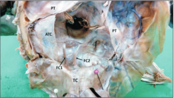
backwards along with falx cerebri. At this stage, a partial du- plication of tentorium cerebelli was observed. Below the left half of the tentorium cerebelli and above the left cerebellar hemisphere, there was a second fold of dura mater. This fold was about 3cm broad and 5 cm wide and extended from the lower lip of left transverse sulcus behind, to the upper border of the left petrous temporal in front. This fold was made up of meningeal layer of dura mater and did not have any dural ve- nous sinus in it. The accessory tentorium cerebelli was slightly thinner than the main tentorium cerebelli. While lifting the cerebellum from the posterior cranial fossa, it was noted that there was a complete duplication of falx cerebelli. Both falx cerebelli were of equal size and they occupied the valleculae of cerebellum. The vermis was situated in between these two folds. There was a single occipital sinus situated close to the internal occipital crest in the midline, between the two falx cerebelli. The falx cerebri and the sinuses related to that were normal. The parts of the brain related to the tentorium cer- ebelli were also normal. The variations observed have been shown in the Figs. 1 and 2.
Discussion
Folds of the meningeal layer of dura mater traverse and divided the cranial cavity into different compartments for dif-
ferent parts of the brain. Thus they play an important role in supporting and protecting the brain. The tentorium cerebelli is absent in amphibians, fish, and reptiles but present in birds and mammals as reported by Klintworth [2] in his study of comparative anatomy. The tentorium cerebelli is found as two separate halves on two sides, unjoined at the midline in some animals like bats, pigs, gerbils, hamsters, opossums, rats, and mice. In mammals like cats, dogs, humans, rhesus monkeys, minks, and goats, the two halves have fused in the midline to give rise to a crescentic partition posterior to the brain stem.
Jeffery [6] has revealed that the tentorium is rotated inferiorly by uneven growth of the cerebrum versus the cerebellum.
In the present case we observed a complete duplication of falx cerebelli. Both folds were of equal size and they occupied the valleculae of cerebellum. In a cadaveric study, D’Costa et al. [7] have observed a duplicated falx cerebelli, which was associated with two distinct occipital sinuses and internal occipital crests. Hasan and Das [8] have also reported the oc- currence of a duplicated falx cerebelli. These folds were asso- ciated with occipital sinuses which drained into the respective transverse sinus. Shoja et al. [9] have reported the duplication of falx cerebelli with two distinct occipital sinuses and inter- nal occipital crests. A case of duplicated falx cerebelli with single occipital sinus and presence of a large meningeal artery in the posterior cranial fossa have been reported by Satheesha Nayak et al. [10].
Falx cerebelli may be absent in some cases of Chiari mal- formation type II. In such cases, even the internal occipital crest might be absent [11]. Possibly, the congested posterior cranial fossa of people with this type of malformation inhibits the growth of these two structures. Another case of duplicated
Fig. 1. Cranial cavity showing complete duplication of falx cerebelli and partial duplication of tentorium cerebelli. ACF, anterior cranial fossa;
ATC, accessory tentorium cerebelli; FC, falx cerebri; FC1 and FC2, duplicated falx cerbelli; FM, foramen magnum; MCF, middle cranial fossa; PT, petrous temporal; TC, tentorium cerebelli (reflected back
wards).
Fig. 2. Closer view of posterior cranial fossa showing complete duplica
tion of falx cerebelli and partial duplication of tentorium cerebelli.
ATC, accessory tentorium cerebelli; FC1 and FC2, duplicated falx cerbelli; PT, petrous temporal; TC, tentorium cerebelli (reflected back
wards).
Partial duplication of tentorium cerebelli
https://doi.org/10.5115/acb.19.017
Anat Cell Biol 2019;52:337-339
339
www.acbjournal.org
falx cerebelli associated with an arachnoid cyst between the two has been reported by Hassler and Schlenker [4]. Shoja et al. [12] have reported a case of a triple falx cerebelli, with one of the folds was much smaller than the other two. In this case, a single occipital sinus was noticed.
Nayak et al. [13] have reported the presence of triple falx cerebelli related to two aberrant venous sinuses; each one joining the ipsilateral sigmoid and transverse sinuses. Some of the previous studies have also reported the absence, duplica- tion or triplication [14] of the occipital sinus. The occipital sinus is always a possible source of trouble in posterior ap- proaches to the posterior cranial fossa despite of its variability [15]. An awareness of regular neuroanatomic variability is significant for understanding pathologic changes.
Reported variations of falx cerebelli include its absence, duplication, and triplication. Though duplication of falx cerebelli is very rare, a few such cases have been reported earlier. However, partial or complete duplication of tentorium cerebelli is an extremely rare event. To the best of our knowl- edge, a concurrent variation of falx cerebelli and tentorium cerebelli has not been reported yet. The partial duplication of tentorium cerebelli might cause confusions in radiologic diagnosis of posterior cranial fossa lesions or tumors. The ad- ditional fold might also push the left cerebellar hemisphere to a slightly lower level than the right.
ORCID
Satheesha B. Nayak: https://orcid.org/0000-0003-1421-8222 Surekha D. Shetty: https://orcid.org/0000-0002-9301-6719
Author Contributions
Conceptualization: SBN. Data acquisition: SBN. Data analysis or interpretation: SDS. Drafting of the manuscript:
SDS, SBN. Critical revision of the manuscript: SBN. Approval of the final version of the manuscript: all authors.
Conflicts of Interest
No potential conflict of interest relevant to this article was reported.
Acknowledgements
The authors acknowledge the help and support rendered by the University and Institutional officials and also the dis- section hall supporting staff in carrying out this research.
References
1. Snell RS. Clinical neuroanatomy. Baltimore, MD: Lippincott Williams & Wilkins; 2010. p.428-31.
2. Klintworth GK. The comparative anatomy and phylogeny of the tentorium cerebelli. Anat Rec 1968;160:635-42.
3. Greenberg RW, Lane EL, Cinnamon J, Farmer P, Hyman RA.
The cranial meninges: anatomic considerations. Semin Ultra- sound CT MR 1994;15:454-65.
4. Hassler W, Schlenker M. Double falx cerebelli. Case report. Acta Neurochir (Wien) 1982;62:265-9.
5. Pang D, Dias MS, Ahab-Barmada M. Split cord malformation:
Part I: a unified theory of embryogenesis for double spinal cord malformations. Neurosurgery 1992;31:451-80.
6. Jeffery N. Differential regional brain growth and rotation of the prenatal human tentorium cerebelli. J Anat 2002;200(Pt 2):135- 44.
7. D’Costa S, Krishnamurthy A, Nayak SR, Madhyasta S, Prabhu LV, J JP, Ranade AV, Pai MM, Vadgaonkar R, Ganesh Kumar C, Rai R. Duplication of falx cerebelli, occipital sinus, and internal occipital crest. Rom J Morphol Embryol 2009;50:107-10.
8. Hasan M, Das AC. A npte on the falx cerebelli. Acta Anat (Basel) 1969;74:624-8.
9. Shoja MM, Tubbs RS, Khaki AA, Shokouhi G. A rare variation of the posterior cranial fossa: duplicated falx cerebelli, occipital venous sinus, and internal occipital crest. Folia Morphol (Warsz) 2006;65:171-3.
10. Satheesha Nayak B, Srinivasa Rao S, Deepthinath R, Kumar N.
Triple falx cerebelli associated with two aberrant venous sinuses in the floor of posterior cranial fossa. Australas Med J 2013;6:
397-400.
11. Tubbs RS, Dockery SE, Salter G, Elton S, Blount JP, Grabb PA, Oakes WJ. Absence of the falx cerebelli in a Chiari II malforma- tion. Clin Anat 2002;15:193-5.
12. Shoja MM, Tubbs RS, Loukas M, Shokouhi G, Oakes WJ. A complex dural-venous variation in the posterior cranial fossa: a triplicate falx cerebelli and an aberrant venous sinus. Folia Mor- phol (Warsz) 2007;66:148-51.
13. Nayak SB, Shetty SD, Kumar N, Sirasanagandla SR. Double falx cerebelli, single occipital sinus and an unusually large meningeal artery in the posterior cranial fossa: a case report. OA Case Rep 2013;2:30.
14. Lang J. Clinical anatomy of the posterior cranial fossa and its foramina. New York: Thieme Medical Publishers; 1991. p.6.
15. Hollinshead WH. Anatomy for surgeons: the head and neck. 3rd ed. Philadelphia, PA: JB Lippincott; 1982. p.269-74.