Brief Report
S44 Ann Dermatol
Received December 23, 2017, Revised March 6, 2019, Accepted for publication March 25, 2019
Corresponding author: Seong Jun Seo, Department of Dermatology, Chung- Ang University Hospital, 102 Heukseok-ro, Dongjak-gu, Seoul 06973, Korea. Tel: 82-2-6299-1525, Fax: 82-2-6299-1718, E-mail: [email protected].
kr
ORCID: https://orcid.org/0000-0003-2915-839X
This is an Open Access article distributed under the terms of the Creative Commons Attribution Non-Commercial License (http://creativecommons.
org/licenses/by-nc/4.0) which permits unrestricted non-commercial use, distribution, and reproduction in any medium, provided the original work is properly cited.
Copyright © The Korean Dermatological Association and The Korean Society for Investigative Dermatology
pISSN 1013-9087ㆍeISSN 2005-3894
Ann Dermatol Vol. 31, Suppl, 2019 https://doi.org/10.5021/ad.2019.31.S.S44
BRIEF REPORT
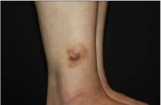
Fig. 1. A protruding solid nodule with a peripheral brownish patch on the lower leg.
Chondroid Syringoma of the Leg with Unusual Presentations
Ji Yeon Hong, Guk Jin Jung, Kapsok Li, Seong Jun Seo
Department of Dermatology, Chung-Ang University College of Medicine, Seoul, Korea
Dear Editor:
Chondroid syringoma (CS) is a rare, benign skin appenda- geal tumor with a reported incidence of less than 0.01%1. Virchow and Minssen also named the entity mixed tumor of the skin due to its histologic appearance showing both epithelial and mesenchymal origins2. CS characteristically presents as a slow-growing, painless subcutaneous or in- tradermal tumor and usually affects the head and neck area, trunk, axilla, inguinal area and rarely on eyelids and ex- ternal ears3.
A 54-year-old female patient presented with a three-year history of a protruding nodule on her right lower leg. She remembered that the nodule had been increasing in size insidiously over the past five years. She did not complain of any subjective symptoms, except aesthetically unpleas- ant brownish pigmentation. Physical examination showed a 1.5×1.5 cm sized firm, non-tender nodule with a sur- rounding 3×3 cm sized brownish patch (Fig. 1). We re- ceived the patient’s consent form about publishing the photograph. There was no localized edema or palpable re- gional lymphadenopathy. The biopsy demonstrated tubular and gland-like structures embedded in an abundant mu- coid stroma (Fig. 2A). The tubular lumina were lined by two layers of luminal cuboidal cells and peripheral flat-
tened cells (Fig. 2B). There was no evidence of necrosis or atypia. The epidermis showed mild hyperkeratosis and in- creased basal pigmentation. The diagnosis of CS was con- firmed, and the patient underwent complete excision.
CS is usually not recognized by physicians due to its rela- tive infrequency and silent clinical course. Since CS is clinically indistinctive, the diagnosis relies largely on his- tologic examination. Histopathologically, CS demonstrates nests of cuboidal or polygonal cells and ducts surrounded by chondromyxoid stroma. It typically shows inter-com- municating tubuloalveolar structures composed of one or two rows of cuboidal cells.
CS usually presents in the head and neck area, except for rare cases involving the trunk, back, or extremities3. Axillar, cerebral, scrotal, or vulvar cases have also been reported as very rare localizations1. The present case was located in the lower leg. To the best of our knowledge, this is only the second of two cases on the lower leg, the first of which was reported by Sulochana et al.4.
Since our case showed peripheral brownish pigmentation
Brief Report
Vol. 31, Suppl, 2019 S45 Fig. 2. (A) Low power view shows nodular dermal mass with overlying epidermis of increased basal pigmentation (H&E, ×40). (B) Low power view shows abundant basophilic stroma (H&E, ×100). (C) High power histologic feature shows a nodular dermal tumor composed of branching tubular epithelial elements embedded in a myxoid stroma (H&E, ×200). No atypical cells are observed.
around the nodule, we worried about possible invasion by malignant transformation5, which was, however, not found in the pathology. Considering that the patient persistently squeezed and irritated the nodule to remove it by herself, the dark pigmentation may be due to postinflammatory hyperpigmentation. Because the lesion was located on an extremity and occurred in a female patient, we recom- mended including a margin of normal tissue with the complete removal of the tumor.
We report this case because CS is an uncommon, confus- ing adnexal tumor. Especially in this case, the nodule showed atypical location on the leg and hyperpig- mentation, which misled us about clinical impression. By pathologically confirming tubular epithelium and baso- philic stroma, CS was finally diagnosed. Due to the poten- tial risk of malignant transformation, surgical excision and close follow-up are recommended.
CONFLICTS OF INTEREST
The authors have nothing to disclose.
ORCID
Ji Yeon Hong, https://orcid.org/0000-0002-5632-8449 Guk Jin Jung, https://orcid.org/0000-0002-2379-0370 Kapsok Li, https://orcid.org/0000-0002-1333-1680 Seong Jun Seo, https://orcid.org/0000-0003-2915-839X
REFERENCES
1. Sungur N, Uysal A, Gümüş M, Koçer U. An unusual chon- droid syringoma. Dermatol Surg 2003;29:977-979.
2. Borman H, Özcan G. Chondroid syringoma at the fingertip:
an unusual localization. Eur J Plast Surg 1998;21:311-313.
3. Yavuzer R, Başterzi Y, Sari A, Bir F, Sezer C. Chondroid sy- ringoma: a diagnosis more frequent than expected. Der- matol Surg 2003;29:179-181.
4. Sulochana S, Manoharan M, Anitha. Chondroid syringoma-an unusual presentation. J Clin Diagn Res 2014;8:FD13-FD14.
5. Sirivella S, Gielchinsky I. Chondroid syringoma: a rare tumor of the chest wall. Ann Thorac Surg 2010;89:983-985.