J Korean Soc Radiol 2016;75(6):466-470 https://doi.org/10.3348/jksr.2016.75.6.466
INTRODUCTION
The lungs are a common site for malignancy, either primary or metastases. Surgical resection is the treatment of choice for lung malignancies. However, some patients are not good candi- dates for lung resection due to various reasons, including inabil- ity to meet the criteria for lung operation, patient’s poor general conditions such as cardiovascular disorders, impaired pulmo- nary function, or even patients’ refusal for surgery.
For cases of inoperable focal malignancies, image-guided per- cutaneous cryoablation is currently evolving as a minimally in- vasive and potentially effective treatment option. Unlike heat- based therapies such as microwave and radiofrequency ablation, cryoablation is based on the therapeutic application of extreme cold to living tissue in order to cause local destruction. Advan- tages of cryoablation include good monitoring by CT during the procedure resulting in minimizing the risk of injury to nearby
structures, sparing collagen containing structures and causing less pain than radiofrequecy ablation (RFA). In this report, we present a case of successful cryoablation which was performed for a small metastatic lesion from surgically resected primary lung cancer.
CASE REPORT
A 76-year-old male patient underwent left upper lobectomy for lung adenocarcinoma in July 2010. The pathologic stage was pT3 (separate nodule in the same lobe) N0M0, stage IIB. He had received adjuvant chemotherapy. Three years after left up- per lobectomy, a metastatic nodule was found in the right upper lobe and wedge resection was performed for the lesion. Then, he underwent radiation therapy. Fifteen months after metastasecto- my of the nodule in the right upper lobe, an approximately 1.5 cm sized subpleural nodule was found on the follow up CT scan
Inducing of Complete Necrosis of Recurred Lung Cancer by Cryoablation: A Case Report
수술 후 재발한 단일 폐암 병변에 대한 성공적인 냉각치료: 증례 보고
Sunhye Lee, MD, Soo-Youn Ham, MD*, Sung Ho Hwang, MD, Yu-Hwan Oh, MD
Department of Radiology, Korea University Anam Hospital, College of Medicine, Korea University, Seoul, Korea
Lung cancer is one of the most commonly diagnosed cancers, and the lungs are a common site of metastasis from extrathoracic malignancies. Surgical resection is the gold standard treatment for lung malignancies. However, some of the patients are poor surgical candidates due to various reasons. Currently, image-guided abla- tion is used as one of the lung cancer treatment modalities. Cryoablation has been adapted as one of the treatments of lung tumors and a growing body of literature has shown that it is a safe and effective option. We report a case of successful cryo- ablation for a metastatic lesion from surgically resected primary lung cancer.
Index terms Cryotherapy Lung Neoplasm Metastasis
Received January 19, 2016 Revised April 3, 2016 Accepted June 7, 2016
*Corresponding author: Soo-Youn Ham, MD Department of Radiology, Korea University Anam Hospital, College of Medicine, Korea University, 73 Inchon-ro, Seongbuk-gu, Seoul 02841, Korea.
Tel. 82-2-920-5657 Fax. 82-2-929-3796 E-mail: [email protected]
This is an Open Access article distributed under the terms of the Creative Commons Attribution Non-Commercial License (http://creativecommons.org/licenses/by-nc/3.0) which permits unrestricted non-commercial use, distri- bution, and reproduction in any medium, provided the original work is properly cited.
(Fig. 1). CT-guided percutaneous needle biopsy was performed for the lesion and it was proved to be another recurred nodule of adenocarcinoma. Through a multidisciplinary approach, the patient was referred to our department for cryoablation of the recurred malignant lesion. Considering that he had already re- ceived chemotherapy and radiation therapy and that he also had impaired pulmonary function, we came to the conclusion that one of the ablation therapies would be the best treatment option. The reason for choosing cryoablation among the possible ablation therapies was the several advantages that it offers, in- cluding that it is minimally invasive, easy to apply, real-time monitoring is feasible, less painful, and spares the adjacent colla- gen containing structures.
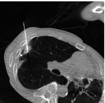
Percutaneous cryoablation was performed under CT guid- ance, in the following manner: first, a limited non contrast low- dose CT scan (80 kV, 30 mAs, 1.2 mm slice) was performed to localize the target lesion. The patient was placed in a supine po- sition with slight right anterior oblique rotation and draped in the usual fashion. Lidocaine solution (1%) was infiltrated subcu- taneously into the soft tissue adjacent to the puncture site. Un- der the guidance by CT fluoroscopy (Siemens Somatom Defini- tion Flash, Forchheim, Germany), the cryoprobe was inserted into the target nodule in the right upper lobe (Fig. 2). The cryo- therapy equipment that we used consisted of an argon: helium gas-based system (SeedNet GoldTM system, Galil Medical Ltd., Yokneam, Israel). Three cycles of freezing and thawing were per- formed. In the first cycle, 3-minute freezing followed by 5-min-
ute thawing, in the second cycle, 7-minute freezing and 10-minute thawing, and in the final cycle, 10-minute freezing and 3-minute active thawing were performed. After removing the probe, the incision site was covered as usual with a proper ointment dress- ing. During the procedure, the patient had a tolerable experi- ence and there was no evidence of any immediate procedure-re- lated complications. After ablation, the patient was monitored by checking his vital signs every 2 hours during the half day.
On the next day of ablation, the patient was discharged without any specific complaints. Follow up chest radiography 4 hours af- ter the procedure and CT scan 1 week after the ablation showed no evidence of procedure-related complications such as pneu- mothorax, hemothorax or pulmonary hemorrhage. Technical success is commonly defined as whether the treatment was per- formed according to the protocol with complete coverage of the tumor. By evaluating the CT scan images acquired during the procedure, we ensured technical success and complete coverage was obtained.
To evaluate the effectiveness of the technique, we compared the CT scan performed before cryoablation with CT scans per- formed one, three, and five months after the procedure. On the follow-up CT scans, the treated lesion showed nearby necrosis and cavitation, suggesting successful ablation (Fig. 3).
Fig. 1. Chest CT with lung window reveals a 1.5 cm sized spiculated nodule with an air bronchogram in the right upper lobe. The nodule was proved to be metastatic adenocarcinoma by percutaneous needle biopsy.
CT = computed tomography
Fig. 2. Under CT fluoroscopy guidance, the cryoprobe was inserted into the target nodule in the right upper lobe. The tip of the cryoprobe transverses the nodule.
CT = computed tomography
DISCUSSION
Image-guided tumor ablation by thermal or non-thermal sources has received increased attention for managing focal ma- lignancies, although the primary management options for lung cancers traditionally depend on the surgical treatment. Due to the increase in the number of screening studies for lung cancer, we can detect small nodules and early stage lung cancers, and nowadays the trend of organ conserving treatment is becoming popular. Hence, the local and less invasive treatment modalities have a high demand.
Cryoablation is usually performed for tumors less than 3 cm in size. In tumors greater than 3 cm in size, there is a higher chance of recurrence than in smaller tumors. Cryoablation is known to be a safer modality for tumors close to large vascular structures as well as tumors located near the chest wall. Percutaneous cryo- ablation enables preservation of the collagenous architecture of the bronchi and the vascular structures, and this advantage al- lows performing cryoablation for tumors located immediately adjacent to the central bronchi. This may be the main advantage of cryoablation, compared to heat-based treatments. Also, it causes less pain than radiofrequency ablation; hence, it is recom- mended for patients with less tolerability. Since about 2–3 years, cryoablation for malignant tumors has been covered by medi- cal insurance in Korea.
Current cryoablation systems use the Joule-Thomson effect to create freeze and thaw cycles. The Joule-Thomson effect de-
scribes the change in temperature of a gas resulting from expan- sion or compression of that gas. Gas expansion occurs in a small chamber inside the distal end of the cryoprobe to create the nec- essary heat sink during freeze cycles and heat source during thaw cycles (1).
During rapid tissue cooling, water is entrapped within the cell membrane, resulting in intracellular ice formation, and can cause recrystallization and extension of the ice within the intracellular matrix. These changes induce enzyme dysfunction and cell mem- brane dysfunction. In tumors with a slowing cooling process, there is ice formation in the extracellular matrix, resulting in os- mosis of water out of the cell. Also, there are many collateral ef- fects of extreme cold, occurring along the blood vessel wall. In blood vessels, direct contact with rapid cooling process, there are similar direct effects. These indirect effects in concert with direct effects result in coagulation necrosis. To achieve an effec- tive cryoablation tissue injury, excellent monitoring of the pro- cess, fast cooling to a lethal temperature, slow thawing and rep- etition of freeze-thaw cycle are the critical factors.
Possible complications after cryoablation range from pneu- mothorax, hemoptysis, pleural effusion, fever, hypertension, sub- cutaneous emphysema, skin injury, infection, to nerve injuries.
The most common complication encountered with cryoablation is pneumothorax, occurring approximately 12–62% of the pa- tients after cryoablation (2-6). The reported rates of hemoptysis after cryoablation range from 0% to 62% (2-6).
CT is a useful modality to evaluate the treated lesion and to monitor the local tumor progression. There is considerable vari- ation in how an ablated zone around the target tumor responds after treatment and progression occurs during follow-up. Im- mediate post-ablation CT scans typically show a definite low at- tenuation area surrounding the ablated foci, suggesting tumor necrosis, termed ice-ball formation. Wang et al. (2) reported that cavitation was the most common postablation finding and approximately 17% of the ablated lesions disappeared at 12 months. The remaining lesions without local recurrence might decrease in size or maintain a stable size. Positron emission to- mography-CT might also have a promising role in the estimation of the therapeutic effects, in the differentiation of tumor necrosis and viable tumor proportion (4).
Yamauchi et al. (7) reported that the overall survival rates af- ter cryoablation for inoperable lung cancer were 95% at 1 year, Fig. 3. On follow up CT scan at 5 months, the ablated nodule has re-
gressed and cavity formation is observed.
CT = computed tomography
88% at 2 years, and 88% at 3 years. The disease-free survival per- centages were 91, 78, and 67% at 1, 2, and 3 years, respectively.
According to a recent report, the efficacy of lung tumor cryoab- lation is comparable with RFA and sublobar resection (3).
There are several reports of cryoablation as an alternative treatment option for lung lesions in Korea. Lee et al. (8) reported a case of successful cryoablation performed for treatment of re- current lung cancer. The patient was not a candidate for surgery because he had already undergone right upper lobe resection and left upper lobe resection due to bilateral lung cancers. Treat- ed lesion showed cavitary necrosis as in our case and maintained stable for 2 years during follow up. However, not all reports showed promising success rates. Park et al. (9) reported the re- sult of cryoablations performed for 14 lung malignancies, with complete ablation achieved in only 4 cases (35.7%). The authors mentioned that the results of percutaneous cryotherapy were not as satisfactory as expected, but still, cryotherapy could be performed safely in inoperable patients with lung malignancy and it might lengthen their survival period. Recently, Kim et al.
(10) reported that cryoablation could be used as a new treat- ment method for ground-glass nodules.
We report a case of complete necrosis of recurrent lung can- cer after resection and radiotherapy. Percutaneous ablation is a minimally invasive therapy and it is generally safe and well toler- ated, with low rates of morbidity and very low procedure-relat- ed mortality. The advantages of cryoablation over radiofrequen- cy ablation include the following: it can achieve larger tumor ablation volumes, allows for the use of multiple applicators, pro- vides a highly visible ablation zone, and causes less procedural pain due to the analgesic effect of freezing. Compared with heat- based thermal ablation therapies, cryoablation preserves the col- lagenous tissue and cellular architecture, which makes it a safer option near vasculature or bronchi. A growing body of literature describes the successful use of cryoablation in the treatment of malignancies in the lung, and application of percutaneous cryo- ablation may represent a new promising effective treatment op- tion for lung tumors in patients who are not candidates of surgi- cal resection.
REFERENCES
1. Ahmed M, Brace CL, Lee FT Jr, Goldberg SN. Principles of and advances in percutaneous ablation. Radiology 2011;
258:351-369
2. Wang H, Littrup PJ, Duan Y, Zhang Y, Feng H, Nie Z. Tho- racic masses treated with percutaneous cryotherapy: ini- tial experience with more than 200 procedures. Radiology 2005;235:289-298
3. Zemlyak A, Moore WH, Bilfinger TV. Comparison of survival after sublobar resections and ablative therapies for stage I non-small cell lung cancer. J Am Coll Surg 2010;211:68-72 4. Zhang X, Tian J, Zhao L, Wu B, Kacher DS, Ma X, et al. CT-
guided conformal cryoablation for peripheral NSCLC: ini- tial experience. Eur J Radiol 2012;81:3354-3362
5. Pusceddu C, Sotgia B, Fele RM, Melis L. CT-guided thin needles percutaneous cryoablation (PCA) in patients with primary and secondary lung tumors: a preliminary experi- ence. Eur J Radiol 2013;82:e246-e253
6. Inoue M, Nakatsuka S, Yashiro H, Ito N, Izumi Y, Yamauchi Y, et al. Percutaneous cryoablation of lung tumors: feasibil- ity and safety. J Vasc Interv Radiol 2012;23:295-302; quiz 305
7. Yamauchi Y, Izumi Y, Hashimoto K, Yashiro H, Inoue M, Na- katsuka S, et al. Percutaneous cryoablation for the treat- ment of medically inoperable stage I non-small cell lung cancer. PLoS One 2012;7:e33223
8. Lee SH, Kim KT, Chung JH, Jo SB, Youn HS, Son HS. Percu- taneous cryoablation of lung cancer in high risk patients.
Korean J Thorac Cardiovasc Surg 2006;39:953-956 9. Park EH, Jin GY, Han YM, Lee YC, Kwon KS. Percutaneous
cryotherapy for inoperable lung malignancy. J Korean Soc Radiol 2012;66:427-435
10. Kim KY, Jin GY, Han YM, Lee YC, Jung MJ. Cryoablation of a small pulmonary nodule with pure ground-glass opacity:
a case report. Korean J Radiol 2015;16:657-661
수술 후 재발한 단일 폐암 병변에 대한 성공적인 냉각치료: 증례 보고
이선혜 · 함수연* · 황성호 · 오유환
폐암은 높은 발생률을 보이는 대표적 종양 중 하나이다. 또한 폐는 폐 외에서 발생한 악성 종양이 흔하게 전이하는 장소이 기도 하다. 이러한 폐 종양에 대해서는 수술적인 제거가 가장 근본적인 치료 방법으로 여겨진다. 하지만 다양한 이유로 인 하여 몇몇 환자들에 있어서는 수술적 치료가 용이하지 않을 수 있다. 최근에는 다양한 영상 유도하의 중재적 치료법이 폐 종양에 대한 치료 방법으로 이용되고 있다. 그중 하나인 냉각치료는 그 안정성과 효과에 대한 문헌적 근거가 점차 증가하고 있다. 이 보고는 수술적으로 폐암을 제거한 뒤 발생한 단일 전이 병변에 대하여 성공적으로 냉각치료를 시행한 사례이다.
고려대학교 의과대학 안암병원 영상의학과