Photodynamic therapy(PDT) is designed to kill targeted tumor cells of various cuta- neous and noncutaneous malignancies by highly reactive oxygen intermediates generat- ed through the interaction of light with a photosensitizer1. It is particularly effective in treating in situ or early invasive tumors.
Several non-neoplastic dermatological condi- tions may also respond to PDT, including psoriasis and alopecia areata2-4. Single lesion of Bowen’s disease can be satisfactorily treated by surgical excision, curettage, cautery, cryotherapy or topical application of 5-fluorouracil5,7. However, these therapies may be impractical for patients with wide- spread or large lesions located on anatomi-
cally difficult areas. PDT has been shown to be effective in these lesions8. It offers the ad- vantages for treating non-invasive, well tol- erated lesions in slow-healing sites, leaving the skin surrounding the tumor intact and functional.
In Korea, Lee et al9 reported six cases of carcinoma in situ treated with PDT, utilizing topical photosensitizer, δ-amino levulinic acid(ALA) in dermatologic literature, which showed complete response9. However, there were no reported cases using systemic photo- sensitizer, Photofrin . We performed PDT using systemic Photofrin in the case of Bowen’s disease arising on the plantar area and 3rd and 4th toewebs of left forefoot. The outcome showed a partial clinical response.
CASE REPORT
A 61-year-old man visited our department with a complaint of a erythematous patch on the plantar area, 3rd and 4th toewebs of left forefoot. It was initially noticed 5-years ago and had slowly developed. The lesion was
A Case of Bowen’s Disease Partially Responded to Photodynamic Therapy
Si Heon Lee, M.D., Byung Cheol Jung, M.D., Min Jung Woo, M.D., Dong Seok Kim*, M.D., Sang Won Kim, M.D.
Goun Skin Clinic*, and Department of Dermatology, Catholic University of Daegu School of Medicine, Daegu, Korea
Photodynamic therapy(PDT) is a treatment modality by highly reactive oxygen intermediates gen- erated through the interaction of light with a photosensiziter. It has been shown to be an effective treatment for various cutaneous and noncutaneous malignancies. It is efficient for the curative and pal- liative treatment of epithelial skin tumor in situ or early invasive lesions. In effect, it is a useful al- ternative treatment for the lesions located on anatomically difficult areas or the large-sized lesions. We treated a case of Bowen’s disease arising on the plantar area and 3rd and 4th toewebs of left fore- foot in a 61-year-old man with PDT using the hematoporphyrin derivative, porfirmer sodium(Photofrin , Russia) as a photosensitizer and gold vapor laser as a visible light source. The outcome showed partial clinical improvement after about 2 months’ follow-up. (Ann Dermatol 14(1) 38-41, 2002).
Key Words : Photodynamic therapy, Porfirmer sodium, Bowen’s disease
Received March 15, 2001.
Accepted for publication October 22, 2001.
Reprint request to : Si Heon Lee, M.D., Department of Dermatology, School of Medicine, Catholic University of Daegu, Daemyung-4-dong, Nam-gu, Daegu 3056-6, Korea, 705-718
Tel. (053) 650-4161, Fax. (053) 650-4891 E-mail. g9563009@cataegu.ac.kr
38
A Case of Bowen's Disease Partially Responded to Photodynamic Therapy 39
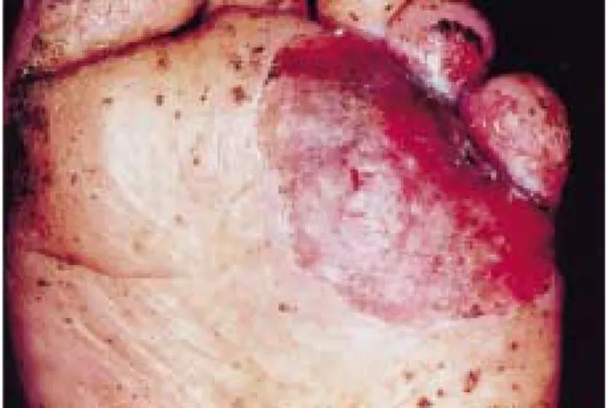
slightly painful and continuously oozing. He was treated with etretinate and topical 5-FU at another skin clinic 4 years ago and with PDT using Photofrin two times in our department about 2 years and 4 years ago, but both approaches were insignificant. He had psoriasis 30 years ago and had diabetes mellitus for about 4 years, but there were no psoriatic lesions at initial visit. He had a his- tory of arsenic intake for 6 months about 30 years ago for the treatment of psoriasis. He had no family history of skin cancer. Exam- ination of the skin revealed a 5.0×4.0 cm sized, well demarcated, round shaped, eroded, erythematous patch on the plantar area
and 3rd and 4th toewebs of left forefoot(Fig. 1). CBC, urinalysis, liver func- tion test, chest X-ray and EKG were within normal limits or negative except elevated serum and urine glucose level. Skin biopsy taken from the erythematous patch revealed scattered dyskeratotic cells with eosinophilic cytoplasm, hyperchromatic nucleus in the epidermis and intact basement membrane consistent with Bowen’s disease(Fig. 2).
Study for visceral involvement was not done because of patient’s refusal. The medical records of the first therapy and progress done about 4 years ago were lost.
He was given an intravenous injection of Fig. 1. A 5.0×4.0 cm-sized, eroded, erythematous
patch on the platar area and 3rd and 4th toewebs of left forefoot.
Fig. 2. Initial biopsy shows dyskeratotic and atypical cells in the epidermal cell layer with moderate infil- trate of lymphohistiocytes in the upper dermis(H&E,
×100).
Fig. 3. Following of 2 months after PDT; the lesion
size was reduced and shows minimal epithelization. Fig. 4. Follow-up of 2 months after PDT; skin lesion showed markedly reduced dyskeratotic cells in epider- mis with granulation tissues in dermis(H&E, ×100).
SH Lee, et al.
Annals of Dermatology Vol. 14, No. 1, January 2002 40
Photofrin via a single bolus at a dose of 2 mg/kg. Approximately 48 hours after injec- tion, the area was treated with 628 nm of laser light at dose of 125 J/cm2 delivered through a flexible 400 um quartz fiber fitted with microlenses. It was used to produce a homogeneous spot. Gold vapor laser was used as a light source. He was treated on the first session and was observed during the fol- low-up period of 1 week, 2 weeks, 1 month and 2 months after treatment.
After about 2 months’ follow-up, the re- sponse was evaluated clinically and histopathologically. The lesion was reduced and showed minimal epithelization, but still remained to be focally ulcerative(Fig. 3).
Biopsy specimen showed markedly destroyed dyskeratotic cells in epidermis but not com- pletely. Diffuse lymphocytic infiltration and granulation tissues with vascular dilatation were prominent in the dermis(Fig. 4).
DISCUSSION
The combination of two individually pho- totoxic light and chemicals are responsible for the PDT-mediated destruction of tissues. In 1978, Dougherty et al6 presented the clinical use in the treatment of cancer as the novel technique. Since then, the modality has been widely practiced in dermatology. It has been proven that PDT is effective in the treatment of cutaneous precancerous lesions and cancers, including Bowen’s dis- ease8,10,11. Robinson et al8 reported a complete response in the treatment of a total of more than 500 lesions in two patients. On- ly less than 10% of the tumors required a second PDT session. Similar results were reported by several investigators12,13. Lee et al9, in Korea, reported excellent clinical re- sults on six carcinoma in situ patients with complete response and cosmetic outcome.
The mechanism of PDT is based on the interaction of three components: photosensi- tizer, light, and oxygen14. The important photosensitizing mechanism is the absorp- tion of a light photon by the sensitizer, causing a promotion of the drug molecule from its ground state to the extremely un-
stable excited singlet state. Subsequently, the singlet excited photosensitizer decays back to the ground state, resulting in deliv- ering singlet oxygen molecule into the tissue.
Singlet oxygen molecule plays the central role in cytotoxicity by means of destroying cellular and organelle membranes14.
The advantages of PDT are the facts that it provides good tumor control with highly selective tissue destruction, excellent cosmetic results, minimal side effects and the ability to treat large areas in a single session. In addition, multiple lesions and anatomically difficult areas can be treated successfully. In our case, the use of PDT therapy on the anatomically inoperable and huge lesion showed to be effective. Erythema and edema of the treated area are common adverse effects and a variety of systemic side effects have also been reported including prolonged photosensitivity, headache, nausea and fever. Blistering, ulceration, or excessive necrosis due to the light overdose rarely de- veloped15. PDT-induced phototoxicity is because of the light within the visible spec- trum lange. Therefore, conventional sun- screens are of no benefit. Use of clothing and protective eyewear and strict avoidance of sunlight and excessive indoor light are rec- ommended. In addition, systemic side effects have been minimized with the use of topical photosensitizer. We performed systemic PDT because topically applied photosensitiz- er showed insufficient penetration at very hyperkeratotic lesions. Initial complaint was painful swelling in the treated area, but swelling gradually subsided after a week, and healing completed within 2 weeks.
There were no other serious systemic compli- cations.
PDT has been applied to almost every type of cutaneous cancers and numerous benign skin disorders in dermatology, in- cluding actinic keratosis, Bowen’s disease, basal cell carcinoma, squamous cell carcinoma, Kaposi’s sarcoma, mycosis fungoides, malig- nant melanoma, metastatic cancer, psoriasis, hemangioma, and so on. However, the sited PDT results only included short follow-up period and scanty histopathologic verifica-
A Case of Bowen's Disease Partially Responded to Photodynamic Therapy 41 tions. The therapeutic effect of deeper skin le-
sions was also limited. Therefore, the thera- peutic evaluation would be needed.
The present case didn’t get a complete response in the case of Bowen’s disease. We presume that this was a result of disturbance of penetration of the light caused by thick keratotic layers at the plantar surface that induce optical scattering of it; otherwise, insufficient concentration of photosensitizer.
Repeated treatment could be attained for better effectiveness.
REFERENCES
1. Bissonnette R, Lui H. Current status of photody- namic therapy in dermatology. Dermatol Clin 1997;15:507-519.
2. Lui H, Anderson RR. Photodynamic therapy in der- matology. Arch Dermatol 1992;128:1631-1636.
3. Berns MW, Rettenmaier M, McCullough J et al.
Response of psoriasis to red laser light following systemic injection of hematoporphyrin derivative.
Lasers Surg Med 1984;4:73-77.
4. Boehncke WH, Sterry W, Kaufmann R. Treatment of psoriasis by topical phytodynamic therapy with polychromatic light. Lancet 1994;343:801.
5. Davies DM. Malignant skin conditions. Br Med J 1985;290:1190-1194.
6. Dougherty TJ, Kaufman JE, Goldfarb A, Weishaupt KR, Boyle D, Mittleman A. Photoradiation therapy for the treatment of malignant tumors. Cancer Res
1978;38:2628-2635.
7. Jansen GT. Use of topical fluorouracil. Arch Der- matol 1983;119:784-785.
8. Robinson PJ, Carruth JAS, Fairris GM. Photody- namic therapy: a better treatment for widespread Bowen’s disease. Br J Dermatol 1988;119:59-61.
9. Lee HN, Lee JD, Baek SC, Byun DG. Houh D.
Clinical effects of photodynamic therapy on carci- noma in situ of the skin. Kor J Dermatol 1998;36:407-414.
10. Roberts DJH, Cairnduff F. Photodynamic therapy in oncology: mechanisms and clinical use. J Natl Can- cer Inst 1993;85:443-456.
11. Rui H. Anderson RR. Photodynamic therapy in der- matology: recent developments. Dermatol Clin 1993;11:1-13.
12. Waldow SM, Lobraico RV, Kohler IK, Wallk S, Fritts HT. Photodynamic therapy for treatment of malignant cutaneous lesions. Lasers Surg Med 1987;7:451-456.
13. Jones CM, Mang T, Cooper M, Wilson BD, Stoll Hl Jr. Photodynamic therapy in the treatment of Bowen’s disease. J Am Acad Dermatol 1992;27:
979-982.
14. Weishaupt KR, Gomer CJ, Dougherty TJ. Identifi- cation of singlet oxygen as the cytotoxic agent in photoinactivation of a murine tumor. Cancer Res 1976;36:2326-2329.
15. McCaughan JS, Guy JT. Hicks W. Laufman L, Nims TA, Walker J. Photodynamic treatment for cutaneous and subcutaneous malignant neoplasms.
Arch Surg 1989;124:211-216.