J Korean Surg Soc 2012;83:30-35
http://dx.doi.org/10.4174/jkss.2012.83.1.30
ORIGINAL ARTICLE
Journal of the Korean Surgical Society
JKSS
pISSN 2233-7903ㆍeISSN 2093-0488
Received December 15, 2011, Revised April 18, 2012, Accepted May 6, 2012 Correspondence to: Sung Su Yun
Department of General Surgery, Yeungnam University Medical Center, 170 Hyeonchung-ro, Nam-gu, Daegu 705-717, Korea Tel: +82-53-620-3580, Fax: +82-53-620-1213, E-mail: [email protected]
cc Journal of the Korean Surgical Society is an Open Access Journal. All articles are distributed under the terms of the Creative Commons Attribution Non-Commercial License (http://creativecommons.org/licenses/by-nc/3.0/) which permits unrestricted non-commercial use, distribution, and reproduction in any medium, provided the original work is properly cited.
Laparoscopic liver resection for malignant liver tumors, why not more?
Ik Soo Kwon, Sung Su Yun, Dong Shick Lee, Hong Jin Kim
Department of General Surgery, Yeungnam University Medical Center, Daegu, Korea
Purpose: The precise role of laparoscopic liver resection in liver malignancies remains controversial despite an increasing number of publications that have used the laparoscopic resection of benign liver tumors. This study was performed to assess the feasibility, safety, and outcome of laparoscopic liver resection for malignant liver tumors. Methods: This study is a retro- spective review of the profiles, pathology, surgery and outcome performed on 61 patients who had undergone laparoscopic liver resection for liver malignancies between January 2004 and March 2011. Results: Among the 61 patients, 34 patients had hepatocellular carcinoma (HCC), 24 patients had liver metastasis. The mean tumor size was 2.8 ± 2.0 cm (mean ± standard de- viation). Tumors located at Couinaud segment number 2 to 8. The resection included 36 anatomical resections, 25 wedge resections. The mean surgical time was 209.7 ± 108.9 minutes. There was one operation that resulted in death. Postoperative complications occurred in 9 patients (14%). There were 2 conversions to laparotomy (3%). The mean postoperative hospital stay was 9.0 ± 4.4 days. Blood transfusion was needed in 11 patients (18%). The mean surgical margin was 1.3 ± 1.2 cm. The mean follow-up period was 18.1 ± 11.1 months. The three-year overall survival rate was 87% for patients with HCC and 95%
for patients having liver metastases from colorectal cancer. Conclusion: Even though laparoscopic liver resection requires a learning curve, it produced acceptable outcomes even in patients who had a malignant liver tumor. This study provides evi- dence to support further investigation and the establishment of laparoscopic liver resection for malignant liver tumors.
Key Words: Laparoscopy, Hepatectomy, Liver neoplasms
INTRODUCTION
Over the past two decades, tremendous achievements have been made in laparoscopic surgery including laparo- scopic liver resection. Some skilled surgeons with ex- tensive experience both in hepatic and laparoscopic sur- gery have demonstrated the feasibility and safety of lapa- roscopic liver resection in selected patients. However, lap- aroscopic liver resection for malignant tumors has not
gained wide spread use due to the long learning curve with technical difficulty and its oncologic safety. Never- theless some experts have reported the usefulness of lapa- roscopic liver resection even in liver malignancy, which is a minimally invasive technique [1-4].
The purpose of the present study was to analyze the fea- sibility, safety, and outcome of 61 patients who underwent laparoscopic resection for liver malignancies in Yeung- nam University Medical Center between January 2004 and
Characteristic Value
Age (yr), mean ± SD 59.1 ± 9.8
Gender (M/F) 43/18
Histology of cancers Primary liver malignancies
Hepatocelluar carcinoma 34
Intrahepatic cholangiocarcinoma 3 Secondary liver malignancies
Liver metastasis from colorectal cancer 23 Liver metastasis form breast cancer 1 Tumor size (cm), mean ± SD
Hepatocelluar carcinoma 2.6 ± 1.4
Metastatic tumor 2.5 ± 1.2
Single/multiple 56/5
Liver function (ICG R15)
Hepatocelluar carcinoma 18.1 ± 16.5
Metastatic tumor 9.9 ± 7.3
Intrahepatic cholangiocarcinoma 28.9 ± 36.1 ICG, indocyanin green.
Table 1. Patients and tumor characteristics
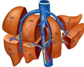
Fig. 1. Schematic illustration of tumor location.
March 2011.
METHODS
Patients
From January 2004 to March 2011, 111 patients under- went laparoscopic liver resection in Yeungnam University Medical Center. Among the 111 patients, 61 patients with malignant liver tumors were included in this study.
Surgical technique
All procedures were performed under general endo- tracheal anesthesia after obtaining informed consent. All procedures were performed under the supine or left later- al decubitus position. The pneumoperitoneum with car- bon dioxide was introduced after the placement of a 12 mm trocar through a subumbilical incision. Intraabdomi- nal pressure was monitored and maintained at less than 14 mmHg. Three or four additional trocars were positioned, depending on the surgical requirements. The trocar in- sertion sites depended on the locations of the lesion.
A flexible laparoscopic ultrasound probe was used to lo- calize the tumor and determine the transection line before parenchymal dissection. One to 2 cm depth of liver paren-
chyma was transected using a Harmonic Scalpel (Ethicon Endo-Surgery Inc., Cincinnati, OH, USA). An ultrasonic dissector (CUSA Excel; Integra Lifesciences Co., Plain- sboro, NJ, USA) was used to dissect deep portions of the liver to skeletonize the portal and hepatic vein [5-7]. An endoclip was used to control larger structures. We did not apply the Pringle maneuver to control blood flow to the liver. The specimen was extracted using a vinyl bag. An ar- gon beam coagulator was used on the cut liver surface to control bleeding with ventilation due to the risk of air embolism. Finally, fibrin glue was applied to the cut surface.
Statistical analysis
Variables analyzed included operation time, surgical margin, blood transfusion, conversion rate to laparotomy, postoperative hospital stay, postoperative complication, operation related death, mean follow-up period, overall survival and disease free survival. Overall and disease free survival rates were calculated using the Kaplan-Meier method. Survival curves were created using the Graph pad prism. Numeric variables were expressed as mean ± standard deviation (SD).
RESULTS
Patient characteristics are summarized in Table 1. There
HCC Metastatic
tumora) ICC Total
Right hemihepatectomy 1 1 2
Left hemihepatectomy 2 2
Right posterior sectionectomy 3 3
Left lateral sectionectomy 8 9 2 19
Segmentectomy 9 1 10
Wedge resection 11b) 13b) 1 25
Total 34 24 3 61
HCC, hepatocellular carcinoma; ICC, intrahepatic cholangiocar- cinoma.
a)Metastasis from colorectal cancer or breast cancer. b)Associated with microwave cauterization in each of HCC and metastasis.
Table 2. Types of operation
HCC (n = 34) Metastatic
tumor (n = 23)a) ICC (n = 3) Operation time
(min)
200.6 ± 106.9 203.9 ± 99.7 265.0 ± 109.6
Resection margin (cm)
1.3 ± 1.3 1.4 ± 1.3 0.7 ± 1.1
Conversion to laparotomy
2 0 0
Hospital stay (day)
9.1 ± 3.8 9.2 ± 5.4 9.7 ± 4.7
Complication 5 3 1
HCC, hepatocellular carcinoma; ICC, intrahepatic cholangiocar- cinoma.
a)Metastasis from colorectal cancer or breast cancer.
Table 3. Surgical outcomes of the laparoscopic resection for liver malignancy
Fig. 2. Three-year overall survival rates were 87% in hepatocellular carcinoma (HCC) and 95% in metastatic tumors.
was a single tumor in 56 patients and multiple tumors in 5 patients. The liver function was assessed by indocyanin green R15. The tumor location according to the Couinaud classification is shown in Fig. 1. Types of laparoscopic liver resection are provided in Table 2.
The mean operating time was 209.7 ± 108.9 (mean ± SD) minutes and mean resection margin was 1.3 ± 1.2 cm.
Blood transfusion was needed in 11 patients (18%). There were two conversions to laparotomy (3%) because of tu- mor rupture and bleeding. The mean postoperative hospi- tal stay was 9.0 ± 4.4 days. Postoperative complications oc- curred in 9 patients (14%), including acute renal failure, wound infection, pleural effusion, etc. However, all prob- lems were resolved with conservative treatment. There was one unexpected operation related death due to esoph-
ageal variceal bleeding at postoperative day 3.
At a mean follow-up period of 18.1 ± 11.1 months, no port site recurrence had developed in any of the 61 patients. During the follow-up period, recurrences were detected in 14 patients. A comparison of surgical outcomes and recurrence for patients with HCC and those with metastasis and those with intrahepatic cholangiocar- cinoma (ICC) is summarized in Table 3. Three ICC were di- agnosed after operation. In one case, the initial diagnosis was a 10 cm sized huge hemorrhagic cyst on the left lobe of the liver. This patient had undergone left lateral sectionec- tomy in 2004 and the tumor recurred in the pancreas at 3 years after the operation. In another case, the initial diag- nosis was HCC. In this patient, transarterial chemo- embolization was performed at first because of severe cir- rhotic liver and then a wedge resection was performed.
The tumor locally recurred after 5 months of operation and the patient died because of esophageal variceal bleeding. In another case, the diagnosis was confused be- tween hepatocellular carcinoma and cholangiocelluar car- cinoma but postoperative histological diagnosis was ICC.
The tumor was located at segment 4 and the tumor size was 2.4 cm and gross type was mass forming. This patient underwent wedge resection and was disease free at 27 months after operation.
The mean follow-up period of HCC was 18.9 ± 11.7 months and that of metastasis was 16.6 ± 7.5 months. The three-year overall survival rates were 87% in HCC and 95% in metastasis (Fig. 2), and the three-year disease free survival rates were 57% in HCC and 90% in metastasis
Fig. 3. Three-year disease free survival rates were 57% in hepatocellular carcinoma (HCC) and 90% in metastatic tumors.
(Fig. 3). The three-year overall and disease free survival rate of metastasis are better than those of HCC (P < 0.05).
DISCUSSION
The widespread success of laparoscopic surgery in many surgical fields has also extended to liver surgery.
Laparoscopic liver resections have several advantages, such as causing minimal damage to the abdominal wall, earlier postoperative recovery and fewer complications.
Furthermore, Simillis and colleagues have found that lap- aroscopic liver resection results in less blood loss and a faster recovery than open procedures [8].
One of the main concerns during hepatectomy is mini- mizing blood loss and avoidance of blood transfusion, which may be achieved by hypotensive anesthesia and vascular clamping, despite the drawback of ischemic in- jury to the liver parenchyma. Intraoperative blood loss was consistently lower in the laparoscopic hepatic re- section group than in the open hepatic resection group, but this finding did not clinically manifest in the need for blood transfusions, which was comparable between the groups. The use of ultrasonic scalpel, and the hemostatic effect of the pneumoperitoneum improved intraoprative blood loss [8].
Another advantage of laparoscopy is lower adhesion af- ter operation. This is advantageous to patients with HCC or liver metastasis who are likely to undergo subsequent abdominal surgery, such as repeat hepatectomy for re-
current cancer or subsequent liver transplantation. Other advantages in cancer patients may include greater preser- vation of the immune function, with some authors advo- cating a possible enhancement in the antineoplasm re- sponse and earlier access to adjuvant treatment for an ear- lier recovery. At the beginning, many surgeons were skep- tical of using this procedure because of concerns regard- ing bleeding, bile leakage and air embolism [9]. However, laparoscopic surgery may provide better visualization of deep vascular structures and possibly a more precise and accurate surgery.
With development of open, laparoscopic surgical skills and instruments, many skilled surgeons are trying laparo- scopic liver resection, although there are still have some limitations associated with this approach. Currently, in a selected group of patients, when liver and laparoscopic surgery is appropriately performed by an experienced surgical team, the technique appears to be safe with an ac- ceptable complication rate and mortality.
The best indication for laparoscopic liver resection for malignancy seems to be solitary lesions, 5 cm or less, lo- cated in the peripheral liver segments (segments: II to VI) [10]. In this study, we included patients with small tumors located in the periphery or left lobe of the liver and ex- cluded patients with poor liver function (child C), lymph node metastasis, large tumors, or patients with tumors lo- cated in the central portion of the right lobe, close to the portal bifurcation or suprahepatic junction.
However, application of this technique to liver malig- nancies remains controversial because of its oncologic safety. In laparoscopic liver resection for liver malig- nancies, the same oncologic surgical principles as used in open surgery should be applied, including tumor free rad- ical resection, anatomical resection or achievement of a 1 cm free resection margin [11]. Although parenchymal- sparing resection is required by the presence of under- lying liver disease, anatomic resection has been shown to reduce local recurrence and improve survival in HCC pa- tients when compared with nonanatomic wedge resec- tions. This attributed to the mode of dissemination of HCC through microvascular portal invasion, which justifies anatomic resection of a portal territory around the tumor.
Based on our experience of laparoscopic liver resection,
Fig. 4. Annual cases of laparoscopic liver resection for benign and malignant liver tumor in our institution.
we could do an anatomical liver resection, if the tumor is located in the left lobe of the liver (segment II to IV).
Otherwise (segment V to VIII), we would attempt to resect it with an adequate resection margin of more than 1 cm, which is difficult because of the loss of tactile sensation. To achieve the 1 cm free surgical margin, we used laparo- scopic ultrasonography.
Regarding air embolism, we fortunately have had no complications related to air embolism. To prevent air em- bolism, we do not try to lower the central venous pressure below 3 cmH2O, as is conducted in open surgery.
Oncologic clearance is also an important issue. Tumor cell seeding and port site metastasis have been addressed by several reports in other types of gastrointestinal malig- nancy [12,13]. We only have one case of tumor seeding be- cause of tumor rupture during surgery. But we believe, we can minimize the tumor seeding and port site metastasis by careful manipulation of tumor and use of a vinyl bag to retrieve the specimen.
Despite the limited number of cases studied, case se- lected and short follow-up period, we have good early sur- gical outcome in HCC and colorectal liver metastasis. In a large series of open resected HCC on cirrhotic liver, the overall 1- and 2-year survival rates were 68 to 80% and 55 to 68% [14-16]. And recent case-matched analysis between laparoscopic and open liver resection in Korea also re- vealed no survival difference in HCC [17]. Choti et al. [18]
and Abdalla et al. [19] also reported that survival rates af- ter laparoscopic liver resection for colorectal liver meta- stasis were similar to those for the open surgery.
In our series of laparoscopically resected HCC, the over- all 3-year survival rate was 87%. For patients with open re- sected colorectal liver metastasis, the overall 1- and 2-year survival rates were 89 to 93% and 62 to 73% [20]. In our ser- ies of laparoscopically resected colorectal liver metastasis, the overall 3-year survival rate was 95%. The complication rate was relatively higher and overall survival rate was lower in HCC than in colorectal metastasis (P < 0.05). It seems to be related to the poor preoperative liver function and natural course of HCC after surgery. R15 ICC has no statistical significance because the case of ICC was too small. In this short-term study, no significant differences between open and laparoscopic surgery in liver malig-
nancies were observed, which indicates that the laparo- scopic approach is oncologically safe. Regarding chol- angiocellular carcinoma, it seems not to be a good in- dication for laparoscopy except small size and mass form- ing type tumors. In this study, only three cases of post- operatively diagnosed cholangiocellular carcinoma were included. One patient who had a small size and mass forming type tumor is 27 months disease free.
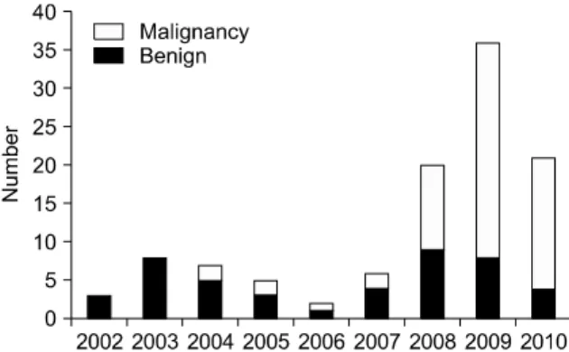
Although laparoscopic liver resection is known to need more learning curve than laparoscopic surgery for the oth- er organs, there are no reports about the learning curve ac- cording to the proficiency [21]. Our institution is not a big center, main purposes of this review were to encourage ourselves for laparoscopic liver resection for malignancy and find rationale to go further in our institution.
Fortunately we have a good result and we want to encour- age the same volume center to start laparoscopic liver re- section for malignancy. Our annual cases of laparoscopic liver resection for benign and malignancy are listed in Fig.
4.
In conclusion, even though laparoscopic liver resection requires a learning curve and only a limited number of pa- tients were included in this study, laparoscopic liver re- section is a feasible approach with reasonable surgical out- come even in patients who had malignant liver tumor es- pecially in HCC and liver metastasis from colorectal cancer. Based on the findings of this study, laparoscopic re- section warrants further study for the treatment of malig- nant liver tumors.
CONFLICTS OF INTEREST
No potential conflict of interest relevant to this article was reported.
REFERENCES
1. Tranchart H, Di Giuro G, Lainas P, Roudie J, Agostini H, Franco D, et al. Laparoscopic resection for hepatocellular carcinoma: a matched-pair comparative study. Surg Endosc 2010;24:1170-6.
2. Croome KP, Yamashita MH. Laparoscopic vs open hepatic resection for benign and malignant tumors: an updated meta-analysis. Arch Surg 2010;145:1109-18.
3. Dagher I, Belli G, Fantini C, Laurent A, Tayar C, Lainas P, et al. Laparoscopic hepatectomy for hepatocellular carcino- ma: a European experience. J Am Coll Surg 2010;211:16-23.
4. Nguyen KT, Laurent A, Dagher I, Geller DA, Steel J, Thomas MT, et al. Minimally invasive liver resection for metastatic colorectal cancer: a multi-institutional, interna- tional report of safety, feasibility, and early outcomes. Ann Surg 2009;250:842-8.
5. Cherqui D, Laurent A, Tayar C, Chang S, Van Nhieu JT, Loriau J, et al. Laparoscopic liver resection for peripheral hepatocellular carcinoma in patients with chronic liver dis- ease: midterm results and perspectives. Ann Surg 2006;
243:499-506.
6. Dagher I, Proske JM, Carloni A, Richa H, Tranchart H, Franco D. Laparoscopic liver resection: results for 70 patients. Surg Endosc 2007;21:619-24.
7. Haghighi KS, Wang F, King J, Daniel S, Morris DL. In-line radiofrequency ablation to minimize blood loss in hepatic parenchymal transection. Am J Surg 2005;190:43-7.
8. Simillis C, Constantinides VA, Tekkis PP, Darzi A, Love- grove R, Jiao L, et al. Laparoscopic versus open hepatic re- sections for benign and malignant neoplasms: a meta- analysis. Surgery 2007;141:203-211.
9. Biertho L, Waage A, Gagner M. Laparoscopic hepatectomy.
Ann Chir 2002;127:164-70.
10. Buell JF, Cherqui D, Geller DA, O'Rourke N, Iannitti D,
Dagher I, et al. The international position on laparoscopic liver surgery: The Louisville Statement, 2008. Ann Surg 2009;250:825-30.
11. Elias D, Cavalcanti A, Sabourin JC, Pignon JP, Ducreux M, Lasser P. Results of 136 curative hepatectomies with a safe- ty margin of less than 10 mm for colorectal metastases. J Surg Oncol 1998;69:88-93.
12. Zmora O, Weiss EG. Trocar site recurrence in laparoscopic surgery for colorectal cancer. Myth or real concern? Surg Oncol Clin N Am 2001;10:625-38.
13. Zmora O, Gervaz P, Wexner SD. Trocar site recurrence in laparoscopic surgery for colorectal cancer. Surg Endosc 2001;15:788-93.
14. Bismuth H, Chiche L, Adam R, Castaing D, Diamond T, Dennison A. Liver resection versus transplantation for hepatocellular carcinoma in cirrhotic patients. Ann Surg 1993;218:145-51.
15. Nagasue N, Kohno H, Chang YC, Taniura H, Yamanoi A, Uchida M, et al. Liver resection for hepatocellular carcinoma. Results of 229 consecutive patients during 11 years. Ann Surg 1993;217:375-84.
16. Franco D, Capussotti L, Smadja C, Bouzari H, Meakins J, Kemeny F, et al. Resection of hepatocellular carcinomas.
Results in 72 European patients with cirrhosis. Gastroen- terology 1990;98:733-8.
17. Kim HH, Park EK, Seoung JS, Hur YH, Koh YS, Kim JC, et al. Liver resection for hepatocellular carcinoma: case- matched analysis of laparoscopic versus open resection. J Korean Surg Soc 2011;80:412-9.
18. Choti MA, Sitzmann JV, Tiburi MF, Sumetchotimetha W, Rangsin R, Schulick RD, et al. Trends in long-term survival following liver resection for hepatic colorectal metastases.
Ann Surg 2002;235:759-66.
19. Abdalla EK, Vauthey JN, Ellis LM, Ellis V, Pollock R, Broglio KR, et al. Recurrence and outcomes following hep- atic resection, radiofrequency ablation, and combined re- section/ablation for colorectal liver metastases. Ann Surg 2004;239:818-25.
20. Scheele J, Stang R, Altendorf-Hofmann A, Paul M.
Resection of colorectal liver metastases. World J Surg 1995;19:59-71.
21. Kim CG, Yoon YS, Han HS, Shin SH, Chun KS, Jang JY, et al. Experience of total laparoscopic hepatectomy. J Korean Surg Soc 2007;73:490-5.