Copyright ⓒ 2017 by Korean Society for Surgery of the Hand, Korean Society for Microsurgery, and Korean Society for Surgery of the Peripheral Nerve. All Rights reserved.
This is an Open Access article distributed under the terms of the Creative Commons Attribution Non-Commercial License (http://creativecommons.org/licenses/by-nc/4.0/) which permits unrestricted non-commercial use, distribution, and reproduction in any medium, provided the original work is properly cited.
Guyon’s canal in the wrist is the passage of the ulnar nerve located at the base of the wrist and it is a struc- ture that extends from the proximal aspect of the palmar carpal ligament to the fibrous bow of the hypothenar muscle1. Because the ulnar nerve is divided into the superficial sensory branch and the deep motor branch within Guyon’s canal, the extent of motor impairment and sensory loss may vary depending on which part of Guyon’s canal is damaged.
Scar formation after tissue injury is an essential part
of wound healing. The injured peripheral nerve often thickens the nerve endothelium, forms scar tissue around the nerve, and scar tissue induces adhesion between the surrounding critical structures. It can then progress to chronic retraction and permanent nerve compression due to peripheral nerve compression and retraction.
In this paper, we report a case where we successfully performed a surgical technique of nerve wrapping using a silicone tube when performing neurorrhaphy in order to prevent neuropathy caused by scar tissue formation after
신경병증을 방지하기 위해 실리콘 튜브를 이용한 척골관 안에서의 신경 접합술: 증례 보고
박 범ㆍ김석원ㆍ김지예
연세대학교 원주의과대학 성형외과학교실
Neurorrhaphy of Ulnar Nerve with Silicon Wrapping in Guyon’s Canal to Prevent Neuropathy: A Case Report
Beom Park, Sugwon Kim, Jiye Kim
Department of Plastic and Reconstructive Surgery, Yonsei University Wonju College of Medicine, Wonju, Korea
An 18-year-old female patient presented with complaints of motor function and sensory impairment of the ulnar nerve on the 9th day after primary repair with a laceration of the right palm. We performed nerve wrapping with silicone tube after primary nerve repair to prevent neuropathy due to scar formation. As a result of surgical treatment, motor function and sensory impairment of ulnar nerve were improved significantly without complication until 2 years postoperatively. Nerve wrapping with silicone tube can be widely used in patients who underwent primary repair of ulnar nerve and patients who have undergone surgical decompression due to other causes of Guyon’s canal syndrome. This surgical technique is ex- pected to prevent neuropathy caused by scar formation.
Key Words: Ulnar tunnel, Ulnar nerve, Ulnar neuropathies
Received June 19, 2017, Revised June 22, 2017, Accepted June 25, 2017 Corresponding author: Sugwon Kim
Department of Plastic and Reconstructive Surgery, Yonsei University Wonju College of Medicine, 20 Ilsan-ro, Wonju 26426, Korea TEL: +82-33-741-0611, FAX: +82-33-741-0001, E-mail: [email protected]
Case Report Microsurgery
performing nerve repair to treat a deep laceration at the area of the Guyon’s canal in the wrist, and we present the case report with a review of the literature.
CASE REPORT
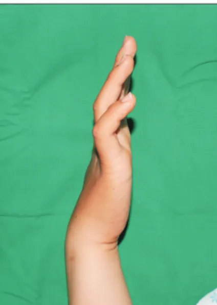
An 18-year-old female patient visited the emergency department after she fell in the bathroom and suffered a deep laceration of the right palm at the sharp part of the tile floor. The patient was discharged after debridement and primary repair. On the 9th day after the injury, she visited the hospital for outpatient treatment and com- plained of sensory impairment in the outer half of the fourth and fifth digits of the hand and motor function dis- turbance. The patient was diagnosed as having clawhand deformity at the time of the physical examination (Fig.
1). Physical examination revealed that the patient did not spread the fingers of the injured hand apart or draw them together well, and the results of Froment’s sign were positive (Fig. 2). The results of simple radiographs of the hand were normal findings. Ulnar nerve injury was suspected in the electromyogram and nerve conduction study performed on the 12th day after the injury. On the 23rd day after the injury, the skin was incised along the sutured wound using general anesthesia and tourniquet, and the Guyon’s canal was exposed, and it was found that the ulnar nerve and ulnar artery were completely cut- ted and adhered to the surrounding tissues (Fig. 3). After finding the severed ulnar nerve and artery, adhesions with
the surrounding tissues were resolved, and neurorrhaphy was performed on the deep and superficial branches of the ulnar nerve. At the same time, a commercially avail- able silicone tube (Silicone tube; Sun Medical Co., Seoul, Korea) was used to wrap the ulnar nerve in the area where there was a possibility of adhesion with surround- ing tissue or compression (Fig. 4). After performing arte- riorrhaphy, the wound was sutured and a short arm splint was applied for 2 weeks after surgery. Before entering operationg room, written informed consent was obtained from the patient after informing rare possibility of com- plications caused by implantation of foreign substances.
Fig. 2. On physical examination, finger abduction and adduction is abnormal on the right hand.
Fig. 1. On physical examination, claw hand deformity is observed.
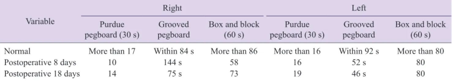
The results of the Hand Grip Power and Dexterity test performed on the 18th postoperative day showed a signif- icant improvement in muscle strength and motor function compared with the results of the same test on the 8th day (Table 1, 2). At 8 months postoperatively, the electro- myogram showed a significant improvement compared to the preoperative level. When the Jebsen-Taylor Hand Function Test was performed 9 months after surgery, the results were similar to the normal values in most of the items (Table 3). The patient did not develop any com- plications including discomfort, which is most common complication of silicone tube during the follow-up period of 2 years and showed continuous improvement of motor
and sensory function through rehabilitation.
DISCUSSION
Because the ulnar nerve is located relatively close to the skin when passing through the elbows and wrists, it is very likely to be damaged in these areas. Shea and McClain2 classified the injuries of the ulnar nerve in the wrist into three categories according to the injury loca- tion. Type 1 is sensory and motor deficit due to the injury of the area proximal to or in the canal of Guyon, Type 2 is motor impairment due to the injury of the deep branch
Fig. 3. In intraoperative exploration, total rupture of ulnar nerve and ulnar artery is observed.
Fig. 4. Neurorrhaphy is performed on the deep branches and superficial branches of the ruptured ulnar nerve. Nerve wrapping with silicone tube after primary nerve repair is performed.
Table 1. Hand grip power test, after postoperative 8 days and 18 days
Variable Right Left
Grip Lateral Palmar Tip Grip Lateral Palmar Tip
Normal (range) 25-27 4-7 4-8 3-5 25-27 3-6 4-8 3-5
Postoperative 8 days 2.0 1.5 0 0 32.0 5.5 6.0 4.5
Postoperative 18 days 11.0 2.5 2.0 2.0 32.0 6.3 7.5 6.0
Table 2. Hand dexterity test, after postoperative 8 days and 18 days Variable
Right Left
Purdue
pegboard (30 s) Grooved
pegboard Box and block
(60 s) Purdue
pegboard (30 s) Grooved
pegboard Box and block (60 s) Normal More than 17 Within 84 s More than 86 More than 16 Within 92 s More than 80
Postoperative 8 days 10 144 s 58 16 52 s 80
Postoperative 18 days 14 75 s 73 19 46 s 80
of the ulnar nerve, and Type 3 is sensory loss due to the injury of the distal part of the ulnar canal where the su- perficial branch of the ulnar nerve is located.
Perineural scar formation and adhesion can be induced by traumatic injury, hemorrhage of the surgical site, chronic inflammation, or a simple surgical operation.
Scar formation after neurorrhaphy is considered to be inevitable3. After undergoing decompression because of carpal tunnel syndrome, 20% of the patients reported recurrence of the symptom because of scar tissue forma- tion, and it has been reported that adhesion of the nerve and surrounding tissue recurs in most patients even after secondary neurolysis4.
If perineural scar formation occurs, the nerve is com- pressed and the compressed nerve causes irreversible damage as well as ischemic changes, resulting in symp- toms such as sensory deficit, muscle atrophy, functional disability, and chronic pain. In addition, nerve regenera- tion is interfered by scar tissue itself 5.
For this reason, previous studies continued to inves- tigate various surgical techniques and pharmacological treatments to minimize scar formation around the nerve after neurorrhaphy. In order to minimize scar formation, methods such as microsurgery, endoscopy, nerve transpo- sition, and fat graft have been performed. In some cases, the operation site was irradiated with radiation after sur- gery, and methods using laser irradiation, fibrin glue, and the like have also been used as an attempt to reduce for- eign body reaction by the suture material itself6,7. In ad- dition, since the use of triamcinolone acetonide and cis- hydroxyproline for suppression of scar formation after neurosurgical operations in the 1960s, various medicines
such as amniotic fluid and hyaluronic acid have been used with the expectation that they would help suppress scar formation after neurorrhaphy8,9. However, previous studies on the surgical procedures and pharmacological treatments described above did not provide a consistent and satisfactory result, and the ultimate treatment for pre- vention of scar formation after neuronal surgery has not been presented yet.
Nerve wrapping was derived from the concept of sup- pressing over-activated fibroblast reaction after a neuro- surgical operation by placing a biologic barrier consisting of fascia, fat, and vein graft around the damaged nerve.
Actually, nerve wrapping using the saphenous vein has been performed in cases of chronic median nerve com- pression. Recently, silicone tubing has been successfully used for peripheral nerve repair and major complications have not been reported10.
In respect of the case described in this paper, we per- formed neurorrhaphy for the patient whose ulnar nerve injury was not detected immediately but found later, and additionally tried the surgical technique of nerve wrap- ping with a commercial silicone tube, employing the concept of nerve wrapping used to prevent neuropathy caused by scar formation after neurorrhaphy. After sur- gical treatment, motor and sensory deficits due to ulnar nerve injury were significantly improved, and the patient recovered without complications until 2 years postopera- tively. Continuous follow-up is needed to confirm that there are no complications, including nerve compression and foreign body reactions. However, wrapping the nerve with a silicone tube is expected to prevent the occurrence of scar-induced neuropathy not only in patients who have Table 3. Jebsen-Taylor Hand Function Test postoprative 9 months
Variable Right Left
Normal (s) Postoperative 9 months (s) Normal (s) Postoperative 9 months (s)
Writing 9.1 7.2 8.5 10.1
Simulated page turning 4.4 6.0 3.4 6.2
Lifting small, common objects 5.8 7.1 5.5 6.2
Simlated feeding 8.2 5.1 9.2 5.3
Stacking checker 2.9 3.1 2.9 3.5
Lifting large, light object 3.5 2.9 3.0 3.0
Lifting large, heavy object 3.5 2.7 3.0 3.2
undergone nerve repair because of ulnar nerve injury as in the case reported in this study but also in patients un- dergoing surgical treatment such as nerve decompression because of Guyon’s canal syndrome due to other causes.
CONFLICTS OF INTEREST
The authors have nothing to disclose.
REFERENCES
1. Depukat P, Mizia E, Klosinski M, et al. Anatomy of Guyon’s canal: a systematic review. Folia Med Cracov.
2014;54:81-6.
2. Shea JD, McClain EJ. Ulnar-nerve compression syn- dromes at and below the wrist. J Bone Joint Surg Am.
1969;51:1095-103.
3. Mackinnon SE, Dellon AL. Experimental study of chronic nerve compression. Clinical implications. Hand Clin.
1986;2:639-50.
4. Cobb TK, Amadio PC, Leatherwood DF, Schleck CD, Ilstrup DM. Outcome of reoperation for carpal tunnel syn- drome. J Hand Surg Am. 1996;21:347-56.
5. Kwann JHM, Rappaport I. Postoperative brachial palsy:
a study on the mechanism. Arch Surg Neurol. 1970;
101:612-5.
6. Görgülü A, Uzal C, Doğanay L, Imer M, Eliuz K, Cobanoğlu S. The effect of low-dose external beam radia- tion on extraneural scarring after peripheral nerve surgery in rats. Neurosurgery. 2003;53:1389-95; discussion 1395- 6.
7. Menovsky T, Beek JF. Laser, fibrin glue, or suture repair of peripheral nerves: a comparative functional, histologi- cal, and morphometric study in the rat sciatic nerve. J Neurosurg. 2001;95:694-9.
8. Ozgenel GY, Filiz G. Effects of human amniotic fluid on peripheral nerve scarring and regeneration in rats. J Neu- rosurg. 2003;98:371-7.
9. Ozgenel GY. Effects of hyaluronic acid on peripheral nerve scarring and regeneration in rats. Microsurgery.
2003;23:575-81.
10. Dahlin LB, Anagnostaki L, Lundborg G. Tissue response to silicone tubes used to repair human median and ul- nar nerves. Scand J Plast Reconstr Surg Hand Surg.
2001;35:29-34.
신경병증을 방지하기 위해 실리콘 튜브를 이용한 척골관 안에서의 신경 접합술: 증례 보고
박 범ㆍ김석원ㆍ김지예
연세대학교 원주의과대학 성형외과학교실
18세 여자 환자가 오른 손바닥의 열상으로 일차 봉합술을 시행받은 후 수상 9일째 척골신경의 운동 기능 및 감각 기능 소실을 호소하며 내원하였다. 절단된 척골신경에 대해 신경 봉합술을 시행하였고, 2차적인 반흔 형성으로 인 한 신경병증을 예방하고자 상용화된 실리콘 튜브를 이용하여 신경 봉합 부위를 감싸주었다. 이후 척골신경 손상으 로 저하되었던 근력 기능 및 감각 기능이 현저히 호전되는 것을 확인할 수 있었다. 향후 척골관 내에 척골 신경을 손상받은 환자에게서 실리콘 튜브를 이용한 신경 감싸기는 반흔 형성으로 인한 신경병증의 발생을 예방할 수 있을 것으로 기대된다.
색인단어: 척골관, 척골 신경, 척골 신경병증
접수일 2017년 6월 19일 수정일 2017년 6월 22일 게재확정일 2017년 6월 25일
교신저자 김석원
26426, 원주시 일산로 20, 연세대학교 원주의과대학 성형외과학교실 TEL 033-741-0611 FAX 033-741-0001 E-mail [email protected]