Customized left-sided hepatectomy and bile duct resection for perihilar cholangiocarcinoma in a patient with left-sided
gallbladder and multiple combined anomalies
Helayel Almodhaiberi1,2, Shin Hwang1, Yoo-Jeong Cho1, Yongjae Kwon1, Bo-Hyun Jung1, and Myeong-Hwan Kim3
Departments of 1Surgery and 3Gastroenterology, Asan Medical Center, University of Ulsan College of Medicine, Seoul, Korea, 2Department of Hepatobiliary and Liver Transplant Unit, General Surgery Department,
Prince Sultan Military Medical City, Riyadh, Saudi Arabia
Left-sided gallbladder (LSGB) is a rare anomaly, but it is often associated with multiple combined variations of the liver anatomy. We present the case of a patient with LSGB who underwent successful resection of perihilar cholangiocarcinoma. The patient was a 67-year-old male who presented with upper abdominal pain and obstructive jaundice. Initial imaging studies led to the diagnosis of Bismuth-Corlette type IIIB perihilar cholangiocarcinoma. Due to the unique location of the gallbladder and combined multiple hepatic anomalies, LSGB was highly suspected. During surgery after hilar dissection, we recognized that the tumor was located at the imaginary hilar bile duct bifurcation, but its actual location was corresponding to the biliary confluence of the left median and lateral sections. The extent of resection included extended left lateral sectionectomy, caudate lobe resection, and bile duct resection. Since some of the umbilical portion of the portal vein was invaded, it was resected and repaired with a portal vein branch patch.
Due to anatomical variation of the biliary system, only one right-sided duct was reconstructed. The patient recovered uneventfully without any complication. LSGB should be recognized as a constellation of multiple hepatic anomalies, and therefore, thorough investigations are necessary to enable the performance of safe hepatic and biliary resections.
(Korean J Hepatobiliary Pancreat Surg 2015;19:30-34)
Key Words: Hepatectomy; Perihilar cholangiocarcinoma; Left-sided gallbladder; Anatomical variation
Received: February 15, 2015; Revised: February 21, 2015; Accepted: February 24, 2015 Corresponding author: Shin Hwang
Department of Surgery, Asan Medical Center, University of Ulsan College of Medicine, 88, Olympic-ro 43-gil, Songpa-gu, Seoul 138-736, Korea Tel: +82-2-3010-3930; Fax: +82-2-3010-6701; E-mail: [email protected]
Copyright Ⓒ 2015 by The Korean Association of Hepato-Biliary-Pancreatic Surgery
This is an Open Access article distributed under the terms of the Creative Commons Attribution Non-Commercial License (http://creativecommons.org/
licenses/by-nc/3.0) which permits unrestricted non-commercial use, distribution, and reproduction in any medium, provided the original work is properly cited.
Korean Journal of Hepato-Biliary-Pancreatic Surgery ∙ pISSN: 1738-6349ㆍeISSN: 2288-9213
INTRODUCTION
Left-sided gallbladder (LSGB) usually means that the gallbladder is located to the left side of the round liga- ment without situs inversus viscerum. In addition, the middle hepatic vein should run to the right of the gall- bladder and the round ligament itself should originate from the left portal vein to meet its definition. The in- cidence of LSGB in Korean population is unknown, but its incidence in patients undergoing hepatobiliary surgery at our institution is estimated to be less than 0.1%.1 The presence of LSGB is often associated with multiple com- bined anomalies of the portal vein, hepatic artery, and bile duct. Such a constellation of combined hepatic anomalies can make resection of hepatobiliary malignancy difficult
or impossible; therefore, it is essential to establish a spe- cial operative plan after thorough evaluation of both com- bined anomaly and tumor extent.2
In review of the English literature, only two Japanese patients with LSGB have been reported to undergo cura- tive surgery for perihilar cholangiocarcinoma.2 Here, we present the case of a patient with LSGB who underwent successful resection of perihilar cholangiocarcinoma.
CASE
This case was of a 67-year-old male patient who pre- sented with upper abdominal pain and obstructive jaundice. Laboratory examination on admission showed serum total bilirubin level of 9.6 mg/dl. Abdominal com-
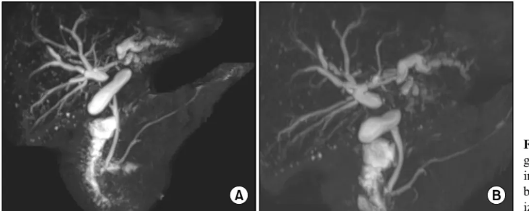
Fig. 1. Magnetic resonance cholan- giopancreatography images show- ing wide separation of the hilar bile ducts (A) without visual- ization of the usual B4 duct (B).
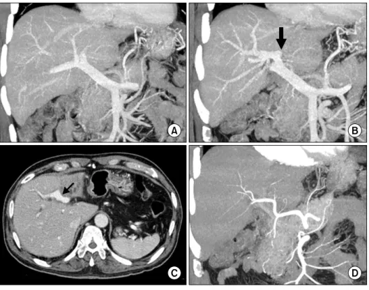
Fig. 2. Computed tomography image indicating the possible presence of the left-sided gallbladder (arrow).
puted tomography (CT) and magnetic resonance imaging revealed Bismuth-Corlette type IIIB perihilar cholan- giocarcinoma with left portal vein and left hepatic artery invasion, type III portal vein variation, dilated right intra- hepatic bile duct, and left liver lobe atrophy (Fig. 1). Due to the unique location of the gallbladder and combined multiple hepatic anomalies, presence of LSGB was highly suspected (Fig. 2). The branching pattern of the portal vein appeared strange compared with the usual type III portal vein variation, in which the liver appeared to be divided into 3 parts; the right posterior section, the right anterior section integrated with the left medial section, and the left lateral section (Fig. 3). For biliary decom- pression, endoscopic nasobiliary drainage was performed following endoscopic retrograde cholangiography. Surgery was performed when the serum total bilirubin level was 1.7 mg/dl.
After dissection of the hilar structures, we recognized that the tumor was located at the imaginary hilar bile duct bifurcation, but its actual location was corresponding to the biliary confluence of the left median and lateral sections. After transecting the distal bile duct at the level of the pancreas, we found that there was no vascular in- vasion of the right-sided liver. Thus, the extent of liver resection included extended left lateral sectionectomy (Figs. 4, 5). Through the anterior transection approach without the hanging maneuver, the liver was transected and the bile duct was resected en bloc. The right-sided bile duct orifice in the liver was 8 mm in size and single, and probing exploration revealed that it was the com- mon-channel bile duct confluence of the above-mentioned three sections in the right-sided liver. The resection mar- gin was tumor-negative on frozen-section biopsy. Since
only the left-sided wall of the umbilical portion of the portal vein was partially invaded by the tumor, the in- volved portion was excised and then covered with a rem- nant branch patch after excision of a small adjacent portal vein branch in the left medial section (Fig. 5).
The caudate duct did not appear to be involved based on the preoperative imaging study findings, but the Spigelian lobe with the paracaval portion was excised to achieve maximal curability according to the general con- cept of surgical resection for perihilar cholangiocarci- noma. One 5-mm-sized bile duct opening was exposed following caudate lobe resection, but it was directly com- municating with the right-sided duct; therefore, it was closed because bilio-enteric reconstruction was not necessary. Locoregional lymph nodes were dissected from the hepatic hilum to the celiac axis because some nodes were tumor-positive on frozen-section biopsy. Biliary re- construction was performed through a single, large hep-
Fig. 3. Computed tomography images showing the hepatic in- flow vessels. Portal vein trifurca- tion is visible with complete oc- clusion of the left portal vein (A);
A black arrow indicates the in- sertion site of the round ligament (B); The umbilical portion of the portal vein is partially invaded by the tumor (arrow, C); The hepatic arteries do not appear to have sig- nificant variation (D).
Fig. 4. Magnetic resonance cholangiopancreatography image superimposed by the pre-planned hepatic transection plane (dotted line).
aticojejunostomy using a Roux-en-Y limb.
Pathological examination revealed that the final diag- nosis was moderately differentiated adenocarcinoma in- volving the liver with vascular invasion of the left hepatic artery and left portal vein, concurrent perineural invasion without lymphovascular invasion, and lymph node meta- stasis in 5 out of 12 nodes (Fig. 6). Thus, tumor staging was T3N1M0, being stage IIIB according the 7th edition of the American Joint Committee on Cancer (AJCC) stag-
ing system.
The patient recovered uneventfully without any compli- cation and was discharged 11 days after surgery. Currently, the patient is receiving concurrent chemoradiation therapy.
DISCUSSION
According to our experience and the literature, the pres- ence of LSGB is often associated with serious combined anomalies of the hepatic vascular and biliary systems.
Such a constellation of combined hepatic anomalies can make liver surgery difficult; therefore, the operation should be prudently planned with accurate preoperative evaluation. Besides complete preoperative evaluation, the absence of anatomical variations contraindicating the pre-planned extent of hepatectomy should be confirmed intraoperatively.
In adult individuals with LSGB, portal anomaly must be the most significant part in the constellation of multiple anomalies.1-6 Nagai et al.3 reviewed collectively the ana- tomical variations combined with LSGB in their own 3 cases of living donors and previously reported 15 cases.
The patterns of intrahepatic portal venous branching were divided into the bifurcation type (5 cases), the trifurcation type (8 cases), and undetermined (5 cases) due to lack of
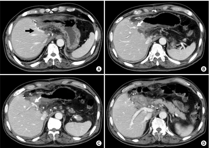
Fig. 5. Computed tomography images taken at 10 days after surgery. Two middle hepatic vein trunks are visible (A); The portal vein branch to the theoretical left medial section is visible (arrow, B); The portal vein branch to the right anterior section is visible and an arrow indicates the site of branch patch repair (C); The portal vein branch to the right posterior section is visible (D).
Fig. 6. Gross photographs of the operative field after resection (A) and the resected specimen (B).
detailed description. We also presented 3 cases of living donors with LSGB, the bifurcation type was found in two cases and the trifurcation type was found in one case [1].
The present case appeared to be of the trifurcation type although there was some dissimilarity.
The anatomy of the hepatic artery in individuals with
LSGB has not been described in detail in the literature, but we presume that it runs along the course of the portal vein branches. Variation of the hepatic artery is often dif- ferent from that of the portal vein. Bile duct anatomy in individuals with LSGB has also not been described in de- tail elsewhere. Based on our experience of 4,000 living
donor hepatectomies, the rarer the type of the portal vein variation was, the more common the combined bile duct variation became. Variant portal vein anatomy such as type III variation is usually accompanied by the un- common types of bile duct variation.7
In patients with LSGB who were diagnosed with peri- hilar cholangiocarcinoma, the anatomy of the biliary sys- tem must be seriously distorted; therefore, it is important to imagine the premorbid variant biliary anatomy and then assess the extent of tumor invasion. If the present patient had usual liver anatomy, it might not have been possible to achieve macroscopic curative resection because the tu- mor might have invaded the portal confluence as well as the right hepatic artery. In fact, the anomalous anatomy in LSGB might be helpful for resecting the tumor because it might displace the location of the tumor towards the peripheral side of the left hepatic duct. Such a tumor loca- tion is rare in the usual patients with perihilar cholangiocarcinoma. Probably due to such unusual loca- tion of the tumor, the caudate duct was free of tumor invasion. From a retrospective viewpoint, concurrent cau- date lobe resection did not appear to be necessary in the present patient. However, during operation, we intended to achieve maximal macroscopic curability; therefore, caudate lobe resection was carried out as it is routinely performed in other patients with perihilar cholangiocar- cinoma.
In conclusion, we experienced a case of a patient with perihilar cholangiocarcinoma and concurrent anomalies of LSGB, and based on the preoperative and intraoperative
assessment of the liver anatomy and tumor extent, a cus- tomized extended liver resection of the extended left later- al section and bile duct resection were accomplished safe- ly without any postoperative complication. LSGB should be recognized as a constellation of multiple hepatic anomalies, and therefore, thorough investigations are nec- essary to enable the performance of safe hepatic and bili- ary resections.
REFERENCES
1. Hwang S, Lee SG, Park KM, Lee YJ, Ahn CS, Kim KH, et al.
Hepatectomy of living donors with a left-sided gallbladder and multiple combined anomalies for adult-to-adult living donor liver transplantation. Liver Transpl 2004;10:141-146.
2. Kaneoka Y, Yamaguchi A, Isogai M, Harada T. Hepatectomy for cholangiocarcinoma complicated with right umbilical portion:
anomalous configuration of the intrahepatic biliary tree. J Hepatobiliary Pancreat Surg 2000;7:321-326.
3. Nagai M, Kubota K, Kawasaki S, Takayama T, BandaiY, Makuuchi M. Are left-sided gallbladders really located on the left side? Ann Surg 1997;225:274-280.
4. Asonuma K, Shapiro AM, Inomata Y, Uryuhara K, Uemoto S, Tanaka K. Living related liver transplantation from donors with the left-sided gallbladder/portal vein anomaly. Transplantation 1999;68:1610-1612.
5. Ogawa T, Ohwada S, Ikeya T, Shiozaki H, Aiba S, Morishita Y. Left-sided gallbladder with anomalies of the intrahepatic por- tal vein and anomalous junction of the pancreaticobiliary ductal system: a case report. Hepatogastroenterology 1995;42:645-649.
6. Hardy KJ, Jones RM. Failure of the portal vein to bifurcate.
Surgery 1997;121:226-228.
7. Hwang S, Lee SG, Lee YJ, Park KM, Kim KH, Ahn CS, et al.
Donor selection for procurement of right posterior segment graft in living donor liver transplantation. Liver Transpl 2004;10:
1150-1155.