ISSN 2234-3806 • eISSN 2234-3814
428 www.annlabmed.org https://doi.org/10.3343/alm.2020.40.5.428 Ann Lab Med 2020;40:428-430
https://doi.org/10.3343/alm.2020.40.5.428
Letter to the Editor
Diagnostic Immunology
Serial Assays of QuantiFERON-TB Gold In-Tube and QuantiFERON-TB Gold-Plus in Subjects Exposed to Patients with Active Tuberculosis
Hee-Won Moon , M.D., Ph.D.1, Ahram Yi , M.D.2, Sumi Yoon , M.D.1, Hanah Kim , M.D., Ph.D.1, Hee-Jung Chung , M.D.1, Mina Hur , M.D., Ph.D.1, Yeo-Min Yun , M.D., Ph.D.1, and Kwang Ha Yoo , M.D., Ph.D.3
1Department of Laboratory Medicine, Konkuk University School of Medicine, Seoul, Korea; 2Department of Laboratory Medicine, Green Cross Laboratories, Yongin, Korea; 3Department of Internal Medicine, Konkuk University School of Medicine, Seoul, Korea
Dear Editor,
The detection and management of subjects with latent tubercu- losis infection (LTBI) are important steps to control and de- crease the incidence of tuberculosis (TB). Exposure to patients with active TB is one of the most important transmission routes of LTBI [1]. Interferon-gamma (IFN-γ) release assays (IGRAs), in particular, the QuantiFERON-TB Gold In-Tube assay (QFT;
QIAGEN, Germantown, MD, USA), have been widely used for investigating subjects exposed to patients with active TB. How- ever, high reversion rates and low reproducibility of QFT have also been documented [2].
The new version of QFT, QuantiFERON-TB Gold-Plus (QFT- Plus, QIAGEN), might provide higher sensitivity for detection of early infection in subjects exposed to patients with active TB, owing to its ability to assess both CD8+ and CD4+ T cell-medi- ated immune responses [3, 4]. Since there is sparse data on QFT and QFT-Plus in subjects exposed to patients with active TB [5, 6], we investigated the results of serial assays of QFT and QFT-Plus in such subjects. From December 2016 to November 2018, 69 subjects exposed to patients with active TB (including 57 households and 12 occupational exposures; median age [in-
terquartile range, IQR], 35 years [27-50 years]) were enrolled at Konkuk University Medical Center (KUMC), Seoul, Korea. We excluded subjects who had a history of a positive QFT or tuber- culin skin test (TST) result. This study was approved by the In- stitutional Review Board of KUMC (KUH1200076), and in- formed consent was obtained from all enrolled subjects.
Both QFT and QFT-Plus were performed within eight weeks of first exposure as the first assessment, with a median (IQR) of 14 (6-30) days after exposure. Follow-up assays were performed in 39 subjects at eight weeks after the first assessment (i.e., sec- ond assessment). If there were discrepancies between the first and second assessments, a third assessment was performed after eight weeks, when possible.
Peripheral blood (1 mL; Nil, QFT TB antigen, QFT-Plus TB1, TB2 antigen [CD4+ T cell response and both CD4+ and CD8+
T cell response, respectively]) was collected in each specialized tube. The samples were immediately incubated at 37°C for 16- 24 hours, and the separated plasma was stored at 4°C. QFT and QFT-Plus were performed according to the manufacturer’s instructions. For QFT, the results are considered positive when the TB antigen minus Nil IFN-γ concentration is ≥0.35 IU/mL
Received: November 29, 2019 Revision received: January 13, 2020 Accepted: March 3, 2020
Corresponding author: Hee-Won Moon, M.D., Ph.D.
Department of Laboratory Medicine, Konkuk University School of Medicine Konkuk University Medical Center, 120-1 Neungdong-ro, Gwangjin-gu, Seoul 05030, Korea
Tel: +82-2-2030-5583, Fax: +82-2-2030-5587, E-mail: [email protected]
© Korean Society for Laboratory Medicine
This is an Open Access article distributed under the terms of the Creative Commons Attribution Non-Commercial License (https://creativecommons.org/licenses/by-nc/4.0) which permits unrestricted non-commercial use, distribution, and reproduction in any medium, provided the original work is properly cited.
1 / 1 CROSSMARK_logo_3_Test
2017-03-16 https://crossmark-cdn.crossref.org/widget/v2.0/logos/CROSSMARK_Color_square.svg
Moon HW, et al.
Serial QFT and QFT-Plus after TB exposure
https://doi.org/10.3343/alm.2020.40.5.428 www.annlabmed.org 429
and ≥25% of the Nil value. The QFT-Plus is interpreted as posi- tive when the TB antigen tube (TB1 or TB2) minus Nil IFN-γ concentration is ≥0.35 IU/mL and ≥25% of the Nil value. Con- cordance between the results of the two assays was measured using Cohen’s Kappa, and statistical analysis was performed us- ing MedCalc Statistical Software (version 17.2; MedCalc Soft- ware, Ostend, Belgium).
The agreement between QFT and QFT-Plus results was strong (110 coupled tests, kappa =0.857, 95% confidence inter- val=0.746-0.967, overall concordance rate=94.5%). The posi- tive rate of QFT and QFT-Plus at the first assessment was 24.6% (17/69) and 26.1% (18/69), respectively. Prophylactic treatment was performed in eight subjects between the first and second assessments. The positive rate of QFT and QFT-Plus at the second assessment was 23.1% (9/39) and 28.2% (11/39), respectively. The IFN-γ concentrations of the TB antigen minus Nil tube in QFT and QFT-Plus are shown in Table 1. The QFT-
Plus TB2 values were significantly higher in the second assess- ment than in the first assessment (P =0.032, Wilcoxon signed rank test). However, the QFT TB and QFT-Plus TB1 values were not significantly different between the first and second assess- ments.
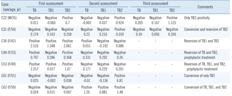
Among the 39 subjects with available serial assay results, the conversion rate (negative to positive) of QFT and QFT-Plus was 3.5% (1/29) and 10.7% (3/28), respectively, and the reversion rate (positive to negative) was 20.0% (2/10) and 27.3% (3/11), respectively. The inconsistent results between QFT and QFT- Plus or between the first and follow-up assessments are shown in Table 2. Samples from two subjects (C22 and C61) showed positivity only in QFT-Plus TB2, suggesting that TB2 might be converted earlier in cases of recent exposure. In particular, samples from C22 showed only TB2 positivity consistently on the first, second, and third assessments, and the TB-Nil, TB1- Nil, and TB2-Nil values showed an increasing trend (except TB
Table 1. Results of QuantiFERON-TB Gold In-Tube (TB), QuantiFERON-TB Gold-Plus TB1 (TB1), and QuantiFERON-TB Gold-Plus TB2 (TB2) minus Nil values in the first and second assessments
First assessment Second assessment
Median Interquartile range Median Interquartile range P*
TB 0.111 0.072-0.884 0.110 0.060-0.779 0.064
TB1 0.011 -0.010-0.286 0.106 0.068-0.477 0.192
TB2 0.029 -0.010-0.700 0.128 0.063-0.863 0.032
*P was determined by the Wilcoxon signed rank test.
Abbreviations: TB, TB minus Nil; TB1, TB1 minus Nil; TB2, TB2 minus Nil.
Table 2. Inconsistent results of QuantiFERON-TB Gold In-Tube (TB), QuantiFERON-TB Gold-Plus TB1 (TB1), and QuantiFERON-TB Gold- Plus TB2 (TB2) or between the first and follow-up assessments
Case (sex/age, yr)
First assessment Second assessment Third assessment
Comments
TB TB1 TB2 TB TB1 TB2 TB TB1 TB2
C22 (M/35) Negative 0.011
Negative -0.066
Positive 0.7
Negative -0.002
Negative 0.037
Positive 0.924
Negative 0.205
Negative 0.167
Positive 1.125
Only TB2-positivity C31 (F/54) Negative
0.174 Negative
0.163 Negative
0.258 Negative
0.23 Negative
0.216 Positive
0.559 Negative
0.34 Negative
0.056 Negative
0.269 Conversion and reversion of TB2 C36 (F/42) Positive
2.516
Positive 1.348
Positive 2.061
Positive 0.651
Negative -0.192
Negative 0.086
- - - Reversion of TB1 and TB2
C44 (F/23) Positive
0.797 Negative
0.286 Positive
0.368 Negative
0.331 Negative
0.292 Negative
0.26 - - - Reversion of TB and TB2,
prophylactic treatment C53 (F/49) Positive
2.257
Positive 0.917
Positive 1.07
Negative 0.177
Negative 0.229
Negative 0.201
- - - Reversion of TB, TB1, and TB2,
prophylactic treatment C61 (F/51) Negative
0.025 Negative
-0.002 Negative
0.038 Negative
-0.01 Negative
-0.136 Positive
6.81 - - - Conversion of only TB2
C62 (F/50) Negative 0.024
Negative 0.015
Negative 0.047
Positive 1.26
Positive 0.881
Positive 1.48
- - - Conversion of TB, TB1, and TB2
Abbreviations: M, Male; F, Female; TB, TB minus Nil; TB1, TB1 minus Nil; TB2, TB2 minus Nil.
Moon HW, et al.
Serial QFT and QFT-Plus after TB exposure
430 www.annlabmed.org https://doi.org/10.3343/alm.2020.40.5.428 at the second assessment), indicating an ongoing conversion
process. Samples from C61 showed an abrupt increase in TB2- Nil; thus, a follow-up assay would be helpful for accurate inter- pretation, although it could not be performed in this study. An- other subject (C62) showed a positive result only at the second assessment; the first assessment was too early (at four days af- ter exposure), and a second assessment was, therefore, neces- sary in this subject. Recent findings suggest more delayed con- version with IGRA than with the TST, whereas QFT-Plus showed earlier conversion than QFT [1, 7, 8]. Reversion was observed in four subjects (C31, C36, C44, and C53), including two with QFT-TB (C44 and C53), two with QFT-Plus TB1 (C36 and C53), and four with TB2 (C31, C36, C44, and C53). Prophylactic treatment might explain the reversion in two subjects (C44 and C53) [9]. Reversion in the other two subjects could be related to natural clearing, an unstable immune response, or assay-related variability [10] due to pre-analytical, analytical, post-analytical, manufacturing, and immunological factors [2]. Since we could not perform a baseline QFT, the possibility that infection in some subjects occurred before exposure to patients with active TB cannot be ruled out. We tried to decrease this possibility by ex- cluding subjects with a history of a positive QFT or TST result.
Despite the limited sample size, we showed serial results of QFT and QFT-Plus in subjects exposed to patients with active TB. Since QFT-Plus TB2 seems to be converted earlier, follow- up assays would be helpful before starting prophylactic treat- ment, owing to the possibility of reversion and delayed conver- sion. Moreover, assessment should be done within enough time to allow for maturation of the immune response, and detailed guidelines should be prepared using additional data.
ACKNOWLEDGEMENTS
QIAGEN provided the QFT-Plus kits for this study.
AUTHOR CONTRIBUTIONS
MHW designed the study, analyzed the data, and wrote the manuscript. YA, YS, and YKH participated in subject enroll- ment, research protocol and analysis. KH, CHJ, HM, and YYM reviewed the manuscript. All authors read and approved the fi- nal manuscript.
CONFLICTS OF INTEREST
None declared.
RESEARCH FUNDING
None declared.
ORCID
Hee-Won Moon https://orcid.org/0000-0001-9509-6073 Ahram Yi https://orcid.org/0000-0002-3107-936X Sumi Yoon https://orcid.org/0000-0001-7529-1613 Hanah Kim https://orcid.org/0000-0002-3266-638X Hee-Jung Chung https://orcid.org/0000-0002-1479-0731 Mina Hur https://orcid.org/0000-0002-4429-9978 Yeo-Min Yun https://orcid.org/0000-0002-5485-8331 Kwang Ha Yoo https://orcid.org/0000-0001-9969-2657
REFERENCES
1. Barcellini L, Borroni E, Brown J, Brunetti E, Campisi D, Castellotti PF, et al. First evaluation of QuantiFERON-TB Gold Plus performance in con- tact screening. Eur Respir J 2016;48:1411-9.
2. Banaei N, Gaur RL, Pai M. Interferon-gamma release assays for latent tuberculosis: what are the sources of variability? J Clin Microbiol 2016;
54:845-50.
3. Rozot V, Patrizia A, Vigano S, Mazza-Stalder J, Idrizi E, Day CL, et al.
Combined use of Mycobacterium tuberculosis-specific CD4 and CD8 T- cell responses is a powerful diagnostic tool of active tuberculosis. Clin Infect Dis 2015;60:432-7.
4. Moon HW, Gaur RL, Tien SS, Spangler M, Pai M, Banaei N. Evaluation of QuantiFERON-TB Gold-Plus in health care workers in a low-incidence setting. J Clin Microbiol 2017;55:1650-7.
5. Gonzślez-Moreno J, García-Gasalla M, Gallego-Lezaun C, Fernández-Ba- ca V, Mir Viladrich I, Cifuentes-Luna C, et al. Role of QuantiFERON(®)- TB Gold In-Tube in tuberculosis contact investigation: experience in a tu- berculosis unit. Infect Dis (Lond) 2015;47:244-51.
6. Ferrarini MA, Spina FG, Weckx LY, Lederman HM, De Moraes-Pinto MI.
Rate of tuberculosis infection in children and adolescents with house- hold contact with adults with active pulmonary tuberculosis as assessed by tuberculin skin test and interferon-gamma release assays. Epidemiol Infect 2016;144:712-23.
7. Lee SW, Oh DK, Lee SH, Kang HY, Lee CT, Yim JJ. Time interval to con- version of interferon-gamma release assay after exposure to tuberculo- sis. Eur Respir J 2011;37:1447-52.
8. Ribeiro-Rodrigues R, Kim S, Coelho da Silva FD, Uzelac A, Collins L, Palaci M, et al. Discordance of tuberculin skin test and interferon gam- ma release assay in recently exposed household contacts of pulmonary TB cases in Brazil. PLoS One 2014;9:e96564.
9. Zhang H, Xin H, Li X, Li H, Li M, Feng B, et al. Reversion of QuantiFER- ON-TB Gold In-Tube test in individuals with and without prophylactic treatment for latent tuberculosis infection: a systematic review and me- ta-analysis. J Infect 2018;77:276-82.
10. Pai M, Denkinger CM, Kik SV, Rangaka MX, Zwerling A, Oxlade O, et al.
Gamma interferon release assays for detection of Mycobacterium tuber- culosis infection. Clin Microbiol Rev 2014;27:3-20.