PGHN
Case Report
Bannayan-Riley-Ruvalcaba Syndrome in a Patient with a PTEN Mutation Identified by Chromosomal Microarray Analysis:
A Case Report
Sun Hwa Lee, Eell Ryoo, and Hann Tchah
Department of Pediatrics, Gachon University Gil Medical Center, Incheon, Korea
Bannayan-Riley-Ruvalcaba syndrome (BRRS) is one of the phosphatase and tensin homolog hamartoma tumor syndrome with a PTEN gene mutation. It is a rare dominant autosomal disorder characterized by cutaneous lipomas, macrocephaly, intestinal polyps, and developmental delay. Diagnosing this syndrome is important, because it may represent the pediatric phenotype of Cowden syndrome, in which there is an increased risk for malignant tumors in children. Until now, the prevalence of BRRS is unknown. Several dozen cases have been reported in the medical literature, but no case has been reported in Korea. Here we report a case of a 19-year-old girl who was diagnosed with BRRS because of macrocephaly, intellectual disability, and intestinal polyps. Her mother had similar findings and a PTEN mutation. Neither patient had mutations detected by conventional mutation-detection techniques, but a PTEN gene deletion was demonstrated by chromosomal microarray analysis.
Key Words: Bannayan-Riley-Ruvalcaba syndrome, Microarray analysis
Received:December 13, 2015, Revised:March 28, 2016, Accepted:April 6, 2016
Corresponding author: Eell Ryoo, Department of Pediatrics, Gachon University Gil Medical Center, 21 Namdong-daero 774beon-gil, Namdong-gu, Incheon 21565, Korea. Tel: +82-32-460-2362, Fax: +82-32-460-3224, E-mail: [email protected]
Copyright ⓒ 2017 by The Korean Society of Pediatric Gastroenterology, Hepatology and Nutrition
This is an openaccess article distributed under the terms of the Creative Commons Attribution NonCommercial License (http://creativecommons.org/licenses/by-nc/4.0/) which permits unrestricted noncommercial use, distribution, and reproduction in any medium, provided the original work is properly cited.
INTRODUCTION
Bannayan-Riley-Ruvalcaba syndrome (BRRS) is one of the phosphatase and tensin homolog ha- martoma tumor syndromes (PHTS). It is a rare domi- nant autosomal disorder characterized by cutaneous lipomas, macrocephaly, intestinal polyps, and devel- opmental delay associated with PTEN gene mutation (tumor suppressor gene deletion on chromosome 10q22-q23) [1]. This disease encompasses three pre- viously described disorders, such as Bannayan-Zonana
syndrome, Riley-Smith syndrome, and Ruvalca- va-Myhre-Smith syndrome [2]. In 1971, Bannayan [3] reported the congenital combination of macro- cephaly with multiple subcutaneous and visceral lip- omas and hamangiomas. Then, in 1980, Ruvalcaba described two males with macrocephaly, harmatom- atous intestinal polyposis, and pigmentary spotting of the penis [2]. The prevalence of BRRS is unknown but several dozen cases have been reported in the medical literature. Researchers suspect that the dis- order is under-diagnosed because its signs and
symptoms vary and some are subtle [4,5].
Although there are no international consensus cri- teria for diagnosing BRRS, several groups of inves- tigators have proposed criteria to facilitate clinical di- agnosis [6]. Marsh et al. [7] defined a clinical diag- nosis of BRRS as meeting three out of four features:
macrocephaly, lipomatosis, hemangiomas and speck- led pigmented maculae on the penis. Parisi et al. [8]
defined the syndrome in patients with two of three of the following features of macrocephaly, hamarto- mas (including at least one lipoma, hemangioma, or intestinal polyp), and penile macules in males.
A chromosomal analysis for PTEN gene mutations also serves as a useful diagnostic tool.
The prevalence of identified germline PTEN muta- tions in PHTS varies widely, with Cowden syndrome (CS) having 80% prevalence of identified intragenic PTEN mutations, BRRS with 65% prevalence, and Proteus syndrome with <20% prevalence [6].
Here, we report a case of a 19-year-old girl who was diagnosed with BRRS because of macrocephaly, intellectual disability and intestinal polyps as well as PTEN gene mutation.
CASE REPORT
A 19-year-old female patient was admitted to Gachon University Gil Medical Center (Incheon, Korea) because of a history of refractory iron defi- ciency anemia (IDA) and recurrent gastroenteritis.
She first visited our outpatient clinic because of re- current gastroenteritis 4 years prior. Laboratory in- vestigation was normal except for IDA. She was put on iron tablets and regular follow-up at our out- patient department, but her anemia did not improve.
She denied any history of skipped medicine. So, we recommended the endoscopic study 3 years ago be- cause of the suspicion of a gastrointestinal bleeding or hamartomatous polyposis syndrome, but she re- fused at that time; however, 3 years later it could be performed.
The patient had a history of mild mental re- tardation at the age of 8 years as her mother. Their intelligence quotient score ranged from 50-75. She
had one sister who was reported as normal.
General physical examination revealed moderate pallor, normal oral cavity. There was no jaundice, cyanosis, edema, thyromegaly, lymphadenopathy, clubbing, or scoliosis. There were no dermatologic abnormalities or any abnormality affecting the ge- nitalia. Anthropometric examination revealed mac- rocephaly with head circumference of 608 mm (for reference, the average and 99th percentile occipi- to-frontal circumferences of 19- and 24-year-old Korean females are 552 and 585 mm, respectively), height 162.8 cm (55-75th percentile), weight 62 kg (90-95th percentile). Her sister and mother showed no macrocephaly.
Lab investigations revealed hypochromic micro- cytic anemia with hemoglobin 8.4 g/dL and mean cell volume 68.8, ferritin 24.8 μg/L, iron/total iron binding capacity ratio 6.6% and stool occult blood testing was negative.
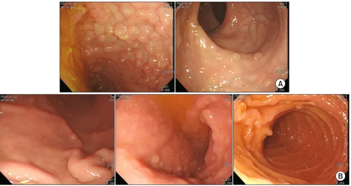
Gastrofibroscopy and colonoscopy revealed multi- ple polyps of different sizes and shapes in the termi- nal ileum, stomach, and duodenum (Fig. 1). Several biopsies confirmed inflammatory and hyperplastic polyps.
In family screening, her sister and mother were called and examined. Her sister was found as normal but her mother revealed multiple polyps on stomach, and duodenum.
The patient also had an intellectual disability and the physical examination revealed a head circum- ference suggestive of macrocephaly.
Considering all symptoms and the results of the examinations, BRRS was the most likely clinical diagnosis. Although imaging studies including brain magnetic resonance imaging were required, they were not conducted because of cost.
PTEN gene mutation analysis was conducted us- ing DNA extracted from peripheral blood leukocytes.
No mutations were detected by conventional poly- merase chain reaction (PCR) mutation-deletion techniques or direct sequencing with reference to the NM_000314.4 m-RNA sequence. Thus, we con- ducted a chromosomal microarray analysis (CMA) using the Affymetrix Cytoscan 750k array with refer-
Fig. 1. (A) Colonoscopy showed multiple polyps on the terminal ileum. Some flat polyps without neck were noted on the rectum.
(B) Gastrofibroscopy showed small multiple polyps on the stomach and duodenum.
ence to the human gene ver. 19 (Affymetrix, Santa Clara, CA, USA). As a result, an approximates 240 kb microdeletion was detected in the 10q23.31 region (Fig. 2).
An approximate 220 kb microdeletion was de- tected in her mother in the 10q23.31 region. Her old- er sister had no such findings.
DISCUSSION
BRRS is associated with germline mutations of the PTEN tumor suppressor gene, which has a sig- nificant role in cellular proliferation, migration, and apoptosis pathways. PTEN mutations are seen in up to 65% of patients with a suspected PHTS diagnosis [9,10].
Three other clinically distinct syndromes are asso- ciated with PTEN mutations and are collectively re- ferred to as PHTS. These allelic disorders include CS, Proteus syndrome, and Proteus-like syndrome, and all have established diagnostic criteria. However, no BRRS diagnostic criteria have been established [2,11].
BRRS can be diagnosed based on clinical ob- servations, including the presence of macrocephaly, lipomas, hamartomatous intestinal polyposis, devel- opmental delay and mental retardation, as well as pigmented macules on the glans penis in males.
At least half of affected patients have macro- cephaly, and many also have a high birth weight.
Growth usually slows during childhood, so affected adults are of normal height and body size. Gontijo et al. [12] reported BRRS with deforming lipomatous hamartomas in infant. Intestinal polyps mostly pres- ent in childhood through chronic anemia, diarrhea or invagination of the small bowel. The signs and symptoms of BRRS are present from birth or become apparent in early childhood. Our patient’s past medi- cal history was significant for recurrent gastro- enteritis from childhood and refractory IDA.
BRRS and CS reportedly share clinical character- istics and represent a single entity. However, almost everyone with CS develops hamartomas. These growths are most commonly found on the skin and mucous membranes (such as the lining of the mouth and nose), but they can also occur in the intestine
Fig. 2. (A) Karyogram of the patient. The deletion in the long arm of chromosome 10 (arrow). Left: the patient, right: her mother.
(B) Chromosomal microarray profile of chromosome 10. The X-axis represents the probe index on chromosome 10, and the Y-axis represents the signal log2 ratio of the probe. The 10q23.31 region showed a 220kb micro-deletion in Chr10. Left: the patient, right:
her mother.
and other parts of the body. The growth of hamarto- mas on the skin and mucous membranes typically becomes apparent by a person’s late twenties [4,5].
No such criteria exist for BRRS, but the syndrome is sus¬pected in the presence of macrocephaly, ha- martomatous intestinal polyposis, and mental re- tardation, all of which were present in our patient. At present, she most closely fits BRRS, although further development of symptoms with time may eventually lead to the diagnosis of CS.
Although an increased risk for malignancies in pa- tients with BRRS has not been documented, some authors recommend that patients with BRRS comply
with the same malignancy screening recommen- dations for patients with CS because of the clinical and genetic similarities between CS and BRRS and because patients with PTEN mutations have in- creased risk for cancers [7,8]. Therefore, early diag- nosis is important.
Genetic testing is available to identify mutations and/or deletions within PTEN. Identification of such alterations provides confirmation of the PHTS diag- nosis and further permits predictive testing and pre- natal diagnosis within affected families [11]. Several methodologies are currently used to detect PTEN mutations [13].
The appropriate order of PTEN testing to optimize yield first includes sequencing all PTEN coding exons 1-9 and flanking intronic regions. If no pathogenic variant is identified, deletion/duplication analysis is recommended [14]. So, we conducted conventional PCR mutation-deletion techniques and direct se- quencing. Since no mutation was identified, we per- formed deletion analysis by microarray. Previous study showed that the detection rates of microarray ranged from 5% to 17% for children undergoing a ge- netics evaluation for a variety of conditions who pre- viously had a karyotype with no chromosome abnor- malities [15]. Menko et al. [16] reported variable phenotypes associated with 10q23 microdeletions detected by microarray.
Recently, some consensus statements have pro- posed utilization of CMA as a first-line test in pa- tients with multiple congenital anomalies not specif- ic to a well delineated genetic syndrome, devel- opmental delay and intellectual disability, or autism spectrum disorders. CMA enables genome-wide de- tection of submicroscopic chromosomal abnormal- ities with greater precision and accuracy [17].
Although CMA has distinct advantages, there are several limitations, including its inability to detect balanced chromosomal rearrangements and low-level mosaicism, its interpretation of copy number var- iants of uncertain clinical significance, and sig- nificantly higher costs. Thus, CMA is not currently a replacement for conventional cytogenetics but can be used as an adjunct to conventional cytogenetics to identify chromosomal abnormalities, leading to a more accurate and comprehensive assessment of chromosomal aberrations [18,19].
In conclusion, our case study suggests that CMA can be used to identify chromosomal abnormalities even if conventional cytogenetics testing was normal in BRRS.
REFERENCES
1. Buisson P, Leclair MD, Jacquemont S, Podevin G, Camby C, David A, et al. Cutaneous lipoma in children:
5 cases with Bannayan-Riley-Ruvalcaba syndrome. J
Pediatr Surg 2006;41:1601-3.
2. Schreibman IR, Baker M, Amos C, McGarrity TJ. The hamartomatous polyposis syndromes: a clinical and molecular review. Am J Gastroenterol 2005;100:
476-90.
3. Bannayan GA. Lipomatosis, angiomatosis, and macrencephalia. A previously undescribed congenital syndrome. Arch Pathol 1971;92:1-5.
4. Genetics Home Reference. Bannayan-Riley-Ruvalcaba syndrome [Internet]. Bethesda: U. S. National Library of Medicine; 2012 [cited 2016 Mar 13]. Available from:
https://ghr.nlm.nih.gov/condition/bannayan-riley- ruvalcaba-syndrome.
5. Genetics Home Reference. Cowden syndrome [Internet].
Bethesda: U. S. National Library of Medicine; 2012 [cited 2016 Mar 13]. Available from: https://ghr.nlm.nih.gov/
condition/cowden-syndrome.
6. Blumenthal GM, Dennis PA. PTEN hamartoma tumor syndromes. Eur J Hum Genet 2008;16:1289-300.
7. Marsh DJ, Coulon V, Lunetta KL, Rocca-Serra P, Dahia PL, Zheng Z, et al. Mutation spectrum and geno- type-phenotype analyses in Cowden disease and Bannayan-Zonana syndrome, two hamartoma syn- dromes with germline PTEN mutation. Hum Mol Genet 1998;7:507-15.
8. Parisi MA, Dinulos MB, Leppig KA, Sybert VP, Eng C, Hudgins L. The spectrum and evolution of phenotypic findings in PTEN mutation positive cases of Bannay- an-Riley-Ruvalcaba syndrome. J Med Genet 2001;
38:52-8.
9. Latiff ZA, Atmawidjaja RW, RajaLope RJ, Syed Omar SA, Syed Zakaria SZ, Jamal RA. Bannayan Riley Ruvalcaba syndrome. Ann Acad Med Singap 2010;39:578-82.
10. Lachlan KL, Lucassen AM, Bunyan D, Temple IK.
Cowden syndrome and Bannayan Riley Ruvalcaba syn- drome represent one condition with variable expression and age-related penetrance: results of a clinical study of PTEN mutation carriers. J Med Genet 2007;
44:579-85.
11. Hobert JA, Eng C. PTEN hamartoma tumor syndrome:
an overview. Genet Med 2009;11:687-94.
12. Gontijo GM, Pinto CA, Rogatto SR, Cunha IW, Aguiar S Jr, Alves CA. Bannayan-Riley-Ruvalcaba syndrome with deforming lipomatous hamartomas in in- fant--case report. An Bras Dermatol 2013;88:982-5.
13. Zhou XP, Waite KA, Pilarski R, Hampel H, Fernandez MJ, Bos C, et al. Germline PTEN promoter mutations and deletions in Cowden/Bannayan-Riley-Ruvalcaba syndrome result in aberrant PTEN protein and dysre- gulation of the phosphoinositol-3-kinase/Akt pathway.
Am J Hum Genet 2003;73:404-11.
14. Eng C. PTEN hamartoma tumor syndrome (PHTS) [Internet]. In: Pagon RA, Adam MP, Ardinger HH, et al., editors. GeneReviews. Seattle: University of Wa- shington; 2001 Nov 29 [updated 2014 Jan 23; cited 2016 Mar 13]. Available from: http://www.ncbi.nlm.nih.gov/
books/NBK1488/.
15. Shaffer LG, Bejjani BA. Using microarray-based molec- ular cytogenetic methods to identify chromosome abnormalities. Pediatr Ann 2009;38:440-7.
16. Menko FH, Kneepkens CM, de Leeuw N, Peeters EA, Van Maldergem L, Kamsteeg EJ, et al. Variable pheno- types associated with 10q23 microdeletions involving the PTEN and BMPR1A genes. Clin Genet 2008;74:
145-54.
17. Seo EJ. Clinical applications of chromosomal micro- array analysis. J Genet Med 2010;7:111-8.
18. Manning M, Hudgins L; Professional Practice and Guidelines Committee. Array-based technology and recommendations for utilization in medical genetics practice for detection of chromosomal abnormalities.
Genet Med 2010;12:742-5.
19. Miller DT, Adam MP, Aradhya S, Biesecker LG, Brothman AR, Carter NP, et al. Consensus statement:
chromosomal microarray is a first-tier clinical diag- nostic test for individuals with developmental dis- abilities or congenital anomalies. Am J Hum Genet 2010;86:749-64.