J Korean Soc Coloproctol Vol. 22, No. 1, 2006
34
Self-expandable Metallic Stent for the Ma- nagement of Acute Malignant Large-bowel Obstruction
Yoon-Ah Park, M.D., Kwang-Hun Lee, M.D.
1, Sun-Il Lee, M.D., Seung-Kook Sohn, M.D.Departments of Surgery and 1Radiology, Yonsei University College of Medicine, Seoul, Korea
Purpose: The purpose of this study was to review our
experience with the use of self-expandable metallic stents as the initial interventional management for acute malignant large-bowel obstruction. Methods: The records of 35 patients who underwent placement of a colonic stent for acute malignant obstruction at our institution between January 2003 and December 2004 were reviewed. Results:Stents were placed for palliation in 19 patients and as bridge to surgery in 16 patients. Technical success of stent placement was achieved in all patients (100%), but clinical failure occurred in two patients due to limited expansion of the metallic stent. One of them who had clinical failure underwent an emergency operation, and the other needed no further procedure because of his death. Complications occurred in 4 patients (12%), including one pelvic abscess associated with colon perforation, two minor bleedings, and one anal pain. All the patients in the bridge-to-surgery group underwent an elective colon resection without stoma. In the palliative group, stent reocclusion was observed in three patients during the follow-up (median:
65 days; range: 27∼440 days), two of which were managed by reinsertion of a stent. In the remaining patients, the stent was patent until death or the last follow up date (median: 65 days). Conclusions: Placement of a self- expandable metallic stent is a safe and effective procedure for immediate decompression of acute malignant large- bowel obstruction. It provides a chance of elective surgery
for patients with resectable disease, as well as relief of symptoms for those with unresectable disease. J Korean
Soc Coloproctol 2006;22:34-40
Key Words: Malignant, Acute malignant large-bowel ob- struction, Self-expandable metallic stent 악성 질환, 급성 대장 폐색, 자가팽창형 금 속스텐트
ꠏꠏꠏꠏꠏꠏꠏꠏꠏꠏꠏꠏꠏꠏꠏꠏꠏꠏꠏꠏꠏꠏꠏꠏꠏꠏꠏꠏꠏꠏꠏꠏꠏꠏꠏꠏꠏꠏꠏꠏꠏꠏꠏꠏꠏꠏꠏꠏꠏ
서 론
급성 대장 폐색은 악성 질환에 의한 경우가 가장 흔 하며, 원발성 및 재발성 대장암 혹은 타장기암의 복막 전이 등에 의해 주로 발생한다.1 현재까지 악성 질환에 의한 좌측 결장 및 직장 폐색의 치료는 하트만 술식과 같은 단계적 수술과, 단일 수술법(single stage opera- tion)으로서 아전결장절제술(subtotal colectomy) 혹은 수술 중 장세척(intraoperative colonic lavage)을 이용한 절제 및 문합술을 시행하였으며,2,3 근치적 절제가 불 가능한 경우에는 영구적인 결장조루술을 시행해 왔다.
그러나 전신 상태가 불량한 고령의 환자를 대상으로 장세척이 되어 있지 않은 진행성 암을 응급 수술하는 데에 따른 높은 유병률과 사망률이 문제점으로 제기 되었다.
최근에 급성 대장 폐색에 대한 초기 치료로서 스텐트 유치술의 이용이 확대되면서 그 효용성에 대한 보고 가 늘고 있다. 계획 수술을 시행하기 전에 일시적 감압
악성 질환에 의한 급성 대장 폐색에서 자가팽창형 금속스텐트를 이용한 치료법
연세대학교 의과대학 외과학교실, 1영상의학과교실
박윤아․이광훈1․이선일․손승국
접수: 2005년 8월 9일, 승인: 2006년 1월 31일
책임저자: 손승국, 135-270, 서울시 강남구 도곡동 146-92 연세대학교 의과대학 외과학교실
Tel: 02-3497-3370, 3372, Fax: 02-3462-5994 E-mail: [email protected]
Received August 9, 2005, Accepted January 31, 2006
Correspondence to: Seung-Kook Sohn, Department of Surgery, Yonsei University College of Medicine, Youngdong Severance Hospital, 146-92 Dogok-dong, Gangnam-gu, Seoul 135-270, Korea.
Tel: +82-2-3497-3370, Fax: +82-2-3462-5994 E-mail: [email protected]
목적뿐만 아니라 근치적 절제가 불가능하여 영구적인 결장조루술이 필요한 환자의 경우에는 최종적인 치료 법으로 이용될 수 있다.4-12 이에 저자들은 악성 질환에 의한 급성 대장 폐색을 자가팽창형 금속스텐트 (self-expandable metallic stent)를 이용하여 치료한 경험 을 보고하는 바이다.
방 법 1) 대상 환자
2003년 1월부터 2004년 12월까지 연세대학교 의과 대학 외과학교실 영동 세브란스 병원에서 악성 질환 에 의한 급성 대장 폐색으로 자가팽창형 금속스텐트 유치술을 시행한 35명(남자 17명, 여자 18명; 평균 연 령 64세, 범위 28∼93세)의 환자를 대상으로 후향적 연 구를 하였다. 급성 대장 폐색은 (1) 복통, 오심 및 구토, 변비; 혹은 (2) 복부 팽만; 그리고 (3) 복부 X-ray 사진 이나 컴퓨터 전산화 단층 촬영 상 근위부 대장의 팽대 를 동반한 폐색성 병변이 의심되는 경우 진단하였다.
우측 결장 및 근위부 횡행 결장의 병변과 장천공을 동 반한 경우는 제외하였다.
복부 전산화 단층 촬영상 병변의 근치적 절제가 가 능하다고 판단되는 경우에는 근치적 수술 전단계(bri- dge to surgery)의 목적으로 스텐트를 유치하였으며 (N=16), 절제 불가능한 국소 진행성 질환 혹은 원격 전 이를 동반한 경우나 내과적인 질환으로 인해 수술의 위험도가 높다고 판단되는 경우에는 고식적 목적으로 시술하였다(N=19).
2) 스텐트의 종류 및 유치 방법
스텐트는 직경 22∼24 mm, 길이 6∼16 cm의 자가팽 창형 금속스텐트(Hanarostent; M.I.Tech, Seoul, Korea) 를 사용하였다. 모든 스텐트 유치술은 한 명의 술자가 방사선 투시 유도하에서 내시경적 보조 없이 진행하 였으며 진통제나 마취 유도제는 사용하지 않았다. 환 자를 좌측와위 혹은 복와위 자세로 위치시킨 후 대장 바륨조영술과 마찬가지로 도뇨관을 직장에 삽입한다.
수용성 조영제와 무균성 식염수를 1:1로 섞은 용액 을 주입한 후에 대장의 주행방향과 폐색 병변의 원위 부를 확인한다. 0.035-inch radiofocus hydrophylic guide wire (Terumo, Tokyo, Japan)와 5-Fr angiographic cath- eter (Cobra, C1/Headhunter, HN1; COOK, Bloomington, IN, USA)를 삽입하여 병변부위를 통과시킨다. Catheter 는 남겨두고 guide wire를 제거한 후 조영제를 주입하 여 폐색 병변의 근위부를 확인하고 병변의 길이를 측 정한 후에 그보다 4∼5 cm 긴 것으로 삽입할 스텐트를 선택한다. 0.035-inch Amplatz super stiff guide wire (Boston Scientific/Medi-tech, Miami, FL, USA)로 교체한 후 5-Fr angiographic catheter는 제거한다. guide wire를 따라 스텐트가 압축 장착된 introducer를 삽입하고 팽 창시킨 후에 guide wire를 제거한다(Fig. 1).
3) 스텐트 유치 후 평가 및 처치방법
스텐트의 팽창과 성공적인 감압 여부를 확인하기 위하여 복부 X-ray 사진을 시술 직후, 24시간, 48시간, 72시간에 촬영하였다. 시술의 성공은 스텐트의 직경이 50% 이상 충분히 확장되고 시술 48시간 이내에 환자
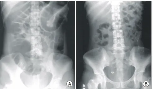
Fig. 1. Plain abdominal radiography before (A) and after (B) stent deployment in sigmoid colon. The proximal colonic distension was relieved after stent insertion.
A B
가 가스 방출, 배변, 복부 팽만의 감소 등 증상의 호전 을 보이면서 구강 섭취가 가능해지는 경우에 판정하 였다. 스텐트를 삽입한 후에는 충분한 수액 공급 및 전 해질 교정을 시행하였고, 유동식 혹은 저잔사 식이를 처방하였다. 근치적 수술 전단계로 시술한 경우에는 수술 하루 전에 장세척을 위해 polyethylene glycol을 2∼4 L 복용하였다.
결 과
총 35예 가운데 34예에서 한 개의 스텐트를 유치하 였으며, 1예에서는 두 군데의 분리된 폐색 병변에 대 하여 각각 한 개의 스텐트를 유치하였다(Fig. 2). 33예 (94%)에서 효과적인 감압에 성공하여 평균 7.0시간(범 위: 1∼33시간) 후에 가스 방출, 배변 및 복부 팽만 감 소 등의 호전을 보였으며 평균 3.0일(범위: 0.2∼6일) 후에 구강 섭취가 가능하였다. 나머지 2예(6%)에서는 성공적으로 스텐트를 유치하였으나 충분히 팽창되지 않아 감압에 실패하였는데, 그 중 재발성 자궁경부암 으로 S자 결장 내에 스텐트를 삽입하였던 1예에서는 시술 후 5일째에 회장 조루술을 시행하였으며, 진행성 근육 이형성증(progressive muscular dystrophy)이 있으 면서 다발성 간 및 폐전이를 동반한 직장암으로 스텐
트를 삽입한 1예에서는 시술 2일 후에 사망하였다. 스 텐트 유치와 관련된 합병증은 4예(12%)에서 발생하였 는데, 1예에서 골반 내 농양으로 시술 후 3일째에 방사 선 투시검사 하에 배액관을 삽입하여 치료하였으며, 나머지 2예에서는 경미한 출혈, 1예에서는 항문통이 발생하여 보존적 치료 후에 호전되었다.
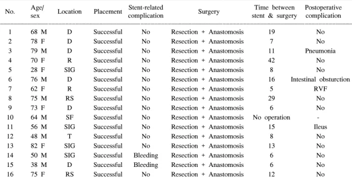
Table 1. Cases summary of patients with malignant colonic obstruction treated by colonic stent for bridge to surgery ꠚꠚꠚꠚꠚꠚꠚꠚꠚꠚꠚꠚꠚꠚꠚꠚꠚꠚꠚꠚꠚꠚꠚꠚꠚꠚꠚꠚꠚꠚꠚꠚꠚꠚꠚꠚꠚꠚꠚꠚꠚꠚꠚꠚꠚꠚꠚꠚꠚꠚꠚꠚꠚꠚꠚꠚꠚꠚꠚꠚꠚꠚꠚꠚꠚꠚꠚꠚꠚꠚꠚꠚꠚꠚꠚꠚꠚꠚꠚꠚꠚꠚꠚꠚꠚꠚꠚꠚꠚꠚꠚꠚꠚꠚꠚꠚꠚꠚꠚꠚꠚꠚꠚꠚ
Age/ Stent-related Time between Postoperative
No. Location Placement Surgery
sex complication stent & surgery complication
ꠏꠏꠏꠏꠏꠏꠏꠏꠏꠏꠏꠏꠏꠏꠏꠏꠏꠏꠏꠏꠏꠏꠏꠏꠏꠏꠏꠏꠏꠏꠏꠏꠏꠏꠏꠏꠏꠏꠏꠏꠏꠏꠏꠏꠏꠏꠏꠏꠏꠏꠏꠏꠏꠏꠏꠏꠏꠏꠏꠏꠏꠏꠏꠏꠏꠏꠏꠏꠏꠏꠏꠏꠏꠏꠏꠏꠏꠏꠏꠏꠏꠏꠏꠏꠏꠏꠏꠏꠏꠏꠏꠏꠏꠏꠏꠏꠏꠏꠏꠏꠏꠏꠏꠏ
1 68 M D Successful No Resection + Anastomosis 19 No
2 78 F D Successful No Resection + Anastomosis 7 No
3 79 M D Successful No Resection + Anastomosis 11 Pneumonia
4 70 F R Successful No Resection + Anastomosis 42 No
5 28 F SIG Successful No Resection + Anastomosis 8 No
6 76 M D Successful No Resection + Anastomosis 16 Intestinal obsturction
7 62 F R Successful No Resection + Anastomosis 5 RVF
8 75 M RS Successful No Resection + Anastomosis 29 No
9 73 F D Successful No Resection + Anastomosis 6 No
10 64 M SF Successful No Resection + Anastomosis No operation -
11 56 M SIG Successful No Resection + Anastomosis 15 Ileus
12 48 M T Successful No Resection + Anastomosis 8 No
13 82 F SIG Successful No Resection + Anastomosis 13 No
14 50 M SIG Successful Bleeding Resection + Anastomosis 6 No
15 38 M D Successful Bleeding Resection + Anastomosis 6 No
16 75 F RS Successful No Resection + Anastomosis 12 No
ꠏꠏꠏꠏꠏꠏꠏꠏꠏꠏꠏꠏꠏꠏꠏꠏꠏꠏꠏꠏꠏꠏꠏꠏꠏꠏꠏꠏꠏꠏꠏꠏꠏꠏꠏꠏꠏꠏꠏꠏꠏꠏꠏꠏꠏꠏꠏꠏꠏꠏꠏꠏꠏꠏꠏꠏꠏꠏꠏꠏꠏꠏꠏꠏꠏꠏꠏꠏꠏꠏꠏꠏꠏꠏꠏꠏꠏꠏꠏꠏꠏꠏꠏꠏꠏꠏꠏꠏꠏꠏꠏꠏꠏꠏꠏꠏꠏꠏꠏꠏꠏꠏꠏꠏ R = rectum; RS = rectosigmoid junction; SIG = sigmoid colon; D = descending colon; SF = splenic flexure; T = transverse colon.
Fig. 2. Resected open specimen shows sigmoid colon con- taining double stents deployed for the two separated ob- structing tumor.
1) 근치적 수술 전단계(bridge to surgery)
16예에서 원발성 대장암에 대한 근치적 수술의 전 단계로서 일시적인 감압을 위해 스텐트를 유치하였다 (Table 1). 시술 전 증상 발현 기간은 평균 7일(범위:
1∼30일)이었으며, 병변의 위치는 하행 결장과 S자 결 장이 각각 6예 및 4예로 가장 많았다. 시술 후 추적 관 찰이 불가능하였던 1예를 제외한 모든 환자에서 시술 후 평균 11일(범위: 5∼42일)에 1회의 계획 수술(elec- tive single stage operation)로서 절제 및 문합을 시행하 였고, 수술 후 사망이나 문합부 유출 등의 합병증은 없 었다.
2) 고식적 치료 목적(palliative aim)
19예에서 대장암 및 타장기암에 의한 대장폐색에 대하여 고식적인 목적의 최종 치료(definitive treat- ment)로서 스텐트를 유치하였다(Table 2). 시술 전 증 상 발현 기간은 평균 3일(범위: 1∼60일)이었고, 병변 의 위치는 에스 결장 및 직장이 14예로 많았다. 고식적 목적으로 스텐트를 유치한 원인으로서 고령이나 내과
적 합병 질환으로 수술의 위험도가 높다고 판단되거 나 절제 불가능한 전이성 병변을 동반한 원발성 대장 암의 경우가 8예로 가장 많았다. 타장기암으로는 위암 의 복막 전이로 발생한 대장 폐색이 5예로 가장 흔했 다. 성공적인 스텐트 유치 후에 추적 관찰이 가능하였 던 15명의 환자 가운데(추적 관찰 기간 65일, 범위 2 7∼440일), 12예에서 사망 당시 혹은 최종 추적일까지 스텐트가 효과적으로 기능하였다. 나머지 3예에서는 시술 후 각각 53일, 125일, 195일에 종양의 성장(tumor ingrowth) 및 고형변에 의한 스텐트의 폐쇄가 발생하였 다. 그 중 2예에서는 막부착형 스텐트(covered metallic stent)로 교체하여 재삽입하였고 1예에서는 직장 튜브 (rectal tube) 삽입 및 관장 등의 보존적 치료 후에 증상 이 호전되었다.
고 찰
악성질환에 의한 급성 대장 폐색을 응급 수술하는 데에 따른 유병률과 사망률은 각각 30%, 10%로 계획 수술의 경우에 비해 높은 것으로 보고된다.13-15 이는
Table 2. Cases summary of patients with malignant colonic obstruction treated by colonic stent with palliative aim ꠚꠚꠚꠚꠚꠚꠚꠚꠚꠚꠚꠚꠚꠚꠚꠚꠚꠚꠚꠚꠚꠚꠚꠚꠚꠚꠚꠚꠚꠚꠚꠚꠚꠚꠚꠚꠚꠚꠚꠚꠚꠚꠚꠚꠚꠚꠚꠚꠚꠚꠚꠚꠚꠚꠚꠚꠚꠚꠚꠚꠚꠚꠚꠚꠚꠚꠚꠚꠚꠚꠚꠚꠚꠚꠚꠚꠚꠚꠚꠚꠚꠚꠚꠚꠚꠚꠚꠚꠚꠚꠚꠚꠚꠚꠚꠚꠚꠚꠚꠚꠚꠚꠚꠚ
Age/ Cause of Stent-related Duration of
No. Cancer type Location Placement Follow up
sex palliation complication patency (d)
ꠏꠏꠏꠏꠏꠏꠏꠏꠏꠏꠏꠏꠏꠏꠏꠏꠏꠏꠏꠏꠏꠏꠏꠏꠏꠏꠏꠏꠏꠏꠏꠏꠏꠏꠏꠏꠏꠏꠏꠏꠏꠏꠏꠏꠏꠏꠏꠏꠏꠏꠏꠏꠏꠏꠏꠏꠏꠏꠏꠏꠏꠏꠏꠏꠏꠏꠏꠏꠏꠏꠏꠏꠏꠏꠏꠏꠏꠏꠏꠏꠏꠏꠏꠏꠏꠏꠏꠏꠏꠏꠏꠏꠏꠏꠏꠏꠏꠏꠏꠏꠏꠏꠏꠏ
1 89/F Primary CRC Operation refused RS Successful No 10 Loss
2 93/F Primary CRC Severe comorbidity R Successful No 1 Loss
3 35/M Primary CRC Distant metastasis R Failed - 2 Patient died
4 52/F Primary CRC Distant metastasis SIG Successful Pelvic abcsess 27 Remained patent 5 36/F Recurrent stomach Distant metastasis R Successful Anal pain 53 Reocclusion/Restenting 6 61/F Recurrent stomach Distant metastasis T Successful No 51 Remained patnet
7 87/M Primary CRC Operation refused D Successful No 287 Remained patent
8 74/M Recurrent CRC Distant metastasis R Successful No 125 Reocclusion/Rectal tube 9 88/F Primary CRC Operation refused SIG Successful No 195 Reocclusion/Restenting 10 62/M Recurrent CRC Distant metastasis SIG Successful No 28 Patent at death 11 83/M Recurrent stomach Distant metastasis RS Successful No 68 Patent at death 12 35/F Recurrent cervix Distant metastasis SIG Failed - 5 Emergent surgery 13 60/F Recurrent CRC Distant metastasis R Successful No 41 Remained patent 14 62/F Primary Cervix Locally advanced SIG Successful No 118 Remained patent 15 63/F Recurrent stomach Distant metastasis T Successful No 52 Remained patent 16 85/F Recurrent stomach Distant metastasis RS Successful No 295 Remained patent 17 51/F Primary CRC Distant metastasis T Successful No 142 Patent at death 18 35/M Primary stomach Distant metastasis SF Successful No 87 Patent at death 19 80/M Primary CRC Distant metastasis SIG Successful No 28 Patent at death ꠏꠏꠏꠏꠏꠏꠏꠏꠏꠏꠏꠏꠏꠏꠏꠏꠏꠏꠏꠏꠏꠏꠏꠏꠏꠏꠏꠏꠏꠏꠏꠏꠏꠏꠏꠏꠏꠏꠏꠏꠏꠏꠏꠏꠏꠏꠏꠏꠏꠏꠏꠏꠏꠏꠏꠏꠏꠏꠏꠏꠏꠏꠏꠏꠏꠏꠏꠏꠏꠏꠏꠏꠏꠏꠏꠏꠏꠏꠏꠏꠏꠏꠏꠏꠏꠏꠏꠏꠏꠏꠏꠏꠏꠏꠏꠏꠏꠏꠏꠏꠏꠏꠏꠏ CRC = colorectal cancer; R = rectum; RS = rectosigmoid junction; SIG = sigmoid colon; D = descending colon; SF = splenic flexure; T = transverse colon.
장폐색으로 인한 전해질 불균형, 패혈증, 그리고 팽대 된 허혈성 대장을 수술하는 데에서 오는 기술적인 어 려움 이외에도 고령의 나이, 내과적 합병 질환, 영양 섭취의 부족, 그리고 진단 당시 병변이 진행된 점 등이 영향을 미치는 것으로 알려져 있다.16-18 또한 전통적인 수술법인 단계적 수술의 경우에 1차 술식으로 형성된 장루를 복원하지 못하는 경우가 40%에 이르고,1 응급 장루 조성으로 인한 장루 관련 합병증이 흔히 발생할 뿐만 아니라 영구적인 장루 보유에 따른 삶의 질의 저 하가 문제점으로 제기되었다.19,20
급성 대장 폐색에 대한 치료로서 자가팽창형 금속 스텐트 유치술은 1990년대에 점차 그 이용이 확대되 면서 최근에 Khot 등10은 58개 문헌 검토를 통하여 598 명 환자들의 치료 성적을 보고한 바 있다. 계획 수술을 시행하기 전에 시술함으로써 감압한 후 장세척을 통 해 병변의 근위부에 대한 평가가 가능하고 정확한 병 기 결정이 용이해질 뿐만 아니라 응급 수술로 인한 합 병증을 줄일 수 있다.4-7,21 진행암으로 인해 근치적 절 제가 불가능하며 특히 전신상태가 불량하여 수술의 위험도가 높다고 판단되는 경우에는 고식적 목적의 최종 치료로서 안전하고 효과적인 비수술적 치료법이 될 수 있다.9,11,12
시술 성공률은 85∼100%로 보고되며,7,9,10 본 연구에 서도 94%로 이와 유사하였다. 스텐트를 삽입하였으나 충분히 확장되지 않아 감압에 실패하였던 2예는 모두 고식적 목적으로 시술한 환자였는데, 골반강 및 복강 내에 암종증을 동반 재발성 난소암 및 진행성 직장암 에 의해 내강이 압박되어 급성 대장 폐색을 유발하였 다. 골반강은 비교적 제한된 공간이라는 점과 골반측 벽 및 복강 내에 광범위하게 침습한 암종증이라는 점 이 스텐트의 확장을 저해하는 요인으로 작용하였을 것으로 생각된다. 이외에도 암종증에 의한 대장 폐색 은 병변이 다발성인 경우가 많아 스텐트 유치술로 충 분한 감압을 유도하기 어려울 수 있다.
시술과 관련된 합병증은 9∼42%로 보고된다.10 그 중 장천공은 가장 심각한 합병증으로서 발생하는 기 전은 다양하다. 첫째, 시술 중에 유도선이나 유치도관 을 삽입하는 중에 발생할 수 있다. 이 경우 시술 중 조 영제가 복강 내로 유출되는 소견으로 확인이 가능하 며 대개는 증상이 없는 경우가 많아 시술을 종료하는 데에 문제가 되지 않는다.22,23 둘째, 스텐트를 삽입하기 전에 협착부위에 풍선확장을 하여 발생할 수 있는데 이 경우 복막염으로 발전할 수 있어 가능하면 풍선확 장은 하지 않을 것을 권하고 있다.10 셋째, 종양 조직이
약한 경우 인공관이 확장되면서 강한 방사력에 의해 발생할 수 있는데 이 경우 복막염으로 발전할 수 있으 므로 시술 후에 주의 깊은 관찰을 요한다.6 넷째, 스텐 트의 선단이 장벽을 파고들어 발생할 수 있는데 이 경 우 천공이 미세하여 증상이 거의 없고 주변 조직으로 의 염증 파급도 심하지 않다.6 본 연구에서는 고식적 목적으로 S자 결장암에 인공관을 유치한 1예(3.0%)에 서 시술 3일 후에 골반강 내 농양이 관찰되었다. 시술 중 조영제의 유출이나 시술 후 복부 X-ray 사진상에서 장외 공기음영의 소견은 없었으나 복부 초음파상에서 골반강 내에 농양이 관찰되어 방사선 투시 하에 배액 관을 삽입하여 치료하였다.
하부 직장의 폐색으로 스텐트를 삽입한 경우 항문 통 및 후중감을 호소하는 것으로 보고된다.24 본 연구 에서는 위암 수술 후 복막 암종증이 발생한 1예에서 직장에 스텐트를 삽입한 후 항문통이 발생하였으며 진통제 투여 등의 보존적 치료 후에 호전되었다. 하지 만 증상이 지속될 때에는 스텐트를 제거해야 하는 경 우도 있으므로 최종 치료로서 하부 직장에 스텐트를 유치하는 경우에는 주의를 요하며 시술 전 환자 및 보 호자에게 충분히 설명하는 것이 필요하다.
스텐트의 폐쇄나 이동은 주로 고식적인 목적으로 스텐트를 유치한 후 추적 관찰 기간 중에 발생할 수 있다. 스텐트 폐쇄의 경우 종양이 스텐트의 격자 사이 로 자라 들어와 발생하거나 고형변이 스텐트를 막아 발생하며 막미부착형(bare stent)에서 흔하다. 대개의 경우 막부착형 스텐트(covered stent)를 재삽입함으로 써 치료할 수 있다.10,25,26 본 연구에서는 최종 치료로서 스텐트를 유치하고 추적 관찰이 가능하였던 15예 가 운데 3예(20%)에서 스텐트 폐쇄가 발생하여 2예에서 는 막부착형 스텐트로 재삽입하였고 1예에서는 보존 적인 치료 후에 호전되었다. 스텐트 이동은 막부착형 에서 흔하며 대개 스텐트 유치 후 수일 내에 발생하지 만 항암약물치료나 방사선치료를 시행하여 종양의 크 기가 감소함으로써 발생할 수도 있다.6,10,25 치료는 이 동한 스텐트를 제거한 후에 재삽입하거나 재폐색의 우려가 적다고 판단되는 경우에는 관찰해 볼 수 있다.
급성 대장 폐색의 치료로서 스텐트 유치술과 응급 수 술의 치료 결과를 비교한 연구를 보면 근치적 수술 전 단계의 목적인 경우 스텐트 유치술이 응급 수술에 비 하여 일차 문합의 가능성이 많고 수술 후 합병증이 적 으며 중환자실 치료 기간 및 총 재원기간이 짧다고 보 고된다.4 고식적 목적의 최종 치료로서 시술한 경우에 도 장루 조성이 필요한 경우가 적고 중환자실 치료 기
간 및 총 재원기간이 짧은 것으로 보고된다.8,12 앞으로 근치적 절제가 가능했던 환자들의 장기간 추적 관찰 을 통하여 단기 치료성적 이외에 스텐트 유치술 후 계 획수술을 시행한 환자와 응급수술을 시행한 환자간의 종양학적 결과를 비교하는 연구가 필요할 것으로 생 각된다. 또한 고식적 목적으로 스텐트를 유치한 환자 와 절제수술을 시행한 환자간의 생존율과 삶의 질에 관한 전향적인 비교 연구가 필요하리라고 생각된다.
결 론
자가팽창형 금속스텐트 유치술은 악성 질환에 의한 급성 대장 폐색을 해소하여 응급 수술로 인한 합병증 을 최소화함으로써 근치적 수술 전단계뿐만 아니라 고식적 목적의 최종 치료로서 이용될 수 있는 비교적 안전하고 유용한 치료법이다.
REFERENCES
1. Deans GT, Krukowski ZH, Irwin ST. Malignant obstruc- tion of the left colon. Br J Surg 1994;81:1270-6.
2. Single-stage treatment for malignant left-sided colonic obstruction: a prospective randomized clinical trial com- paring subtotal colectomy with segmental resection following intraoperative irrigation. The SCOTIA Study Group. Subtotal Colectomy versus On-table Irrigation and Anastomosis. Br J Surg 1995;82:1622-7.
3. Torralba JA, Robles R, Parrilla P, Lujan JA, Liron R, Pinero A, et al. Subtotal colectomy vs. intraoperative colonic irrigation in the management of obstructed left colon carcinoma. Dis Colon Rectum 1998;41:18-22.
4. Martinez-Santos C, Lobato RF, Fradejas JM, Pinto I, Ortega-Deballon P, Moreno-Azcoita M. Self-expandable stent before elective surgery vs. emergency surgery for the treatment of malignant colorectal obstructions: com- parison of primary anastomosis and morbidity rates. Dis Colon Rectum 2002;45:401-6.
5. Mainar A, De Gregorio Ariza MA, Tejero E, Tobio R, Alfonso E, Pinto I, et al. Acute colorectal obstruction:
treatment with self-expandable metallic stents before scheduled surgery-results of a multicenter study. Radio- logy 1999;210:65-9.
6. Camunez F, Echenagusia A, Simo G, Turegano F, Va- zquez J, Barreiro-Meiro I. Malignant colorectal obstruc- tion treated by means of self-expanding metallic stents:
effectiveness before surgery and in palliation. Radiology 2000;216:492-7.
7. Watson AJ, Shanmugam V, Mackay I, Chaturvedi S,
Loudon MA, Duddalwar V, et al. Outcomes after place- ment of colorectal stents. Colorectal Dis 2005;7:70-3.
8. Law WL, Choi HK, Chu KW. Comparison of stenting with emergency surgery as palliative treatment for obstructing primary left-sided colorectal cancer. Br J Surg 2003;90:1429-33.
9. Law WL, Choi HK, Lee YM, Chu KW. Palliation for advanced malignant colorectal obstruction by self-ex- panding metallic stents: prospective evaluation of out- comes. Dis Colon Rectum 2004;47:39-43.
10. Khot UP, Lang AW, Murali K, Parker MC. Systematic review of the efficacy and safety of colorectal stents. Br J Surg 2002;89:1096-102.
11. Bhardwaj R, Parker MC. Palliative therapy of colorectal carcinoma: stent or surgery? Colorectal Dis 2003;5:
518-21.
12. Carne PW, Frye JN, Robertson GM, Frizelle FA. Stents or open operation for palliation of colorectal cancer: a retrospective, cohort study of perioperative outcome and long-term survival. Dis Colon Rectum 2004;47:1455-61.
13. Tobaruela E, Camunas J, Enriquez-Navascues JM, Diez M, Ratia T, Martin A, et al. Medical factors in the morbidity and mortality associated with emergency colorectal cancer surgery. Rev Esp Enferm Dig 1997;
89:13-22.
14. Lee YM, Law WL, Chu KW, Poon RT. Emergency surgery for obstructing colorectal cancers: a comparison between right-sided and left-sided lesions. J Am Coll Surg 2001;192:719-25.
15. Scott NA, Jeacock J, Kingston RD. Risk factors in patients presenting as an emergency with colorectal cancer. Br J Surg 1995;82:321-3.
16. Mainar A, Tejero E, Maynar M, Ferral H, Castaneda- Zuniga W. Colorectal obstruction: treatment with metal- lic stents. Radiology 1996;198:761-4.
17. Runkel NS, Schlag P, Schwarz V, Herfarth C. Outcome after emergency surgery for cancer of the large intestine.
Br J Surg 1991;78:183-8.
18. Wholey MH, Levine EA, Ferral H, Castaneda-Zuniga W.
Initial clinical experience with colonic stent placement.
Am J Surg 1998;175:194-7.
19. Makela JT, Turku PH, Laitinen ST. Analysis of late stomal complications following ostomy surgery. Ann Chir Gynaecol 1997;86:305-10.
20. Nugent KP, Daniels P, Stewart B, Patankar R, Johnson CD. Quality of life in stoma patients. Dis Colon Rectum 1999;42:1569-74.
21. 이강영, 김남규, 박준성, 박재균, 이용찬, 민진식. 좌측 대 장암에 의한 장폐색에서 근치적 절제술을 위한 stent 삽 입술. 대한외과학회지 2001;60:667-70.
22. Binkert CA, Ledermann H, Jost R, Saurenmann P, Decurtins M, Zollikofer CL. Acute colonic obstruction:
clinical aspects and cost-effectiveness of preoperative and palliative treatment with self-expanding metallic stents-a preliminary report. Radiology 1998;206:199-204.
23. Saida Y, Sumiyama Y, Nagao J, Takase M. Stent endo- prosthesis for obstructing colorectal cancers. Dis Colon Rectum 1996;39:552-5.
24. Tejero E, Fernandez-Lobato R, Mainar A, Montes C, Pinto I, Fernandez L, et al. Initial results of a new procedure for treatment of malignant obstruction of the
left colon. Dis Colon Rectum 1997;40:432-6.
25. Baron TH, Dean PA, Yates MR 3rd, Canon C, Koehler RE. Expandable metal stents for the treatment of colonic obstruction: techniques and outcomes. Gastrointest En- dosc 1998;47:277-86.
26. Repici A, Reggio D, De Angelis C, Barletti C, Marchesa P, Musso A, et al. Covered metal stents for management of inoperable malignant colorectal strictures. Gastrointest Endosc 2000;52:735-40.