https://doi.org/10.14734/PN.2017.28.2.69 pISSN 2508-4887•eISSN 2508-4895
Jisoo Kim, MD1, Young Ju Hong, MD2, Hyun-hae Cho, MD, PhD3, So-Yeon Shim, MD, PhD1, Eun Ae Park, MD, PhD1, Su Jin Cho, MD, PhD1
1Department of Pediatrics, Ewha Womans University School of Medicine, Seoul, 2Department of General Surgery, Ewha Womans University School of Medicine, Seoul, 3Department of Radiology, Ewha Womans University School of Medicine, Seoul, Korea
We report a case of ileal atresia presenting with massive chylous ascites and hydrocele in a neo
nate. A male neonate was born at 38 weeks of gestation with a weight of 3,360 grams. The antenatal ultrasonography performed at 37+6 weeks of gestation showed ascites and huge bila teral hydrocele. At birth, he manifested hypotonia, a massively distended abdomen and huge bilateral hydrocele. Exploratory laparotomy was performed on the day of birth. A large amount of chyle (approximately 200 mL) was found in the peritoneal cavity between the bowel loops. An additional 120 mL of chyle was removed from both scrotal sacs. The color of chyle was ivory yellow with whitish debris. Ileal atresia had occurred 7 cm above the ileocecal valve. The proximal small bowel was disconnected, and the distal small bowel loop ended in a blind loop. Lymphatic drainage from that cross section of the proximal small bowel might be a cause of chylous ascites. Segmental small bowel resection and double barrel ileostomy was performed. We described this case with a review of the literature.
Key Words: Intestinal atresia, Ascites, Chyle, Newborns
Introduction
Neonatal chylous ascites is a very rare condition; the exact incidence of neonatal chylous ascites is unknown.1 Congenital chylous ascites can occurred from a variety of reasons. The most common cause (45–60% cases) is the malformation of the lymphatic vessels.2 Half of neonates, not mentioned above, the chylous ascites is thought to be due to delayed maturation of the lacteals, called “leaky lymphatics.”2 Another reason (20–25%
cases) is obstruction of the lymphatics due to external compression, such as malrotation, intussusception, incarcerated hernia, enlarged lymph nodes and malig nancy.2 In some cases (15–20% cases), it is caused by trauma during surgery, accidents or child abuse.2 We reported a rare case of ileal atresia presenting with massive chylous ascites and bilateral hydrocele at birth.
Case
We report a male neonate with ascites and huge bilateral hydrocele who was born by caesarean section at 38 weeks of gestation with a weight of 3,360 g (50–75 percentile), height of 51.0 cm (75–90 percentile), and head circumference of 33.5 cm (50–75 percen
tile). The antenatal ultrasonography performed at 37+6 weeks of gestation showed ascites Received: 1 February 2017
Revised: 10 March 2017 Accepted: 10 April 2017 Correspondence to Su Jin Cho, MD, PhD
Department of Pediatrics, Ewha Womans University School of Medicine, 1071 Anyangcheonro, Yangcheongu, Seoul 07985, Korea Tel: +82226502859
Fax: +82226533718 E-mail: sujin[email protected] Copyright© 2017 by The Korean Society of Perinatology
This is an Open Access article distributed under the terms of the Creative Com
mons Attribution NonCommercial License (http://creativecommons.org/
license/bync/4.0/), which permits unrestricted noncommercial use, distribution, and reproduction in any
Ileal Atresia Presenting with Massive Chy lous Ascites and Hydrocele in a New
born
ultrasonography and ascites was not turbid. (Fig. 1) The Pleural effusion, pericardial effusion, or edema of subcutane
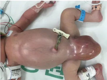
ous tissue was absent. The antenatal ultrasonography showed no evidence of hydrops fetalis. Fe tal heart rate decreased, the baby was born by emergency caesarean section. At birth, the patient manifested hypotonia, massively distended abdomen and bilateral hydrocele (Fig. 2). In addition, he had no sponta
neous breathing and no crying. The patient was warmed and secretions were cleared. After positive pressure ventilation, the pulse rate was under 100 beats per minute, without res
piratory effort and cyanosis persisted. The 1minute Apgar score was 2, and 5minute Apgar score was 3. Endotracheal
intubation was performed, and the baby was transferred to the neonatal intensive care units (NICU). On the physical exami
nation, there was no meconium staining on the newborn’s skin, no hepatosplenomegaly, nor palpable mass. The neonate had a white blood cell count of 12.66×10⁹/L with 62% neutrophils, and 29% lymphocytes, and platelet count of 139×109/L. In a blood sample, sodium was 137 mEq/L, potassium 4.8 mEq/L, chloride 101 mEq/L, aspartate transaminase 106 IU/L, alanine aminotransferase 16 IU/L, cholesterol 27 mg/dL, triglyceride 15 mg/dL, total protein 4.8 g/dL, and al bumin was 2.5 g/dL. C
reactive protein was 0.32 mg/dL in a first laboratory test on the first day of life. From the venous blood gas analysis (VBGA), venous blood pH was checked as 7.160, PCO2 was 41.0 mmHg, base excess was 14 mmol/L. Seven hours after birth, abdo
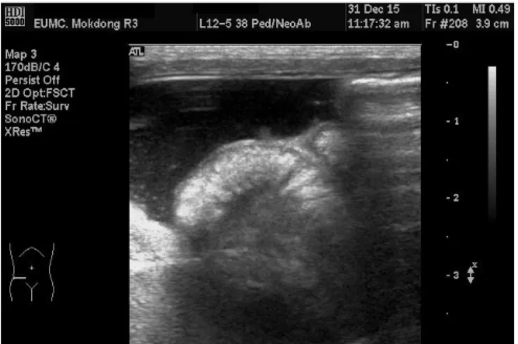
minal distension worsened. On the abdomen xray, the bowel gas was located centrally (Fig. 3). Abdominal ultrasonography revealed meconium peritonitis with a large amount of compli
cated ascites suggestive of probable bowel perforation. Thick
ening of the small bowel loop was seen especially from the bowel loops in the right lower quadr ant area (Fig. 4). The possibility of underlying bowel pathology like atresia or internal hernia was mentioned from that abdo minal ultrasonography.
There was no evidence of midgut volvulus such as the
‘whirlpool’ or ‘coffee bean’ sign. Large amount of complicated ascites was seen in both scrotal sac areas. Both kidneys
Fig. 2. At birth, the patient showed a massively distended abdomen and huge bilateral hydrocele.
Fig. 1. The ascites depth of 2.3 cm was detected by antenatal ultra- sonography.
Fig. 3. On the abdomen x-ray, the bowel gas was located centrally.
small bowel loop ended in a blind loop (Fig. 5). There was no evidence of malrotations or dilatation of lymphatics. Laparoto
my with subsequent histopathology confirmed the diagnosis.
Anatomical origin was the small intestine, which showed ne
crotic tissue and scanty amount of viable small intestine wall (Fig. 6). The pathologic findings from the segmental resection of small intestine indicated submucosal and subserosal conge
stion with chronic inflammation, fibrinous exudate and micro calcifications at serosal surface with meconium peritonitis.
Segmental small bowel resection and double barrel ileostomy was performed, and the patient was transferred to the NICU.
Bowel motility was observed on postoperative day 7 (post
natal day 8), and feeding was initiated. He was weaned off oxygen on postnatal day 15. Gastrograffin enema for evalu
ation of the proximal and the distal bowel loops was done on postnatal day 22. The diameter of rectum was intact; whereas, the diameter of colon loop above rectum was decreased, con
sistent with an underused micro colon.
The level of conjugated bilirubin increased to 4.2 mg/dL on postnatal day 6. The hepatobiliary scan showed delayed he
patic excretion. Ursodeoxycholic acid and phenobarbital were administered.
Brain magnetic resonance imaging was taken on postnatal day 19. On the diffusion weighted image, there are lesions with high signal intensity and diffusion restriction at splenium and posterior body of corpus callosum. It could be possible from the hypoxic ischemic injury. There are T1 high, T2 low punctate lesions at white matter of the both frontoparietal appeared normal in size with normal echotexture. Otherwise,
no definite abnormality was detected in both kid neys. The urinary bladder showed normal contour and wall thickness.
Exploratory laparotomy was performed on the day of birth.
A large amount of chyle (approximately 200 mL) was found in the peritoneal cavity between the bowel loops. An additional 120 mL of chyle was removed from both scrotal sacs. The color of chyle was ivory yellow with whitish debris. The analysis of fluid from peritoneal cavity and both scrotal sacs was not conducted. The flow of chyle from the peritoneal cavity through the inguinal canal had resulted in huge bilateral hydrocele.
Ileal atresia had occurred 7 cm above the ileocecal valve.
The proximal small bowel was disconnected, and the distal
Fig. 4. In the ultrasonography, large amount of ascites and thickening of the small bowel loop were seen.
Fig. 5. The proximal small bowel was disconnected, and the distal
small bowel ended in a blind loop. Fig. 6. Few necrotic tissues with scanty amount of viable small intes- tine wall were observed.
lobes. It could be thought petechial hemorrhage.
The patient was discharged from the hospital on postnatal day 46 with a weight of 3,450 grams (<3 percentile), height of 53.0 cm (3–5 percentile), and head circumference of 35.0 cm (<3 percentile). The level of direct bilirubin normalized at 3 months after birth. The patient was readmitted for repair of ileostomy on postnatal day 86 with a weight of 5,100 grams (3 percentile), height of 58.5 cm (10–25 percentile). Closure of stoma of small intestine was done and he has been doing well at outpatient followup.
Discussion
Fetal ascites is defined as the accumulation of fluid in the peritoneal cavity. It is caused by immune hydrops fetalis, non
immune hydrops fetalis, cardiac malformations, congeni tal infections, gastrointestinal anomalies and genitourinary ano
malies.3 Ascites can be accompanied by other abnorma lities, such as structural malformations or genetic syndromes.4 On ultrasonography examination, fluid accumulation appears as echofree fluid outlining the falciform ligament, umbilical vein, be tween the bony rib cage and viscera, and in the peritoneal cavity between the bowel loops.4
Neonatal chylous ascites is a very rare condition.1 Con
genital chylous ascites can occurred from a variety of reasons.
The 1st most common cause is the malformation of the lym
phatic vessels.2 The 2nd most common cause is delayed maturation of the lacteals, called “leaky lymphatics.”2 Another reason is obstruction of the lymphatics due to external com
pression, such as malrotation, intussusception, incarcerated hernia, en larged lymph nodes and malignancy.2 In some cases, it is caused by trauma during surgery, accidents or child abuse.2
Paracentesis is both a diagnostic and therapeutic step, and lymphoscintigraphy is used to reveal the lymphatic anatomy.1 Enteral feeding with a medium chain triglyceride formula can be started, but nil per os (NPO) and total parenteral nutrition may be necessary to reduce chylous flow in severe or compli
cated cases.1 The goal of the surgical management is to find and fix the lymphatic leak.1 Patients with chylous ascites have a risk for recurrence, but there were no recurrences in our
case.
The proximal small bowel was disconnected, and the distal small bowel loop ended in a blind loop (Fig. 5). Lymphatic drainage from that cross section of the proximal small bowel might be a cause of chylous ascites. The color of chyle was ivory yellow with whitish debris. There was no meconium stained ascites or soiling of the peritoneal cavity, it was thought that the disconnection of proximal small bowel was happened just before the surgery.
In the republic of Korea, there was one similar case report of a neonate with massive chylous ascites and hydrocele.5 In the report, a male neonate was born at 36 weeks of gesta tion with a weight of 2,990 grams.5 Ascites was detected on an
tenatal ultrasonography at 35 weeks.5 On the day of birth, paracentesis was done and the ascites was chyle.5 Gastro
graffin enema was done on postnatal day 4.5 From that study congenital ileal atresia was seen, and then segmental small bowel resection and ileocolostomy was done.5
From the case we mentioned above, paracentesis was done on the day of birth.5 In my case, paracentesis was not con
ducted, but exploratory laparotomy was performed on the day of birth. From the gastrograffin study, congenital ileal atresia was seen in the case we mentioned above, but in my case gastrograffin study was done after exploratory laparotomy.
We had few information from the study except underused micro colon.
Congenital small bowel atresia of the duodenum, jejunum or ileum is the most common cause of neonatal bowel obstruc
tion, with incidence ranging from 1.3 to 2.8 of 10,000 births.6 Congenital small bowel atresia occurs mostly in the duodenum and less frequently in the jejunum or ileum.7 The signs of jejunoileal atresia include abdominal distension, bilious vo
miting, jaundice, and no passing of meconium on the first day of life.7 The differential diagnosis of gastrointestinal obstruc
tion in neonates includes esophageal atresia, pyloric stenosis, malrotation with midgut volvulus, intestinal stenosis, intestinal atresia, duodenal duplication, annular pancreas, foreign body obstruction, and Hirschsprung disease.7
If bowel dilatation and ascites are detected, small intestinal obstruction must be considered in the diagnosis. Prenatal dia
gnosis of gastrointestinal anomalies is very important to peri
natal management, allowing delivery in centers in which radio
logic imaging and surgical intervention are available. Collabo
rative effort of obstetricians, radiologists, neonatolo gists and pediatric surgeons could lead to early intervention, which can reduce morbidity and improve outcomes.
References
1) Held JM, Restrepo R, Ricca R. Chylous ascites in a neonate with hydrops fetalis. Am Surg 2016;82:783-4.
2) Mouravas V, Dede O, Hatziioannidis H, Spyridakis I, Filippopoulos A.
Diagnosis and management of congenital neonatal chylous ascites.
Hippokratia 2012;16:175-80.
3) Nigam A, Kumar M, Gulati S. Fetal ascites and hydrometrocolpos due to persistent urogenital sinus and cloaca: a rare congenital anomaly and review of literature. BMJ Case Rep2014;2014:bcr2013202231.
4) Baccega F, de Lourdes Brizot M, Jornada Krebs VL, Vieira Francisco RP, Zugaib M. Nonimmune fetal ascites: identification of ultrasound findings predictive of perinatal death. J Perinat Med 2016;44:195-200.
5) Moon SK, Kim JN, Choi MJ, Seo JS, Kim JS, Kim HY, et al. A case of chylous ascites secondary to congenital ileal atresia. Korean J Perinatol 1997;8:309-14.
6) Virgone C, D'antonio F, Khalil A, Jonh R, Manzoli L, Giuliani S. Accuracy of prenatal ultrasound in detecting jejunal and ileal atresia: systematic review and meta-analysis. Ultrasound Obstet Gynecol 2015;45:523-9.
7) Chen HD, Jiang H, Kan A, Huang LE, Zhong ZH, Zhang ZC, et al. Inte- stinal obstruction due to dual gastrointestinal atresia in infants: diag- nosis and management of 3 cases. BMC Gastroenterol 2014;14:108.