1Department of Radiology, Seoul St. Mary’s Hospital, College of Medicine, The Catholic University of Korea
2Department of Radiology, Gyeongsang National University Hospital
3Department of Radiology, St. Vincent Hospital, College of Medicine, The Catholic University of Korea
4Department of Thoracic Surgery, Seoul St. Mary’s Hospital, College of Medicine, The Catholic University of Korea
5Department of Nuclear Medicine, Seoul St. Mary’s Hospital, College of Medicine, The Catholic University of Korea Received June 22, 2009 ; Accepted September 1, 2009
Address reprint requests to : Jung Im Jung, M.D., Department of Radiology, Seoul St. Mary’s Hospital, College of Medicine, The Catholic University of Korea, 505 Banpo-dong, Seocho-gu, Seoul 137-701, Korea.
Tel. 82-2-2258-1435 Fax. 82-2-599-6771 E-mail: [email protected]
Purpose: To investigate the size criteria of multidetector computed tomography (MD- CT) for the evaluation metastatic lymph nodes (LNs) for potentially operable squa- mous esophageal cancer, and to compare this information with the results of positron emission tomography-CT (PET-CT).
Materials and Methods: Twenty-four patients who underwent radical esophagectomy for esophageal cancer were studied. All patients had preoperative MDCT and PET-CT.
The MDCT findings were compared with those of PET-CT and were correlated with the surgical records. The receiver operating characteristic (ROC) curve method was used to determine the appropriate cut-off value to distinguish benign from metastatic LNs.
Results: The size of metastatic LNs (9.35 3.41 mm) was significantly larger than that of benign LNs (5.74 1.64 mm) (p<0.001). The best cut-off value was 7 mm (81.8% sensitivity, 80.8% specificity). PET-CT detected all metastatic LNs except for four in the peritumoral region. The sensitivity and specificity of metastatic LN evalua- tion on PET-CT were 82.6% and 99.4%, respectively. Only one LN without metastasis showed increased fluoro-2-deoxy-D-glucose uptake on PET-CT.
Conclusion: Size of metastatic LNs can typically be < 10 mm. For MDCT, the short di- ameter of 7 mm may be the optimal criterion. PET-CT is very accurate for the assess- ment of metastatic LNs except for those in the peritumoral region.
Index words :Esophageal Neoplasms
Tomography, X-Ray Computed Positron-Emission Tomography Lymph nodes
Multidetector CT Assessment of Lymph Node Size for Nodal Staging in Patients with Potentially Operable Squamous Esophageal Cancer and the
18F-FDG Positron
Emission Tomography CT Correlation
1Soo Kyung Yoon, M.D., Jung Im Jung, M.D., Mi Jung Park, M.D.2, Hyun Jin Park, M.D.3, Myeong Im Ahn, M.D., Jae Gil Park, M.D.4, Ie Ryung Yoo, M.D.5, Seog Hee Park, M.D.
In esophageal cancer patients, determination of the disease stage is important for the selection of therapeutic protocols and for predicting a patient’s prognosis. In par- ticular, the lymph node (LN) stage is an important inde- pendent prognostic factor (1). In addition to the disease stage classified simply based on the presence (N1) or ab- sence (N0) of local LN involvement, the number of in- volved LNs as well as their locations are also important prognostic factors (2-4).
In esophageal cancer patients, chest computed tomog- raphy (CT) is commonly used as a pre-operative test. LN metastasis is evaluated by size, in most cases by refer- ence to lung cancer LN metastasis cases, with the stan- dard of LN metastasis being a diameter ≥ 10 mm.
Therefore, cases with LN metastasis < 10 mm can be overlooked, and LNs that become enlarged due to reac- tive hyperplasia or granulomatous inflammation may not be distinguished from LNs enlarged by tumor spread (5, 6).
In the evaluation of disease stage in esophageal cancer patients, assessment of LN metastasis using positron emission tomography (PET) using 18F-fluorodeoxyglu- cose (FDG) is more accurate than CT (5-7). Recently, with the application of PET-CT to the disease stage eval- uation in esophageal cancer patients, high sensitivity as well as specificity have been reported. However, as CT is a presurgical test commonly used for esophageal can- cer patients, standardization of the size of LN metastasis of esophageal cancer patients is required.
Presently, we evaluated the size criteria of metastatic LN using multidetector computed tomography (MDCT) for squamous esophageal cancer patients and then com- pared the results with those of PET-CT.
Materials and Methods
Patients
From November 2003 to June 2007, 29 patients with biopsy-proven squamous cell carcinoma of the esophga- gus underwent preoperative CT and PET-CT. Of these patients, five were excluded in the study; two did not undergo esophagectomy due to other advanced primary cancers, one received preoperative adjuvant chemother- apy, one underwent endoscopic mucosal resection, and one had simultanoeus esophagectomy for esophageal cancer and gastrectomy for primary gastric cancer. A to- tal of 24 patients (22 males and two females, ranging in age from 50-76 years, with a mean age of 63.5 years) were included in our study. Our local ethics committee
approved this retrospective study, and the requirement for written informed consent was waived for this retro- spective analysis.
MDCT
MDCT scans were obtained from the neck to the mid- dle portion of the both kidneys after intravenous auto- mated injection of contrast medium (100 mL) at a rate of 2 mL/s. CT was done using several scanners including a Lightspeed Volume CT apparatus (GE Healthcare, Milwaukee, WI, USA) for eight patiates, Volume Zoom (Siemens, Forchheim, Germany) for 12 patients, and Sensation16 (Siemens, Forchheim, Germany) for four patients. CT was obtained at 120-140 kVp; 100-250 mA; 1.25-2.5 mm collimation; scan speed 0.5-0.75 s/cy- cle; and a reconstruction interval of 2.5-3 mm. Two ra- diologists reviewed all measurable LNs and recorded the size, number, and sites on an American Joint Committee on Cancer (AJCC) nodal station. The LN size was measured on the basis of the short-axis diameter.
PET-CT
The time interval between MDCT and PET-CT was 1- 25 days (average; 6.3days). All patients fasted for at least 6 h before the PET-CT study. None of the patients had blood glucose levels exceeding 130 mg/dL. Sixty minutes after intravenous injection of 370-550 MBq 18F-FDG,
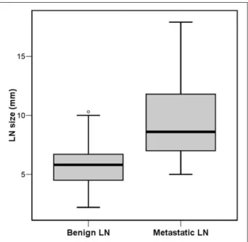
Fig. 1. The sizes of benign and metastatic LNs on the basis of MDCT are significantly different. Benign LN averaged 5.74 ± 1.64 mm (2.2-9.84 mm) and metastatic LN averaged 9.35 ± 3.41 mm (5.0-17.9 mm). Size was evaluated by the Student’s T-test.
PET-CT scans were obtained using a combined PET-CT system (Biograph DUO; Siemens Medical Solutions, Knoxville, TN, USA). CT scanning began at the orbito- meatal line and progressed to the upper thigh (30 mAs;
130 kV; 5 mm slice thickness). PET imaging followed im- mediately over the same body region from the upper thigh and progressing caudally. The CT data were used for attenuation correction, and images were reconstruct- ed using the standard ordered subset expectation maxi- mization (OSEM) algorithm. When an area of presumed primary tumor or a LN showed prominent FDG uptake on visual assessment compared to the background activi- ty, the area or LN was considered to be positive for ma- lignancy. An experienced nuclear medicine radiologist
reviewed the PET and fused PET-CT images and record- ed the presence, number, maximum standardized up- take value (SUVmax), and location of presumed primary tumors and LNs with metastases.
Data Analyses
MDCT and PET-CT findings were compared and cor- related with the surgical records and pathology results.
Descriptive analyses were used to characterize the groups investigated. Continuous variables were ex- pressed as mean ± SD, and differences were analyzed using the Student’s t-test. Fisher’s exact test was used to compare the accuracy of PET-CT. A p value < 0.05 was considered to represent a statistically significant differ- ence. The appropriate cut-off value to distinguish benign from metastatic LNs with the highest sensitivity and specificity was determined using the receiver operating characteristic (ROC) curve method.
Results
Primary Tumors
All patients were diagnosed with squamous cell esophageal cancer, with well-differentiated cancer in three patients, moderately differentiated cancer in 18 patients, and poorly differentiated cancer in three pa- tients. Primary tumors were located in the upper tho- racic esophagus (n=2), midthoracic esophagus (n=5), lower thoracic esophagus (n=16), and in the mid-to-low- er thoracic esophagus (n=1). Regarding the postopera- tive T disease stages, T1 tumor was present in six pa- tients (T1a in two patients and T1b in four patients), T2 tumor in four patients, T3 tumor in 13 patients, and T4 tumor in one patient. Among the 24 study patients, pri- mary tumor could be readily detected by MDCT in 22 patients (91.7%) and by PET-CT in 23 patients (95.8%).
Fig. 2. ROC curve analysis performed by computing the sensi- tivity and specificity of the LN dimensions for determining be- nign and metastatic LNs at various cut-off levels. Area under the ROC curve (AUC): 0.848
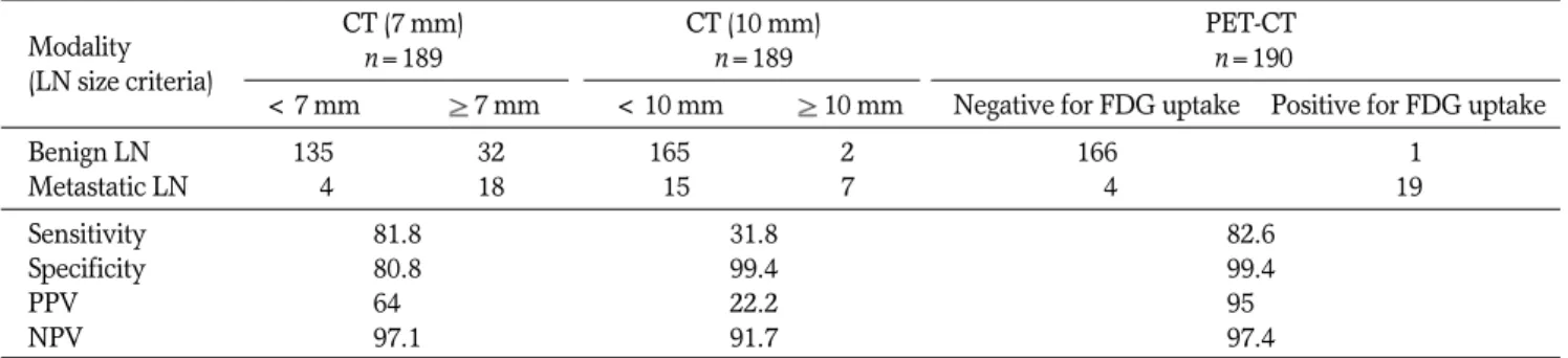
Table 1. Sensitivity, Specificity, PPV, and NPV of CT and PET-CT
Modality CT (7 mm) CT (10 mm) PET-CT
(LN size criteria) n=189 n=189 n=190
< 7 mm 7 mm < 10 mm 10 mm Negative for FDG uptake Positive for FDG uptake
Benign LN 135 32 165 2 166 01
Metastatic LN 004 18 015 7 004 19
Sensitivity 81.8 31.8 82.6
Specificity 80.8 99.4 99.4
PPV 640. 22.2 950.
NPV 97.1 91.7 97.4
PPV: positive predictive value NPV: negative predictive value n=number of detected LNs
In two cases whose tumors were not detected by MD- CT, there were only T1 stage tumors (One T1a and the other T1b); one T1a tumor did not show the FDG up- take, and another T1b tumor showed weak FDG uptake (SUVmax, 2.7).
Lymph Nodes
In 24 patients, a total 203 LNs were detected by MD- CT. A total of 190 LNs were resected by surgery. In the 190 surgically resected LNs, one metastatic LN not de- tected by MDCT was included. Fourteen unresected LNs were excluded from the study. Among the 190 re- sected LNs, 23 LNs in 11 patients were pathologically confirmed as metastasis.
The average size of LNs without metastasis detected by MDCT was 5.74 ± 1.64 mm (2.2-9.84 mm), while the average size of metastatic LNs detected by MDCT was 9.35 ± 3.41 mm (5-17.9 mm); the difference was significant (p<0.001) (Fig. 1). Among the 22 metastatic
LNs detected by MDCT, there were 17 regional LNs, one M1a LN, and four M1bs. The size of the regional LNs averaged 8.79 mm (5-17.9 mm), that of M1a LN was 8.7 mm, and that of M1b LNs averaged 11.93 mm (8.3-15.6 mm). Among the 22 metastatic LNs detected by MDCT, there were 15 LNs with a diameter < 10 mm (68%), and two M1 LNs with diameters < 10 mm (50%).
Considering 7 mm as the optimal cut-off value suggest- ing malignancy, good discriminatory power was shown when the area under the ROC curve was 0.848 (95%
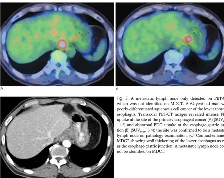
confidence level, 0.756-0.940) (Fig. 2) and the sensitivi- ty, positive predictive value, and negative predictive val- ue were significantly higher than considering 10 mm as the standard value (Table 1). One metastatic LN unde- tected by MDCT was located in the esophago-gastric junction in a patient with lower esophageal cancer.
Although MDCT could not detect this LN, PET-CT showed strong FDG uptake (Fig. 3).
Among the 23 metastatic LNs, 19 were detected by
A B
C
Fig. 3. A metastatic lymph node only detected on PET-CT, which was not identified on MDCT. A 64-year-old man with poorly-differentiated squamous cell cancer of the lower thoracic esophagus. Transaxial PET-CT images revealed intense FDG uptake at the site of the primary esophageal cancer (A) (SUVmax, 11.2) and abnormal FDG uptake at the esophago-gastric junc- tion (B) (SUVmax, 5.4); the site was confirmed to be a metastatic lymph node on pathology examination. (C) Contrast-enhanced MDCT showing wall thickening of the lower esophagus as well as the esophago-gastric junction. A metastatic lymph node could not be identified on MDCT.
A B
C D
E F
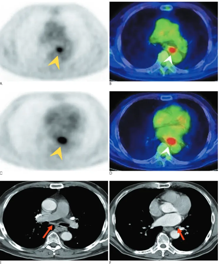
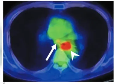
Fig. 4. False-negative loco-regional metastatic lymph nodes on PET-CT. A 66-year-old man with poorly differentiated squamous cell cancer of the lower esophagus. (A-D) Transaxial PET and PET-CT images show intense FDG uptake in the primary tumor (ar- rowhead, SUVmax, 6.23), without other abnormal FDG uptake. (E, F) Axial contrast-enhanced MDCT images obtained at the levels of the subcarinal region (E) and the inferior pulmonary ligament region (F), show marked esophageal wall thickening, which corre- spond to distal esophageal cancer. There were approximately 5.2 mm (arrow in E) and 5.0 mm (arrow in F) lymph nodes, which were proven to be a metastatic lymph nodes on pathology examination.
PET-CT. Their SUVmaxwas 2-10.1, and was was higher than 2. Four metastatic LNs that were not detected by PET-CT were located in the vicinity of the primary tu- mors (Fig. 4). The mean size of these undetected LNs was 5.8 mm. Among the 18 metastatic LNs detected by both MDCT and PET-CT, the average size was 10.38 mm (7-15.6 mm) and 10 were < 10 mm.
One LN with FDG uptake on PET-CT revealed no metastasis on pathology examination, and the SUVmaxof this LN was 2.6; on MDCT, it was seen to be a regional LN located in the subcarinal region and measured 10.3 mm (Fig. 5).
The sensitivity, specificity, positive predictive value, and negative predictive value of PET-CT were 82.6%, 99.4%, 95%, and 97.4%, respectively (Table 1).
Discussion
CT is a common and non-invasive modality used to
evaluate the disease stage of esophageal cancer.
However, it has substantial limitations regarding its ac- curacy in evaluating disease stage. In particular, in LN metastasis, patients with LNs < 10 mm with tumor in- filtration are excluded and the sensitivity is lowered (9, 10).
Schro¨der et al. (11) reported that among 1,196 surgi- cally resected LNs in primary esophageal squamous cell cancer patients, metastasis could be confirmed in 129 LNs (10.8%) by histopathologic analysis. Among these 129 LNs, there were only 15 with a diameter > 10 mm (12%). They also observed that the average size of non- metastatic LNs was 5.1 ± 3.8 mm in maximum diame- ter, the average size of metastatic LNs was 6.7 ± 4.2 mm, and that there was a statistically significant differ- ence (p=0.00006); however, the LN size was not signifi- cantly correlated to the frequency of LN metastasis (p=0.33). Funai et al. (12) also suggested that discount- ing nodes < 10 mm can lead to an underestimation of
A B
C
Fig. 5. False-positive metastatic lymph node on PET-CT in a 60- year-old male with poorly differentiated squamous cell carcino- ma of the mid- to lower esophagus. (A, B) PET and PET-CT re- vealing abnormal increased uptake in the subcarinal lymph node (arrow, SUVmax, 2.6) and primary cancer (arrowhead, SUVmax, 10.1). (C) Concomitant mediastinal window of con- trast-enhanced MDCT showing an approximately 10.3-mm sub- carinal lymph node (arrow) that was proven to be negative for malignancy on pathology examination.
the stage of esophageal cancer during pre-operative eval- uation. Another study reported that the mean size of the metastatic LN was 4.8 mm; the authors suggested that extensive LN dissection is appropriate in esophageal cancer surgery (13). However, most reports regarding metastatic LN size of esophageal cancer have involved histopathologic results of dissected LNs, and little is known about CT criteria of metastatic LN in esophageal cancer patients. So, we evaluated the cut-off value of metastatic LN in esophageal cancer patients.
The average size of LNs without metastasis was 5.74 mm in short-axis diameter and that of metastatic LNs was 9.35 mm, representing a significant difference (p<0.01). Among the 22 metastatic LNs detected on MDCT, the size of LNs was larger than 10 mm as mea- sured by pre-surgical MDCT in only seven cases (32%).
Considering 7 mm as the optimal cut-off value, a sub- stantial discriminatory power was shown, (sensitivity of 81.8%, specificity of 82.2%), and the area under the ROC curve was 0.848 (95% confidence level, 0.756- 0.940). Therefore, we presume that, for esophageal can- cers, it can be assumed that the size of metastatic LNs is smaller. Our results correspond to the previous histopathologic reports (12, 13).
To determine the stage of esophageal cancer, the prog- nosis of cases with non-regional LN metastasis is less fa- vorable than that for cases with locoregional LN metas- tasis, and thus non-regional LN metastasis was classified as M1a (14). In midesophageal cancer, as nonregional lymph node metastasis also shows a poor prognosis sim- ilar to that for distant organ metastases due to the limit- ed lymphatic drainage, they were therefore classified as M1b (14). In esophageal cancer, if metastasis develops only in regional LNs, successful surgery is possible;
however, in cases with non-regional LN metastasis, surgery is not useful, and, therefore, detection of non-re- gional LNs is very important in imaging studies. In our study, as the subjects were only patients who had had surgery, the number of non-regional LN was small; nev- ertheless, even in non-regional LN metastasis cases, LNs
< 10 mm were detected in 40% (2/5) of the patients.
In many studies, PET has been used to evaluate the disease stage of esophageal cancer; the sensitivity and accuracy of PET is higher than that of MDCT for detect- ing LN metastasis (5, 15-17). However, as the spatial resolution of PET is poor, it is limited regarding its abili- ty to detect the false negative component of metastatic LNs located in the vicinity of primary tumors (7). The evaluation of LN metastasis using PET-CT fusion im-
ages improves the sensitivity, accuracy, and the nega- tive predictive value (18). In one study, PET-CT could distinguish the FDG uptake by primary tumors and the FDG uptake by adjacent LNs in 64% of sites located in the proximity to the primary tumor, allowing the locore- gional involvement to be accurately evaluated (19). In another study, among 28 metastatic LNs located in the vicinity of primary tumors, six were false negative ac- cording to PET and two were false-negative according to PET-CT (18).
Similarly, in our study, PET-CT detected metastatic LNs accurately in esophageal cancer patients, regardless of the size of the nodes. In particular, LN metastasis presently located in the esophago-gastric junction in a patient with lower esophageal cancer and which was not identified on MDCT, could be detected by PET-CT.
However, in our study, four metastatic LNs located in the vicinity of the primary tumor were missed, which suggests a limitation of PET-CT for detecting locoregion- al LN metastasis. This might be due to the poor distinc- tion of adjacent LN uptake from the strong primary tu- mor uptake or to the small sized of LN, which prevents its uptake of FDG. The mean size of LNs that could not be detected by PET-CT in our study was 5.8 mm. In ad- dition, one of our cases diagnosed with metastatic LN on PET-CT, was negative for metastasis on pathology. We suggest that careful analysis of the results of PET-CT is required.
It has been reported that in all esophageal cancer pa- tients, abnormal FDG accumulation is detected in pri- mary tumors, with the average detection rate exceeding 90% (15, 20-23). However, as the spatial resolution of PET or PET-CT is poor, it is limited in its ability to detect small tumors; in addition, the adjacent invasion of a pri- mary tumor is difficult to assess in most cases (7, 14).
One study reported that in primary esophageal cancer, PET could not detect T1a stage tumors, however, it could detect T1b stage tumors or higher stage tumors (23). In our study, one case of T1a was not detected by either MDCT or PET-CT and one case of T1b tumor was not detected by MDCT; nonetheless, weak PET-CT up- take was shown on PET-CT.
Our study has several limitations. First, there were on- ly a small number of sample groups. Second, as the sub- jects were operable patients, advanced stage patients were excluded, lowering the prevalence of metastatic le- sions. Third, the total number of resected LNs was small. In addition, as it was a retrospective study, corre- lation of the location of LNs was assessed by surgical
records and so may not have provided very accurate matching.
In summary, in operable esophageal cancers, the size of metastatic LNs can be < 1 cm in many cases. None- theless, the size of metastatic LNs can be significantly larger than LNs without metastasis and, on MDCT, the short diameter of 7 mm may be the optimal diagnostic criterion. As PET-CT was very accurate for assessing metastatic LNs in our study, it is anticipated that it will have a decisive role in determining the esophageal dis- ease stage. But, it is limited in esophageal cancer cases with metastatic LNs in the vicinity of the primary tu- mors and, although rare, it may show false positives, so results should be interpreted with extreme caution.
References
1. Roder JD, Busch R, Stein HJ, Fink U, Siewert JR. Ratio of invaded to removed lymph nodes as a predictor of survival in squamous cell carcinoma of the oesophagus. Br J Surg 1994;81:410-413 2. Ellis FH Jr, Watkins E Jr, Krasna MJ, Heatley GJ, Balogh K. Staging
of carcinoma of the esophagus and cardia: a comparison of differ- ent staging criteria. J Surg Oncol 1993;52:231-235
3. Lieberman MD, Shriver CD, Bleckner S, Burt M. Carcinoma of the esophagus: prognostic significance of histologic type. J Thorac Cardiovasc Surg 1995;109:130-139
4. Korst RJ, Rusch VW, Venkatraman E, Bains MS, Burt ME, Downey RJ, et al. Proposed revision of the staging classification for esophageal cancer. J Thorac Cardiovasc Surg 1998;115:660-669 5. Choi JY, Lee KH, Shim YM, Lee KS, Kim JJ, Kim SE, et al.
Improved detection of individual nodal involvement in squamous cell carcinoma of the esophagus by FDG PET. J Nucl Med 2000;41:
808-815
6. Kato H, Kuwano H, Nakajima M, Miyazaki T, Yoshikawa M, Ojima H, et al. Comparison between positron emission tomogra- phy and computed tomography in the use of the assessment of esophageal carcinoma. Cancer 2002;94:921-928
7. Kato H, Miyazaki T, Nakajima M, Takita J, Kimura H, Faried A, et al. The incremental effect of positron emission tomography on di- agnostic accuracy in the initial staging of esophageal carcinoma.
Cancer 2005;103:148-156
8. Halvorsen RA JR, Daffner R, Thompson WM, The esophagus. In:
Godwin JD. Computed tomography of the chest. Philadelphia:
Lippincott, 1984;247-291
9. Vilgrain V, Mompoint D, Palazzo L, Menu Y, Gayet B, Ollier P, et al. Staging of esophageal carcinoma: comparison of results with en- doscopic sonography and CT. AJR Am J Roentgenol 1990;155:277- 281
10. Goei R, Lamers RJ, Engelshove HA, Oei KT. Computed tomo- graphic staging of esophageal carcinoma: a study on interobserver variation and correlation with pathological findings. Eur J Radiol 1992;15:40-44
11. Schro¨der W, Baldus SE, Mo¨nig SP, Beckurts TK, Dienes HP, Ho¨lscher AH. Lymph node staging of esophageal squamous cell carcinoma in patients with and without neoadjuvant ra- diochemotherapy: histomorphologic analysis. World J Surg 2002;
26:584-587
12. Funai T, Osugi H, Higashino M, Kinoshita H. Estimation of lymph node metastasis by size in patients with intrathoracic oesophageal cancer. Br J Surg 2000;87:1234-1239
13. Yosiaki K, Yoshimi I, Natsumi T, Takayuki A, Fuyumi I, Toshiharu M, et al. Size analysis of lymph node metastasis in esophageal cancer: diameter distribution and assessment of accu- racy of preoperative diagnosis. Esophagus 2006;3:189-195 14. Bruzzi JF, Munden RF, Truong MT, Marom EM, Sabloff BS,
Gladish GW, et al. PET/CT of esophageal cancer: its role in clinical management. Radiographics 2007;27:1635-1652
15. Flanagan FL, Dehdashti F, Siegel BA, Trask DD, Sundaresan SR, Patterson GA, et al. Staging of esophageal cancer with 18F-fluo- rodeoxyglucose positron emission tomography. AJR Am J Roentgenol 1997;168:417-424
16. Kim KM, Park SJ, Kim BT, Lee KS, Shim YM. Evaluation of lymph node metastases in squamous cell carcinoma of the esophagus with positron emission tomography. Ann Thorac Surg 2001;71:290- 294
17. Yoon YC, Lee KS, Shim YM, Kim BT, Kim K, Kim TS. Metastasis to regional lymph nodes in patients with esophageal squamous cell carcinoma: CT versus FDG PET for presurgical detection-prospec- tive study. Radiology 2003;227:764-770
18. Yuan S, Yu Y, Chao KS, Fu Z, Yin Y, Liu T, et al. Additional value of PET/CT over PET in assessment of locoregional lymph nodes in thoracic esophageal squamous cell cancer. J Nucl Med 2006;47:
1255-1259
19. Bar-Shalom R, Guralnik L, Tsalic M, Leiderman M, Frenkel A, Gaitini D, et al. The additional value of PET/CT over PET in FDG imaging of oesophageal cancer. Eur J Nucl Med Mol Imaging 2005;
32:918-924
20. Rankin SC, Taylor H, Cook GJ, Mason R. Computed tomography and positron emission tomography in the pre-operative staging of oesophageal carcinoma. Clin Radiol 1998;53:659-665
21. McAteer D, Wallis F, Couper G, Norton M, Welch A, Bruce D, et al. Evaluation of 18F-FDG positron emission tomography in gas- tric and oesophageal carcinoma. Br J Radiol 1999;72:525-529 22. Jager PL, Que TH, Vaalburg W, Pruim J, Elsinga P, Plukker JT.
Carbon-11 choline or FDG-PET for staging of oesophageal cancer?
Eur J Nucl Med 2001;28:1845-1849
23. Himeno S, Yasuda S, Shimada H, Tajima T, Makuuchi H.
Evaluation of esophageal cancer by positron emission tomogra- phy. Jpn J Clin Oncol 2002;32:340-346
대한영상의학회지 2010;62:235-243
수술 가능한 편평상피식도암 환자의 림프절 병기판정을 위한 MDCT에서의 림프절의 크기 기준 결정과 PET-CT와의 상관관계1
1가톨릭대학교 의과대학 서울 성모병원 영상의학과
2경상대학교병원 영상의학과
3가톨릭대학교 의과대학 성빈센트병원 영상의학과
4가톨릭대학교 의과대학 서울성모병원 흉부외과
5가톨릭대학교 의과대학 서울성모병원 핵의학과
윤수경∙정정임∙박미정2∙박현진3∙안명임∙박재길4∙유이령5∙박석희
목적: 수술 가능한 편평상피식도암 환자의 MDCT에서의 전이 림프절의 크기 기준을 결정하고 PET-CT의 결과와 비교해 보았다.
대상과 방법: 식도암으로 수술전 MDCT와 PET-CT를 시행하고 근치적 식도절제술을 받은 24명의 환자를 대상으 로 하였다. MDCT의 소견을 PET-CT와 비교하였으며, 이후 수술 기록과 비교하였다. 정상 림프절과 전이 림프절을 구별할 수 있는 차단 값을 결정하기 위해 receiver operating characteristic (ROC) curve 방법을 이용하였다.
결과: 전이 림프절의 크기는 9.35 ± 3.41 mm 로 정상 림프절 5.74 ± 1.64 mm 보다 통계적으로 유의하게 컸다 (p < 0.001). 최적의 차단 값을 7 mm 로 하였을 때 변별력이 가장 높게 나타났고, 민감도, 특이도는 각각 81.8%, 80.8%였다. PET-CT에서는 종양주위에 있는 4개의 림프절은 발견하지 못했으며, PET-CT의 민감도, 특이도는 각각 82.6%, 99.4%였다. 1개의 정상 림프절이 PET-CT에서 가양성을 보였다.
결론: 많은 경우 MDCT에서 전이 림프절의 크기가 10 mm 보다 작으며, 단경 7 mm 는 전이 림프절의 적합한 기 준이 될 수 있다. PET-CT는 종양주위에 있는 전이 림프절을 제외하고는 전이 림프절 진단에 매우 정확하다.