Intradural disc herniation is a very rare condition with an incidence that ranges from 0.26% to 0.30% of all her- niated discs (1). Myelography, computed tomography (CT) and magnetic resonance imaging (MRI) are general- ly used to diagnose intradural lumbar disc herniation (IDLDH), but there can be some differences according to each case. Several studies have reported of a potential association between intradural disc herniation and gas within the spinal canal. However, to our knowledge, there are no reports of IDLDH together with intradural vacuum (IDV). This report presents three cases of IDLDH that were diagnosed by a radiological examina- tion with a review of the relevant literature.
Case Reports
Case 1
A 54-year-old man with a 15-year history of chronic
back pain was admitted with complains of severe back pain and right radiculopathy of a three-day duration. A physical examination showed no reflex in the right knee. The CT scan revealed a right paracentral soft tis- sue lesion that contained intradural gas at the L3-4 lev- el (Fig. 1A). The sagittal T2-weighted image showed an intradural iso-intensity mass at the L3-4 level with a pe- ripheral lower-intensity rim signal (Fig. 1B). The axial T2-weighted image demonstrated that the Rt. intradural herniated disc material contained gas that had passed through a Rt. posterior annular tear (Fig. 1C). The pa- tient was preoperatively diagnosed with Rt. intradural herniated disc material that contained gas at the L3-4 level, and he underwent an open lumbar microdiscecto- my. On the surgical field, the right L4 nerve root was se- verely compressed by the transligamentously-extruding disc, and there were dense adhesions between the PLL and the dura. A longitudinal incision was made in the dura mater and the intradural disc fragment was re- moved. He showed a marked improvement in his symp- toms on the follow-up examinations.
Case 2
A 62-year-old female with a 10-year history of right buttock and leg pain was admitted to the hospital due to
Intradural Lumbar Disc Herniation with Intradural Gas: Report of Three Cases1
Seung-Eun Chung, M.D., Sang-Ho Lee, M.D.2, Tae-Hong Kim, M.D.3, Byung-June Jo, M.D.
1Department of Diagnostic Radiology, Wooridul Spine Hospital
2Department of Neurosurgery, Wooridul Spine Hospital
3Department of Neurosurgery, Sanggyepaik Hospital Inje University College of Medicine
Received August 8, 2005 ; Accepted September 28, 2005
Address reprint requests to : Seung-Eun Chung, M.D., Department of Diagnostic Radiology, Wooridul Spine Hospital, 47-7 Chungdam-dong Gangnam-gu Seoul 135-100, Korea.
Tel. 82-2-513-8197 Fax. 82-2-513-8175 E-mail: [email protected]
This paper reports on three cases of an intradural lumbar disc herniation (IDLDH) that were diagnosed by a radiological examination. In all cases, an intradural vacuum (IDV) was detected on the CT scans, and the IDLDH showed iso- or lower signal inten- sity on the T2-weighted images. Enhanced MRI of one case revealed a small amount of air, but this was without enhancement. All the cases showed definite IDV on the CT scans, and this was an important clue for diagnosing IDLDH.
Index words :Intradural disc herniation Dural vacuum
Spine
the sudden exacerbation of symptoms one month before her admission. The physical examination showed that both the ankle dorsiflexion and ankle plantar flexion were 4/4. The dorsiflexion strength of the great toes had decreased to 3/4 with a hallux flexion of 3/4 (Rt/Lt). In addition, her reflexes showed as 2+ at the knees, and 3+ and 2+ at the right and left ankle, respectively.
The CT scan revealed a lesion of the left paracentral soft tissue with an encircling gas density (CT number;
-250~-350 HU) (Fig. 2A). The sagittal T2-weighted MR images showed a Lt. anterior iso- intensity lesion, as compared with the spinal cord, behind the posterior lon-
gitudinal ligament at the L4-5 disc level (Fig. 2C). She was preoperatively diagnosed with a left intradural disc herniation that contained gas, and she underwent an open lumbar microdiscectomy at the left L4-5 level.
The dural opening was not found on the surgical field.
However, the sequestrated disc material was removed after incising the dura. Postoperatively, the pain in her right buttock and leg were relieved.
Case 3
A 59-year-old female with 20-year history of lumbar discomfort complained of a one month duration of back
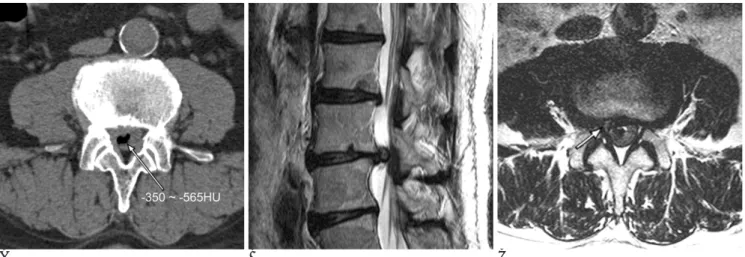
A B C
Fig. 1. L3-4 intradural disc herniation.
A. The CT image showed a Rt. anterior intradural mass-like lesion that contained gas (arrow, CT number; -350~ -565 HU).
B. The sagittal T2-weighted MR image demonstrated an isointense intradural lesion with a peripheral lower signal rim.
C. The axial T2-weighted image showed the Rt. intradural herniated disc material containing gas that had passed through the Rt.
posterior annular tear (arrow).
and posterior thigh pain (the pain on the right side was greater) in both legs. The neurological examination re- vealed weakness in her lower extremities and a de- creased right ankle jerk. The CT scan demonstrated an intradural mass-like lesion surrounded by a small amount of gas (Fig. 3A). The T2-weighted sagittal MRI demonstrated an anterior intradural mass-like lesion at the L2-3 disc level with an iso- or slightly lower signal intensity, and this intensity appeared similar to that of the intervertebral disc (Fig. 3B). The post-contrast T1 sagittal and axial scans showed a peripherally enhanced transligamentous lesion, but there was no definitely en- hanced intradural lesion at the L2-3 disc level (Fig. 3C, D). The preoperative diagnosis was a transligamentous disc herniation with an associated intradural disc herni- ation at the L2-3 level. Therefore, the patient under- went an open lumbar microdiscectomy at the L2-3 lev- el. A small extradural disc herniation was revealed dur- ing surgery. The dural opening could not be found on the surgical field, but a disc rupture was observed after making an incision into the dura. The subsequent
histopathological examination confirmed the diagnosis of a herniated nucleus pulposus. The patient totally re- covered after surgery with no recurrence of her preoper- ative symptoms.
Discussion
Dandy first reported on IDLDH in 1942 (2). Kataoka et al. reported that intradural disc herniation are most commonly found in the lumbar area, and the incidence of this condition ranges from 0.04% to 0.33% in lumbar herniated disc patients (3). The authors experienced three cases of IDLDH out of 9285 lumbar spine disc her- niations, which is a 0.032% incidence.
The pathogenesis of intradural disc herniation is un- clear. However, several theories have been proposed (4).
1. There is adhesion formation between the dura and the posterior longitudinal ligament secondary to chronic inflammation, with the resulting sponta- neous perforation, although not through propulsion
A B
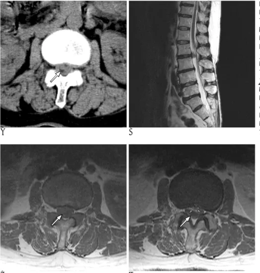
Fig. 3. L2-3 intradural disc hernia- tion.
A. The CT image showed an intradur- al mass-like lesion (large arrow) sur- rounded by a subtle small amount of gas.
B. The T2-weighted sagittal MRI showed an anterior intradural mass- like lesion at the L2-3 disc level.
C, D. The T1-weighted axial image without contrast (C), and the post-con- trast T1 axial image (D). The enhanced T1 axial image showed the marginated enhancing extradural lesion (small ar- rows), but not the enhanced intradural lesion (large arrow).
C D
of the disc material.
2. There is a congenital union between the dura mater and the common posterior vertebral ligament.
3. There are secondary changes resulting from a previ- ous surgery.
In our cases, the dense adhesions were prominent, but there was no dural opening found on the operative field.
Our patients reported that there were no traumatic events or previous surgery. It is believed that the theory pertaining to the adhesions might have led to the IDLDH.
Our patients’ clinical histories consisted of chronic low back pain, acute radicular pain and progressive neu- rological deficits. This type of lesion often presents with cauda equina syndrome, but the symptoms in our pa- tients were not specific.
A diagnosis of intradural disc herniation is difficult, but it can be made by using several imaging techniques.
The myelographic findings of intradural disc herniation are not specific. Myelography shows a complete block in approximately 65% of such cases. However, it is not always possible to determine the intra- or extradural ori- gin (4). Hodge et al. reported that myelogram-CT effec- tively showed the presence of intradural disc material (5). Epstein et al. showed that a myelogram-CT study easily defined an irregular intradural mass that was con- sistent with a disc fragment, whereas the contrast-en- hanced MRI could not readily distinguish a herniated disc fragment from a neurofibroma (6). Hidalgo et al. re- ported that 2 out of 118 intradural disc herniations con- tained air in the spinal canal. This percentage of 1.7% is six times higher than that of disc herniations without spinal gas (7). They proposed that the presence of air within the spinal canal and an intradural mass-like le- sion on the CT scans and MRI, respectively, were al- most certainly evidence of a herniation rather than a tu-
rectly connected to the intradural mass, and a diagnosis of an intradural disc herniation can be made from such a finding (8). Wasserstrom et al. reported on IDLDH with a ring enhancement pattern on a contrast MRI study (9). The ring enhancement pattern on the contrast T1-weighted image was attributed to the granulation tis- sue surrounding the lesion. Our third case showed a low air density lesion in the spinal canal. Therefore, a con- trast MRI study was attempted so as to provide a more precise examination. This case had a transligamentous disc herniation with peripheral enhancement and a nonenhancing intradural lesion at the L2-3 disc level.
The posterior enhancement of the transligamentous disc herniation might have been the result of the mechanical irritation from the chronic disc herniation against the ventral wall of the dura. Considering that the symptoms had been aggravated one month earlier, the nonenhanc- ing intradural lesion was regarded as an unvascularized disc fragment rather than a tumor. Whittaker et al. re- ported the homogenous enhancement of the IDLDH in a 66-year-old-man (10). They suggested that the en- hancement is likely to depend on the age of the intradur- al disc herniation. Our cases showed iso- or slightly low- er signal intensity on the T1- and T2-weighted images.
Almost all the intradural extramedullary tumors were hyperintense on the T2-weighted scan.
IDLDH should be treated by the prompt surgical re- moval of the ruptured disc fragment because the neuro- logical prognosis is closely linked to the preoperative du- ration of the neurological symptoms. An IDLDH can of- ten be overlooked during surgery, and this means that a second and possibly difficult surgical procedure will be needed.
In conclusion, our cases showed intradural gas trapped within the herniated disc material on the CT scan with an iso- or slightly lower-signal-intensity in-
diagnosis and review of the literature. Neurosurg Rev 2004;27:75- 80
5. Hodge CJ, Binet EF, Kieffer SA. Intradural herniation of lumbar in- tervertebral discs, Spine 1978;3:436-450
6. Epstein NE, Syrquin MS, Epstein JA, Decker RE. Intradural disc herniations in the cervical, thoracic, and lumbar spine: report of three cases and review of the literature. J Spinal Disord 1990;3:396- 403
7. Hidalgo-Ovejero AM, Garcia-Mata S, Gozzi-Vallejo S, Izco- Cabezon T, Martinez-Morentin J, Martinez-Grande M. Intradural
disc herniation and Epidural Gas: something more than a casual association? Spine 2004;29:E463-E467
8. Holtas S, Nordstrom CH, Larsson EM. MR Imaging of Intradural Disk Herniation. J Comput Assist Tomogr 1987;11:353-356 9. Wasserstrom R, Mamourian AC, Black JF, Lehman RA. Intradural
lumbar disk fragment with ring enhancement on MR. AJNR Am J Neuroradiol 1993;14:401-404
10. Whittaker CK, Bernhardt M. Magnetic resonance imaging shows gadolinium enhancement of intradural herniated disc. Spine 1994;
19:1505-1507
대한영상의학회지 2005;53:445-449
경막내 가스를 동반한 요추의 경막내 추간판 탈출증: 증례 보고1
1우리들병원 진단방사선과
2우리들병원 신경외과
3인제대학교 상계백병원 신경외과
정승은・이상호2・김태홍3・조병준
경막내 추간판 탈출증은 드문 질환으로 그 발생기전은 분명치 않다. 저자들은 3예의 요추의 경막내 추간판 탈출 증을 경험하였고, 3예 모두 단층촬영에서 경막내 가스(Intra-Dural Vacuum, IDV)을 보였고, T2 강조 시상 자기 공명영상에서 저신호강도를 보였다. 저자들은 이러한 영상 소견이 수술전 다른 질환과의 감별진단에 있어 중요한 단서가 될 것으로 생각되어 문헌 고찰과 함께 영상 소견을 보고하는 바이다.