Spontaneous dissection of the abdominal aorta is a rare malady and it has been only infrequently reported on; its typical course and optimal management are not clearly defined (1). It is usually limited to the infrarenal aorta (1, 2). Isolated abdominal aortic dissection may be classified on the basis of its etiology as iatrogenic, trau- matic or spontaneous (2). Primary abdominal aortic dis- sections comprise less than 2% of all aortic dissections, compared to 70% for ascending aortic dissections, 20%
for descending thoracic aortic dissections and 7% for aortic arch dissections (3). We report here on a case of chronic SIAAD that caused stenotic change of the in- frarenal abdominal aorta along with progressive lower extremity pain and claudication in both legs. The patient was treated with successful interventional management.
Case Report
A 52-year-old female presented with progressive low- er extremity pain and claudication that had persisted for more than 6 months, and this occurred after walking about 200 meters. Her past history included hyperten- sion that had been diagnosed 7 years ago, and this had been treated with taking antihypertensive drugs for the past 6 years. There was no history of trauma, diabetes or any surgery. The patient also had no connective tissue disorders or any other systemic anomalies, and there was no significant family history of disease. However, she was a smoker.
On physical examination, the patient was found to have a height of 157 cm, a weight of 58 kg and a blood pressure of 140/90 mmHg. Her ankle-brachial indexes (ABI) were 0.6 on the right side and 0.7 on the left side.
Computed tomography (CT) detected an aortic dissec- tion that extended from the infrarenal aorta to both proximal common iliac arteries, and there was no in- volvement of the suprarenal aorta. Multiple prominent lumbar artery collaterals were noted as well. Multiple
Primary Stent Placement for Chronic Spontaneous Infrarenal Abdominal Aortic Dissection: A Case Report1
Se Hwan Kwon, M.D., Joo Hyeong Oh, M.D.
1Department of Diagnostic Radiology, Kyung Hee University Hospital Received April 22, 2006 ; Accepted August 28, 2006
Address reprint requests to : Joo Hyeong Oh, M.D., PhD, Department of Diagnostic Radiology, Kyung Hee University Hospital,
Hoeki-dong 1, Dongdaemun-gu, Seoul 130-702, Korea
Tel. 82-2-958-8622 Fax. 82-2-968-0787 E-mail: [email protected]
Spontaneous infrarenal abdominal aortic dissection (SIAAD) is a rare entity with var- ious clinical presentations. We recently encountered the even rarer condition of a fe- male patient suffering from chronic SIAAD with multiple intimal flaps and prominent lumbar artery collaterals; this all caused stenotic changes of the infrarenal abdominal aorta and produced progressive lower extremity pain and claudication in both her legs.
This patient’s condition was successfully managed by primary stent placement fol- lowed by balloon angioplasty.
Index words :Aorta, dissection
Aorta, interventional procedures Stents and prostheses
intimal flaps and septations were shown and these caused stenotic changes of the infrarenal abdominal aor- ta (Fig. 1). However, the arteries in both lower extremi- ties were normal.
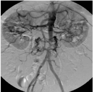
Diagnostic angiography was performed with using a marker pigtail catheter (Cook, Bloomington, IN, U.S.A.) in order to evaluate the dissection (Figs. 2, 3). The left in- ternal iliac artery was occluded. The collaterals from the median sacral artery, lumbar arteries and inferior mesenteric artery (IMA) supplied the left pelvic cavity.
Primary stent deployment was chosen as the best thera- peutic method for managing the dissection and the com- bined stenotic changes of the infrarenal abdominal aorta because the infrarenal aortic stenotic changes were the most problematic findings and there were no complicat- ing aneurysmal changes.
Both femoral accesses with local anesthesia were used for the procedure. After the placement of both 6 Fr in- troducer sheaths, a 0.035 inch guidewire (Radiofocus M;
A B
C D
Fig. 1. A-D. The abdominal CT images reveal multiple intimal flaps and septations, which caused stenotic changes of the infra-re- nal abdominal aorta. The dissections were limited to the infrarenal aorta and to both common iliac arteries.
Fig. 2. Abdominal aortic angiogram shows stenotic changes of the infra-renal abdominal aorta with prominent lumbar artery collaterals that were due to the chronic SIAAD. Both the renal arteries and the supra-renal aorta are normal.
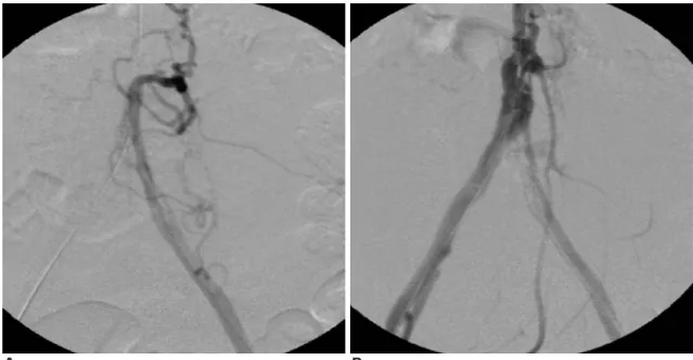
Terumo, Tokyo, Japan) was inserted into the suprarenal aorta from the right femoral artery. Multiple selective angiograms of the dissected lumens were performed on the left side with using a 5 Fr cobra catheter (Cook, Bloomington, IN, U.S.A.) because the right side iliac artery and IMA were not seen on the angiograms through the presumed false lumens. However, the right iliac artery and IMA were visible on the angiogram of the presumed true lumen (Fig. 3). Another guide wire was inserted through this lumen into the suprarenal aor- ta. Intravenous heparin (5,000 IU) was administered fol- lowing the placement of both femoral introducer sheaths.
Next, two 8 mm-8 cm Zilver stents (Cook, Bloomington, IN, U.S.A.) were separately inserted from both sides into the infrarenal abdominal aortic dissec- tion in order to treat the stenotic changes of the dissec- tions with using the “kissing” technique. The right side stent was deployed in the presumed false lumen and the left side stent was deployed in the presumed true lu- men. Post-stent ballooning was performed using a 6 mm balloon and a 4 cm balloon (Boston Scientific, Watertown, MA, U.S.A.) (Fig. 4). The two balloons were inflated simultaneously with use of the kissing balloon technique. The pressure gradients were measured across the lesion before and after stent placement. The measured systolic pressure gradients between the suprarenal aorta and the two external iliac arteries be-
fore and after treatment were 26 and 2 mmHg on the right side and 27 and 3 mmHg on the left side, respec- tively.
A well-perfused aorta and both the common and ex- ternal iliac arteries were seen on the final angiogram and the follow-up CT scan. A decreased number of lum- bar collaterals was noted as well (Fig. 5). Three days af-
A B
Fig. 3. A. The right iliac artery and inferior mesenteric artery are not seen on the angiogram through the presumed false lumen from the left femoral artery approach. B. However, the right iliac artery and inferior mesenteric artery are seen on the angiogram of the presumed true lumen.
Fig. 4. Two 8 mm-8 cm Zilver stents were inserted into the stenotic portion of the infra-renal abdominal aorta with using the “kissing” technique, and post-stent ballooning was per- formed simultaneously with using a 6 mm balloon and a 4 cm balloon.
ter treatment, the patient was discharged from the hos- pital in good condition, and her lower extremity pain and claudication had all resolved. The patient has been followed up for 5 months until now and she is in good physical condition with no complaints of any pain or claudication in both of her lower extremities.
Discussion
The diagnosis of abdominal aortic dissection can be significantly delayed because of its symptoms are char- acteristically non-specific. The clinical findings at pre- sentation may indeed vary greatly as back and abdomi- nal pain, lower extremity ischemia, hypertension, ab- dominal tenderness, absence of the femoral pulses, hematuria, melena and shock. In contrast, some pa- tients remain completely asymptomatic (1, 4-7).
Isolated dissection of the abdominal aorta is a rare mala- dy and it’s usually limited to the infrarenal aorta (1, 2).
In a review of 398 cases of patients with aortic dissec- tion, only 10 cases (2.5%) were isolated to the abdominal aorta (6). In an autopsy study of 182 patients with spon- taneous aortic dissection, only 1% was noted to have dissection limited to the abdominal aorta (3). However, the recent widespread use of CT scanning for cases of non-specific abdominal pain has begun to reveal this condition with increasing frequency.
Farber et al. (2) reported on 10 patients who suffered with isolated dissecting abdominal aortic aneurysms
during a 6 year period. In their series, the dissection flap originated below the renal arteries in 9 cases and at the level of the superior mesenteric artery in 1 case. Their treatment consisted of aortic stent graft deployment in one patient, direct aortic reconstruction in three patients and observation for the remaining six patients. In our patient, conservative therapy was not indicated because of her clinical symptoms and the apparent stenotic changes of the infrarenal abdominal aorta.
Similar findings were reported by Becquemin et al. (5) in a series of seven patients who were affected by acute or chronic dissection of the abdominal aorta. In this study, six of the seven patients had infrarenal dissection and they were treated by replacement of the aorta with a Dacron prosthesis, while the seventh patient, who had a suprarenal dissection, was treated conservatively.
During follow-up for a mean of 3 years, all the patients were alive and free from symptoms. The authors con- cluded that their results favor graft replacement in cases of infrarenal aortic dissection and more selective surgi- cal procedures in cases of suprarenal aortic dissections.
However, for our present case, her infrarenal aortic stenotic changes due to the chronic, spontaneous ab- dominal aortic dissections were the most problematic findings. Therefore, we considered that managing the stenotic changes could solve the patient’s symptoms be- cause previous studies have reported that primary stent placement for the treatment of infrarenal aortic stenosis appeared to be both safe and effective (8, 9).
Focal stenosis of the abdominal aorta most often in- volves the infrarenal aorta and its bifurcation (8, 9).
Localized aortoiliac stenosis is relatively infrequent and it occurs predominantly in young women who are heavy smokers (8). Sheeran et al. (9) recommended stent placement as an adjuvant therapy to angioplasty or as a primary method of treatment in properly selected pa- tients who suffer with focal mid-abdominal aortic steno- sis. Schedel et al. (8) reported that primary stent place- ment for the treatment of calcified infrarenal aortic stenosis proved to be safe and it also provided lasting long-term clinical improvement. Furthermore, our pre- sent case of focal infrarenal abdominal aortic stenotic changes due to chronic SIAAD was successfully man- aged with primary stent placement.
Primary stent placement means implantation of the stent without any the patient having undergone previ- ous intervention or percutaneous transluminal angio- plasty (PTA). Although the success rate of aortic PTA is high, there is a lack of scientific data regarding the safety Fig. 5. The follow-up 3D reconstruction CT image shows a
well-perfused lower abdominal aorta, both iliac arteries, the inferior mesenteric artery and the median sacral artery. A de- creased number of lumbar artery collaterals are noted as well.
and efficacy of PTA for treating complex aortic lesions like eccentric, calcified, ulcerative, multiple and long stenoses (8, 9). Primary stent placement followed by bal- loon angioplasty provides the more durable, long-term therapeutic effect than does PTA alone.
In 2004, Farber et al.(10) conducted a Medline English language literature search for all case reports or series of SIAAD between 1953 and 2003. They described 52 pa- tients who were diagnosed with SIAAD, and their dis- sections were diagnosed by CT scan, magnetic reso- nance imaging (MRI), ultrasound or aortography. A mi- nority of cases were diagnosed in the operating room and at autopsy. In their report, the distal extent of the dissection was found to be in the iliac or femoral artery in 80% of the patients in the ischemia group, in 40% of the patients in the pain group and in 50% of the patients in the asymptomatic group (p<0.05) An associated in- frarenal abdominal aortic aneurysm was found in 20%
of the patients in the ischemia group, in 40% of the pa- tients in the pain group and in 75% of the patients in the asymptomatic group (p<0.05). Most of the patients were treated with aortic, aortoiliac and aortofemoral grafting, as well as with direct aortic repair. There were only 3 cases of endovascular treatment using a stent or stent graft.
In summary, chronic SIAAD is a rare entity with vari- ous presentations, and interventional or surgical repair as treatment has been used with good results. We report here on a case of chronic SIAAD that caused stenotic changes of the infrarenal abdominal aorta and progres-
sive lower extremity pain and claudication in both legs.
In this case, primary stent placement followed by bal- loon angioplasty successfully managed the patient’s con- dition.
References
1. Graham D, Alexander JJ, Franceschi D, Rashad F. The manage- ment of localized abdominal aortic dissections. J Vasc Surg 1988;8:582-591
2. Farber A, Wagner WH, Cossman DV, Cohen JL, Walsh DB, Fillinger MF, et al. Isolated dissection of the abdominal aorta: clini- cal presentation and therapeutic options. J Vasc Surg 2002;36:205- 210
3. Roberts CS, Roberts WC. Aortic dissection with the entrance tear in abdominal aorta. Am Heart J 1991;121:1834-1835
4. VanMaele RG, De Bock L, Van Schil PE, Ysebaert DK, Willocx PM, Couttenye MM, et al. Limited acute dissections of the abdom- inal aorta. Report of five cases. J Cardiovasc Surg 1992;33:298-304 5. Becquemin JP, Deleuze P, Watelet J, Testard J, Melliere D. Acute
and chronic dissections of the abdominal aorta: clinical features and treatment. J Vasc Surg 1990;11:397-402
6. Hirst AE Jr, Johns VJ Jr, Kime SW Jr. Dissecting aneurysms of the aorta: a review of 505 cases. Medicine 1958;37:217-279
7. Borioni R, Garofalo M, De Paulis R, Nardi P, Scaffa R, Chiariello L. Abdominal aortic dissections: anatomic and clinical features and therapeutic options. Tex Heart Inst J 2005;32:70-73
8. Schedel H, Wissgott C, Rademaker J, Steinkamp HJ. Primary stent placement for infrarenal aortic stenosis: immediate and midterm results. J Vasc Interv Radiol 2004;15:353-359
9. Sheeran SR, Hallisey MJ, Ferguson D. Percutaneous transluminal stent placement in the abdominal aorta. J Vasc Interv Radiol 1997;
8:55-60
10. Farber A, Lauterbach SR, Wagner WH, Cossman DV, Long B, Cohen JL, et al. Spontaneous infrarenal abdominal aortic dissec- tion presenting as claudication: case report and review of the liter- ature. Ann Vasc Surg 2004;18:4-10
대한영상의학회지 2007;56:321-325
만성적 콩팥하방 복부대동맥의 자발적 박리에서 일차적 스텐트 설치술: 증례 보고1
1경희대학교 경희의료원 영상의학과
권 세 환・오 주 형
콩팥하방 복부대동맥의 자발적 박리는 임상적으로 증상발현이 다양한 드문 질환이다. 최근 저자들은 여러 개의 내 막피판을 가지며 매우 발달한 허리동맥으로부터 곁순환을 가지는 만성적 콩팥하방 복부대동맥의 자발적 박리 1예를 경험하였는데 환자는 복부대동맥의 협착성 변화로 인한 진행되는 양쪽 하지의 통증과 파행을 주소로 내원하였다. 저 자들은 일차적 스텐트 설치술과 풍선혈관성형술로 성공적으로 치료하였기에 문헌고찰과 함께 보고하는 바이다.