INTRODUCTION
Postoperative leakage is a serious complication in patients after gastric surgery. It can lead to the progressive deteriora- tion in the patient's condition and quality of life. Most patients require a drainage procedure with long-term parenteral nutri- tion. Surgical palliation is indicated in selected cases with peritonitis and paralytic ileus, but because of the invasiveness of the intervention and the poor general condition of these patients, it is associated with significant complications. Al- though there was no report for postoperative leakage after gas- tric surgery, metallic stent placement has shown encouraging results for the palliation of malignant gastrointestinal tract obstruction and fistula (1-13). In this report, we present a case of the postoperative leakage after total gastrectomy which was successfully treated with temporary placement of a cov- ered metallic stent.
CASE REPORT
A 61-yr-old male was admitted to the hospital because of nausea, vomiting, and fever for 1 day. Two weeks prior to this admission, he underwent total gastrectomy with esophago- jejunostomy for high body gastric cancer and was discharged
without complication. The pathologic stage was T2N2M0 (stage IIIA). Immediately after discharge, he developed nau- sea, vomiting, and high fever. On this admission, physical examination revealed left upper quadrant pain with decreased bowel sound. Fever (38.5 ), tachycardia (100/min), and elevated white blood cell count (17,900/ ) were also noted.
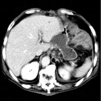
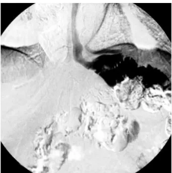
Abdomen-pelvis computed tomography demonstrated about 8 4 cm sized fluid collection in the esophagojejunostomy site suggesting intraabdominal abscess (Fig. 1). Computed tomography-guided 10-Fr locking pig-tail catheter was in- serted via transhepatic approach. Fistulography was obtained via the pig-tail catheter, which showed a fistula from the ab- scess cavity to the esophagus (Fig. 2A). Endoscopy revealed one opening in the jejunal end of Roux limb (Fig. 2B). At this point, it was believed that the best treatment avoiding surgery or long-term parenteral nutrition would be temporary place- ment of a covered metallic stent in this fistula.
Seven days after pig-tail catheter insertion, peroral place- ment of a covered metallic stent was performed. Initially the fistula was demonstrated with a small amount of contrast (Ultravist, Hankook Schering Inc., Ansan, Korea) swallow- ing. Under endoscopic guidance, a long hydrophilic guide wire (Terumo Radiofocus, Terumo Inc., Tokyo, Japan) was inserted via the oral route through the esophagus to negoti- ate the esophagojejunostomy site following oropharyngeal
*
Department of Surgery and Diagnostic Radiology*, University of Ulsan Medical College, Gangneung Asan Hospital, Gangneung-si; Department of Surgery , University of Ulsan Medical College, Seoul Asan Hospital, Seoul, Korea
Yong Pil Cho, M.D.
Department of Surgery, University of Ulsan Medical College, Gangneung Asan Hospital, 415 Bangdong- ri, Sacheon-myeon, Gangneung 210-711, Korea Tel : +82.33-610-3229, Fax : +82.33-641-8120 E-mail : [email protected]
437 J Korean Med Sci 2003; 18: 437-40
ISSN 1011-8934
Copyright The Korean Academy of Medical Sciences
Postoperative leakage is a serious complication in patients after gastric surgery.
It can lead to a rapid deterioration in the patient's condition and quality of life. Treat- ment is guided by the type of anastomosis and the patient's clinical status. The role of interventional radiology in gastrointestinal tract is evolving. Metallic stent placement has shown encouraging results for the palliation of gastrointestinal tract obstruction and fistula in malignant patients. We encountered a case of the leak- age of jejunal end of Roux limb after total gastrectomy. This patient required a drainage procedure with long-term parenteral nutrition. We performed peroral placement of a covered metallic stent to avoid surgery and long-term parenteral nutrition, and he resumed adequate oral intake immediately after stent placement.
This minimally invasive procedure is very promising for the treatment of a gastroin- testinal fistula to avoid surgery and long-term parenteral nutritional support in selected cases.
Key Words : Stents; Postoperative Complications; Gastrectomy
Received : 23 May 2002 Accepted : 15 July 2002
438 Y.P. Cho, D.H. Lee, H.J. Jang, et al.
anesthesia, which was achieved successfully with lidocaine gel. The stent (Niti-S Esophageal Covered Stent, Taewoong Medical Inc., Kyunggi-Do, Korea) and stent introducer were inserted over the guide wire. The metallic stent mesh was cov- ered with polyurethane membrane to prevent leaking through the mesh. The length of the stent body was 6 cm with a diameter of 16 mm. There were 2 cm long shoulders at each end of the stent for the prevention of stent migration. The
outer diameter of the flexible introducer catheter was 20-Fr.
The stent was fully expanded after deployment. On post- stenting contrast study, there was no significant leaking of the contrast suggesting successful sealing of the fistula (Fig.
3A). Three days after the stent placement, a follow-up con- trast study demonstrated slight migration of the stent with- out leakage of the contrast (Fig. 3B). Immediately after a fol- low-up contrast study, he resumed adequate oral intake without parenteral nutritional support. After 10 days of fol- low-up contrast study, he was discharged without complica- tion.
One month after the stent placement, follow-up abdomen- pelvis computed tomography showed no fluid collection with- out migration of the stent, and the pig-tail catheter was re- moved. Three months after the stent placement, esophago- graphy was done for the removal of the stent. However, the stent was passed out spontaneously, and esophagography demonstrated no leakage of the contrast (Fig. 4).
DISCUSSION
Postoperative leakage is a serious complication in patients after gastric surgery and can lead to a rapid deterioration in the patient's condition and quality of life. Most patients require a drainage procedure with long-term parenteral nutrition to relieve the symptoms of leakage and to improve their nutri- tional status. Treatment is guided by the type of anastomo- sis and the patient's clinical status. With a silent or localized leak, oral feeding is discontinued, and the patient is fed par- enterally or by a jejunal tube. Although minimally invasive,
Fig. 1. Initial abdomen-pelvis computed tomography demon- strates about 8 4 cm sized fluid collection in the esophago- jejunostomy site.
Fig. 2.(A) Fistulography shows a fistula from the abscess cavity to the esophagus. (B) Endoscopy reveals one opening in the jejunal end of Roux limb. (E; efferent loop, A; jejunal end of Roux limb, closed arrow; one opening in the jejunal end of Roux limb).
A B
E
A
A Covered Metallic Stent for Postoperative Leakage 439
it is often an inadequate palliative measure, because it does not allow oral intake and a longer hospital stay with a high- er overall treatment cost is required. Peritonitis and paraly- tic ileus are the decisive criteria for surgical palliation. Because most patients have an advanced disease, other unfavorable medical conditions, or advanced age, surgical palliation may be associated with significant morbidity and mortality.
The role of interventional radiology in the gastrointestinal
tract is evolving. It is increasingly supplanting surgical pal- liation as a less invasive and often more definitive modality.
Although there was no report for postoperative leakage after gastric surgery, metallic stent placement has shown encour- aging results for the palliation of malignant gastrointestinal tract obstruction and fistula (1-13). Gastrointestinal stent placement for malignant obstruction or fistula creates a more physiologic conduit than does surgical bypass, resulting in earlier resumption of oral intake, a shorter hospital stay, a lower overall treatment cost, and minimal morbidity (2, 3, 5). Furthermore, recent improvements in the design of longer and more flexible introducing systems have permitted a high- er technical success in 94-100% of the cases with malignant obstruction of the gastrointestinal tract (2). However, the rates of delayed complications including hemorrhage, stent migra- tion, perforation or fistula formation, granulomatous obstruc- tion, and stent covering disruption were substantial. Wang et al. (8) reported that the overall incidence of delayed com- plications was 64.6% after esophageal stent placement for treatment of inoperable malignant esophageal obstructions and fistulas in 82 consecutive patients. In 16 patients with malignant gastroduodenal obstruction, Lopera et al. (1) report- ed that the technical success rate was 94% with no major complications. But long-term results are not available yet and further evaluation is needed, because all patients died 1-48 weeks after stent placement.
Temporary placement of a covered metallic stent is a feasi- ble and effective method of palliation in a patient with the leakage of jejunal end of Roux limb after total gastrectomy.
In our case, the patient resumed adequate oral intake imme- diately after stent placement. Three months after the stent
Fig. 3.(A) Post-stenting contrast study shows a fully expanded stent without significant leakage of the contrast. (B) Three days after the stent placement, a follow-up contrast study demonstrates slight migration of the stent without leakage of the contrast.
A B
Fig. 4.Follow-up contrast study demonstrates that the stent has been passed out spontaneously without leakage of the contrast.
440 Y.P. Cho, D.H. Lee, H.J. Jang, et al.
placement, the stent was passed out spontaneously without complications. Although he was not inoperable, metallic stent placement resulted in earlier resumption of oral intake and a shorter hospital stay. Furthermore, it avoided surgery and surgery associated morbidity and mortality. In conclusion, this minimally invasive procedure is very promising for the treatment of a gastrointestinal fistula to avoid surgery and long-term parenteral nutritional support in selected cases.
REFERENCES
1. Lopera JE, Alvarez O, Castano R, Castaneda-Zuniga W. Initial expe- rience with Song's covered duodenal stent in the treatment of malig- nant gastroduodenal obstruction. J Vasc Interv Radiol 2001; 12:
1297-303.
2. Morgan R, Adam A. Use of metallic stents and balloons in the esoph- agus and gastrointestinal tract. J Vasc Interv Radiol 2001; 12: 283-97.
3. Yim HB, Jacobson BC, Saltzman JR, Johannes RS, Bounds BC, Lee JH, Shields SJ, Ruymann FW, Van Dam J, Carr-Locke DL. Clinical outcome of the use of enteral stents for palliation of patients with malig- nant upper GI obstruction. Gastrointest Endosc 2001; 53: 329-32.
4. Jung GS, Song HY, Kang SG, Huh JD, Park SJ, Koo JY, Cho YD.
Malignant gastroduodenal obstruction: treatment by means of a cov- ered expandable metallic stent-initial experience. Radiology 2000;
216: 758-63.
5. Mauro M, Koehler RE, Baron TH. Advances in gastrointestinal inter- ventions: the treatment of gastroduodenal and colorectal obstructions with metallic stent. Radiology 2000; 215: 659-69.
6. de Baere T, Harry G, Ducreux M, Elias D, Briquet R, Kuoch V, Roche A. Self-expanding metallic stents as palliative treatment of malignant gastroduodenal stenosis. AJR Am J Roentgenol 1997; 169: 1079-83.
7. Feretis C, Benakis P, Dimopoulos C, Georgopoulos K, Milas F, Manouras A, Apostolidis N. Palliation of malignant gastric outlet obstruction with self-expanding metal stents. Endoscopy 1996; 28:
225-8.
8. Wang MQ, Sze DY, Wang ZP, Wang ZQ, Gao YA, Dake MD.
Delayed complications after esophageal stent placement for treatment of malignant esophageal obstructions and esophagorespiratory fistu- las. J Vasc Interv Radiol 2001; 12: 465-74.
9. Dumonceau JM, Cremer M, Lalmand B, Deviere J. Esophageal fis- tula sealing: choice of stent, practical management, and cost. Gas- trointest Endosc 1999; 49: 70-8.
10. Morgan RA, Ellul JP, Denton ER, Glynos M, Mason RC, Adam A.
Malignant esophageal fistulas and perforations: management with plastic-covered metallic endoprostheses. Radiology 1997; 204:
527-32.
11. Song HY, Yang DH, Kuhn JH, Choi KC. Obstructing cancer of the gastric antrum: palliative treatment with covered metallic stents. Radi- ology 1993; 187: 357-8.
12. Strecker EP, Boos I, Husfeldt KJ. Malignant duodenal stenosis: pal- liation with peroral implantation of a self-expanding nitinol stent.
Radiology 1995; 196: 349-51.
13. Park HS, Do YS, Suh SW, Choo SW, Lim HK, Kim SH, Shim YM, Park KC, Choo IW. Upper gastrointestinal tract malignant obstruc- tion: initial results of palliation with a flexible covered stent. Radiol- ogy 1999; 210: 865-70.