INTRODUCTION
The incidence of distant metastases in non-small cell lung cancer (NSCLC) is reported in 40% or more of patients with the disease at the time of diagnosis (1, 2). In spite of the high percentage of extra-thoracic metastases, there has been no consensus recommendation for extra-thoracic metastasis eval- uation using various imaging modalities. International med- ical associations have different opinions about the use of imag- ing (3, 4). One society suggests full scanning for metastatic disease, even if patients lack symptoms and signs of metastat- ic disease (3). Another society advocates no preoperative imag- ing of the brain or skeleton in NSCLC patients without symp- toms or signs of a distant metastasis (4).
The use of integrated fluorine 18fluorodeoxyglucose (FDG) positron emission tomography (PET)/computed tomogra- phy (CT) is more efficient to assess the presence of a metas- tasis of both the mediastinal and extra-thoracic regions than the use of standard methods (5, 6). However, FDG PET or PET/CT has some limitation for brain metastasis evaluation, because it is difficult to differentiate FDG-avid metastases from the normal surrounding hyper-metabolic parenchyma in brain (7). Metastatic lesion to the brain may present in up
to 18% of patients with NSCLC (8, 9). Particularly in ade- nocarcinoma of the lung, the brain metastasis rate has been reported to be 43% (58 of 136 patients) in one study (10).
Considering the high percentage of the brain metastasis rate in lung adenocarcinoma patients and the limited efficacy of PET or PET/CT for detecting brain metastasis, PET or PET/
CT plus an additional brain imaging modality (magnetic resonance imaging [MRI]or CT) may be needed for staging of lung adenocarcinoma. The purpose of our study was to evaluate prospectively the efficacy of PET/CT plus 3T brain MRI for detecting extrathoracic metastases in lung adeno- carcinoma.
MATERIALS AND METHODS
The Institutional Review Board approved this prospective study and informed consent was obtained from all patients (2008-07-019).
Patients
From November 2003 to December 2006, 520 consecu-
1132
Ho Yun Lee1, Kyung Soo Lee1, Byung-Tae Kim2, Young-Seok Cho2, Eun Jeong Lee2, Chin A Yi1, Myung Jin Chung1, Tae Sung Kim1, O Jung Kwon3, and Hojoong Kim3
Department of Radiology and Center for Imaging Science1; Department of Nuclear Medicine2, and Division of Pulmonary and Critical Care Medicine;
Department of Internal Medicine3, Samsung Medical Center, Sungkyunkwan University School of Medicine, Seoul, Korea
Address for correspondence Kyung Soo Lee, M.D.
Department of Radiology, Samsung Medical Center, Sungkyunkwan University School of Medicine, 50 Irwon-dong, Gangnam-gu, Seoul 135-710, Korea Tel : +82.2-3410-2511, Fax : +82.2-3410-2559 E-mail : [email protected] DOI: 10.3346/jkms.2009.24.6.1132
Diagnostic Efficacy of PET/CT Plus Brain MR Imaging for Detection of Extrathoracic Metastases in Patients with Lung Adenocarcinoma
We aimed to evaluate prospectively the efficacy of positron emission tomography (PET)/computed tomography (CT) plus brain magnetic resonance imaging (MRI) for detecting extrathoracic metastases in lung adenocarcinoma. Metastatic evalua- tions were feasible for 442 consecutive patients (M:F=238:204; mean age, 54 yr) with a lung adenocarcinoma who underwent PET/CT (CT, without IV contrast medi- um injection) plus contrast-enhanced brain MRI. The presence of metastases in the brain was evaluated by assessing brain MRI or PET/CT, and in other organs by PET/CT. Diagnostic efficacies for metastasis detection with PET/CT plus brain MRI and with PET/CT only were calculated on a per-patient basis and compared from each other. Of 442 patients, 88 (20%, including 50 [11.3%] with brain metas- tasis) had metastasis. Regarding sensitivity of overall extrathoracic metastasis detec- tion, a significant difference was found between PET/CT and PET/CT plus brain MRI (68% vs. 84%; P=0.03). As for brain metastasis detection sensitivity, brain MRI was significantly higher than PET/CT (88% vs. 24%; P<0.001). By adding MRI to PET/CT, brain metastases were detected in additional 32 (7% of 442 patients) pati- ents. In lung adenocarcinoma patients, significant increase in sensitivity can be achieved for detecting extrathoracic metastases by adding dedicated brain MRI to PET/CT and thus enhancing brain metastasis detection.
Key Words : PET/CT Scan; Lung Neoplasms; Neoplasm Metastasis; Brain; Magnetic Resonance Imaging;
Neoplasm Staging
Received : 20 November 2008 Accepted : 16 January 2009
tive patients with histopathologically (cytology or biopsy) proven adenocarcinoma of the lung (except for bronchioloalve- olar cell carcinoma which mediastinal nodal or extra-thoracic metastasis is unusual) were enrolled. All patients were referred for cancer staging of lung adenocarcinoma. Of these 520 pa- tients, 78 patients were excluded for the following reasons:
58 patients were unable to undergo brain MRI for the ini- tial staging, and 20 patients were lost to follow-up. Ultimate- ly, 442 patients (M:F=238:204; mean age, 54 yr) were includ- ed in this study, in whom both FDG PET/CT and dedicated brain MRI were performed for the initial staging and meta- static evaluations of M staging were feasible with pathologi- cal results or follow-up imaging studies.
Imaging and interpretation
The presence of metastases in the brain was evaluated by using brain MRI (Achieva, Philips Medical Systems, Best, The Netherlands) and PET/CT (Discovery LS; GE Medical Systems, Milwaukee, WI, U.S.A.), and the presence of metas- tases in other organs was determined by the use of PET/CT.
PET/CT and MRI were performed within a one-week time interval (average time interval, 3.1 days; range, 0-7 days).
Detailed imaging methods of integrated PET/CT have been described in previous reports (5, 11). Briefly, glucose level in peripheral blood was ≤150 mg/dL in all patients. The patients received an intravenous injection of 370 MBq (10 mCi) of FDG and then rested for over 45 min before scan- ning. PET/CT device consisted of a PET scanner (Advance NXi; GE Healthcare) and an eight-slice CT scanner (Light- Speed Plus; GE Healthcare). Immediately after taking nonen- hanced CT, emission PET was performed in the identical transverse field of view. Co-registered images were displayed by Xeleris software (GE Healthcare). It took approximately 40 min to complete a PET/CT study. All brain MRI studies were performed by using a 3-Tesla scanner with a standard head coil. Brain MR images were obtained in the axial, sagit- tal, and coronal planes by using three sequences including a T2-weighted axial turbo spin-echo pulse sequence (repetition time 3,000 ms, echo time 80 ms) with fat suppression, a fluid- attenuation inversion-recovery (FLAIR) spin-echo sequence (repetition time 11,000 ms, echo time 125 ms, inversion time 2,800 ms) and a non-contrast enhanced and a contrast-en- hanced T1-weighted spin-echo sequence (repetition time 500 ms, echo time 10 ms). The contrast-enhanced sequence was obtained after bolus injection of a dose of 0.2 mM/kg paramagnetic contrast agent (Magnevist, Schering, Berlin, Germany).
A chest radiologist with 18 yr of experience in CT inter- pretation and a nuclear medicine physician with four years of PET/CT interpretation jointly evaluated the integrated PET/
CT images. Both clinicians were unaware of the brain MRI findings or the clinical and pathological evaluation results.
An abnormal focal FDG uptake that accompanied a corre-
sponding anatomic alteration was considered as the indica- ton of a metastasis (5, 11). The maximum standardized up- take value (SUVmax) of the focal metastatic lesion was mea- sured. All lymph nodes with abnormal FDG uptake (greater than mediastinal blood pool uptake) in the extrathoracic regions were considered as metastatic, unless they showed high attenuation over 70 HU or benign calcification (cen- tral nodular, laminated, popcorn-like and diffuse) at unen- hanced CT (12). Two chest radiologists with three years of experience in whole-body MRI analysis, who were unaware of the clinical, PET/CT findings or histologic diagnoses, inter- preted the brain MR images and decisions on findings were made by consensus. When the two chest radiologists had different opinions on the presence of brain metastasis, they sought for the third opinion from neuro-imaging radiologists and by a majority the presence or absence of brain metasta- sis was determined.
The probability of the presence of metastases on a per-patient basis was then evaluated by the use of a five-point visual scor- ing system: 0, definitely absent; 1, probably absent; 2, pos- sibly present; 3, probably present; 4, definitely present. Before interpreting the images, the reviewers were informed that the categorization of confidence levels of 2 or higher belonged to a category of positive diagnosis for a metastasis. The largest diameter of the detected metastatic nodules on brain MR images and CT component images of PET/CT was measured and recorded.
Reference standard for metastases
The final decisions on the presence of extra-thoracic metas- tases in each patient were reached based on the results of ded- icated standard imaging, a pathologic examination, or a fol- low-up examination.
Metastatic lesions were confirmed at 121 organ sites from 88 (20%) of the 442 patients. Sixteen lesions of metastases were pathologically proven. Ninety-four metastatic lesions were confirmed at the time of initial staging by using the use of specific organ-dedicated imaging studies, i.e., dedicated MRI at the time of staging for 58 lesions, dedicated CT for 21 lesions, and bone scan for 15 lesions. The remaining 11 lesions were detected by follow-up dedicated imaging studies in six months or more, and the lesions were regarded as meta- static (growth from a microscopic metastatic focus) because most of the primary cancers in these patients were resected surgically after the initial staging work-up.
The absence of a metastasis at 18 sites (from 18 patients) which were suspected as metastatic at the initial clinical-imag- ing studies was proven by following methods: negative biopsy results for metastasis (n=5); surgical excision (n=1) of a sus- pected adrenal lesion; organ-specific multiphase CT (n=2) for adrenal lesions; negative bone scan results (n=2); and nega- tive results for brain metastasis (n=2) by the use of dedicat- ed conventional MRI and three times of cerebrospinal fluid
tapping. In the remaining six patients, the findings of clini- co-laboratory follow-up studies served as reference standards for determining the absence of a metastasis. The mean clini- cal follow-up time for these six patients was 36 months (range, 30-41 months).
Statistical analysis
The diagnostic efficacy of PET/CT plus brain MRI was cal- culated on a per-patient basis. Receiver operating character- istic (ROC) curve analysis was used to compare diagnostic capabilities and it was compared by using a Z test, as describ- ed by Hanley and McNeil (12). Sensitivity, specificity, and accuracy of the two methods were statistically compared by use of the McNemar’s test. Statistical differences in the size of PET-detected and PET-missed metastatic lesions were com-
pared by use of the Mann-Whitney U test. A P value <0.05 was considered as a significant difference.
RESULTS Patient demographics
Details of the patient characteristics are summarized in Table 1. Extrathoracic metastatic lesions were confirmed at 121 organ sites for 88 (20%) of the 442 patients. Of the 50 patients with a brain metastasis, 30 (60%) had one organ (brain) metastasis and 35 (70%) patients had a single lesion metastasis. Sixteen (53%) of 30 patients with single organ metastasis to brain had early stage of cancer (stage I or II ex- cluding brain evaluation) (Table 2). Seven (14%) patients had neurologic symptoms or signs (headache, dizziness, vomiting or a neurological deficit) at the time of initial presentation.
Efficacy of MRI and PET/CT for metastasis detection The diagnostic efficacies of the two modalities are sum-
Parameters Numbers
Age (yr)
Mean 54
Range 23-88
Sex (cases)
Male 238
Female 204
Stage (cases)
I 94
II 67
III 157
IV 124
Site-specific metastases (sites)
Brain 50 (11%)
Bone 35 (8%)
Extrathoracic lymph nodes 14 (3%)
Adrenal 10 (2%)
Liver 5 (1%)
Soft tissue 5 (1%)
Kidney 1 (0.2%)
Bowel 1 (0.2%)
Table 1. Characteristics of subjected patients
TN stage Clinical stage
excluding brain evaluation n (%)
T1N0 IA 4 (13)
T2N0 IB 8 (27)
T1N1 IIA 2 (7)
T2N1 IIB 2 (7)
T1N2 IIIA 2 (7)
T2N2 IIIA 2 (7)
T3N1 IIIA 1 (3)
T3N2 IIIA 2 (7)
T4 or N3 IIIB 7 (23)
Total 30
Table 2. Tumor and nodal stage in patients with single organ metastasis to brain
Variables Brain MRI PET/CT P values
Az value <0.001*
Value 0.935 0.619
95% CI 0.881, 0.989 0.525, 0.713 Sensitivity (%)�
No neurologic 86 (37/43) 19 (8/43) <0.001� sign or symptom
Total 88 (44/50) 24 (12/50) <0.001� Specificity (%)� 98 (385/392) 100 (391/392) 0.26� Accuracy (%)� 97 (429/442) 91 (403/442) 0.12� Table 3. ROC analysis and comparison of capability of detec- tion of brain metastases on a per-patient basis
*P values were calculated by using the Z test; �Data in parentheses are the values used to calculate percentages; �P values were calculated by using McNemar’s test.
Az, area under the ROC curve; CI, confidence interval.
Diagnostic
values PET/CT alone PET/CT plus
Brain MRI P values
Az value 0.106*
Value 0.830 0.896
95% CI 0.769, 0.890 0.850, 0.94
Sensitivity (%)� 68 (60/88) 84 (74/88) 0.03� Specificity (%)� 98 (346/354) 95 (337/354) 0.22� Accuracy (%)� 92 (406/442) 93 (411/442) 0.80� Table 4. ROC analysis and sensitivity and specificity in detec- tion of all extrathoracic metastases
*P values were calculated by using Z test; �Data in parentheses are the values used to calculate the percentage; �P values were calculated by using McNemar’s test.
Az, area under the ROC curve; CI, confidence interval.
marized in Tables 3, 4. By adding contrast enhanced MRI to PET/CT, brain metastases were detected in additional 32 (7.2% of 442 patients; 44 in MRI plus PET/CT vs. 12 in PET/CT only) patients, in whom the treatment modality should have been changed. For the assessment of brain metas- tases on a per-patient basis, brain MRI showed a significantly greater area under the ROC curve (Az) than PET/CT (0.985
vs. 0.619; P<0.001) did. In terms of sensitivity for the detec- tion of brain metastasis, a significant difference was found between brain MRI and PET/CT (88% vs. 24%; P<0.001).
For the neurologically asymptomatic cases, the sensitivity of brain MRI was significantly higher than that of PET/CT (86%
vs. 19%; P<0.001). Sizes (measured on enhanced T1-weight- ed images of brain MRI) of PET-detected brain metastases
Numbers are per patient.
*Maximal size of the brain metastatic lesions.
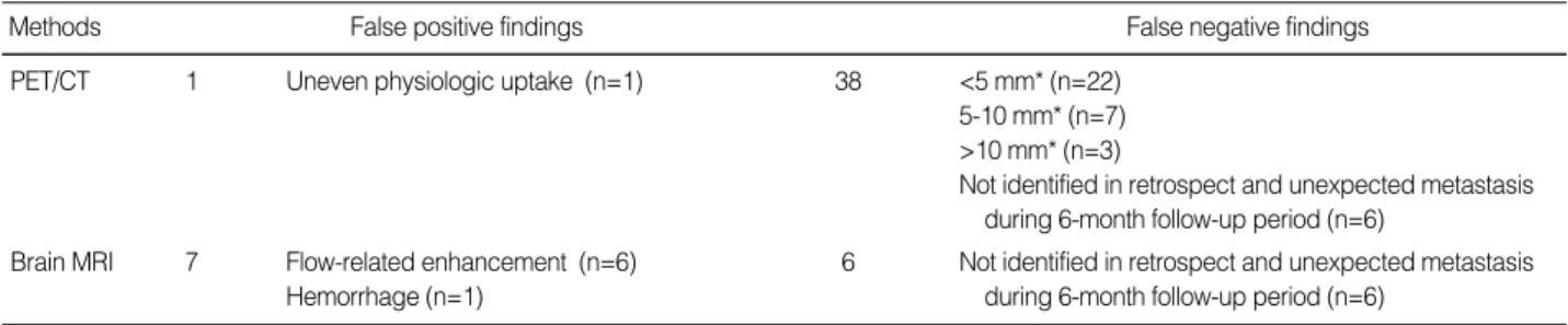
False positive findings
Methods False negative findings
PET/CT 1 Uneven physiologic uptake (n=1) 38 <5 mm* (n=22)
5-10 mm* (n=7)
>10 mm* (n=3)
Not identified in retrospect and unexpected metastasis during 6-month follow-up period (n=6)
Brain MRI 7 Flow-related enhancement (n=6) 6 Not identified in retrospect and unexpected metastasis
Hemorrhage (n=1) during 6-month follow-up period (n=6)
Table 5. Causes of false-positive and false-negative lesions as determined by brain MRI and PET/CT
Fig. 1. A 42-yr-old woman with lung adenocarcinoma and brain metastases. Contrast-enhanced T1-weighted brain MR image (A) clearly demonstrates the presence of cerebellar metastasis (arrow). PET/CT and PET images (B, C) show decreased FDG-uptake.
A B C
Fig. 2. A 27-yr-old man with lung adenocarcinoma and multiple brain metastases. Contrast-enhanced T1-weighted brain MR image (A) shows the presence of multiple small metastases less than 5 mm in diameter (arrows). However, axial images (B, C) of PET/CT and PET images do not indicate the abnormal FDG-uptaking lesion.
A B C
were significantly larger (mean, 27 mm; range, 10-47 mm) than those of PET-missed lesions (mean, 6 mm; range, 1-28 mm) (P=0.002).
ROC analysis determined that PET/CT plus brain MRI was superior to PET/CT for the detection of overall extrathoracic metastases, although statistical significance was not reached (0.896 vs. 0.830; P=0.106). In terms of sensitivity for the detection of overall extrathoracic metastases, a significant dif- ference was found between PET/CT and PET/CT plus brain MRI (68% vs. 84%; P=0.03).
False-negative and false-positive
A listing of false-positive and false-negative cases for brain metastases of lung adenocarcinoma patients identified by means of the two methods is shown in Table 5. Representative cases are shown in Figs. 1, 2. PET/CT could not depict 22 metastat- ic foci of <5 mm in diameter. At brain MRI, all false nega- tive lesions were noted during the follow-up period and were not identified at the initial imaging study, retrospectively.
DISCUSSION
Preoperative evaluation with diagnostic imaging for brain metastases in NSCLC patients remains controversial issue (14, 15). One meta-analysis in patients with no neurologi- cal abnormality on a clinical examination demonstrated the negative predictive value in this setting as 95% (16). In con- trast, a recent report by Jena et al. (17) stated that an asymp- tomatic brain metastasis occurs in 17% (29 of 175; 158 [90%] of 175 patients had stage IV cancer) of lung cancer patients with almost equal number of symptomatic (33 of 62 [53%] patients with a brain metastasis) and asymptomatic (29 of 62 [47%]patients) metastases. In the current study (relatively even distribution of patient cancer stages, 124 [28%]of 442 patients had stage IV cancers), 50 (11%) of the 442 lung ade- nocarcinoma patients had a brain metastasis, and of these 50 patients, 30 (60%) patients had one organ (brain) metastasis only and 35 (70%) patients had a single lesion metastasis.
Only seven (14%) patients complained of any neurological symptoms and signs.
The brain is often the only site of distant metastatic disease and approximately one-third of patients will have limited, potentially resectable lung tumors without distant metastases other than a brain metastasis (18). In the most recent ACCP guidelines (19), the use of routine staging of the central ner- vous system (CNS) system is considered in level 2C for stage III malignancies. However, 53% of the patients with single metastasis to brain in our study had stage I or II lung ade- nocarcinoma. The prognosis in patients with a brain metas- tasis who are untreated is extremely poor (about one month survival after diagnosis), whereas patients with NSCLC who were treated with local radiation therapy survived for about
eight months (20, 21). Therefore, early and accurate diagno- sis of a brain metastasis is crucial to improve quality of life and the poor survival rates of lung cancer patients.
For the assessment of brain metastases on a per-patient basis, our results showed that the Az value of brain MRI is significantly larger than that of PET/CT. In addition, the sen- sitivity of brain MRI is significantly higher than that of PET/
CT. This result is in accord with findings of recent studies by Ohno et al. (21) and Yi et al. (22), which has found higher sensitivity of MRI than FDG PET or PET/CT for the detec- tion of brain metastasis is determined. The disparity in sen- sitivity between brain MRI and PET/CT can be caused by the extremely high level of physiological tissue accumula- tion of FDG in the cerebral cortex as depicted at PET, which hinders the identification of metastatic disease in the brain, and by the insufficient spatial resolution of PET for the detec- tion of a metastasis less than 5 mm.
On a per-patient basis, all false negative lesions on brain MRI were also assessed as false negative on PET, and there was no lesion which was detected on PET. The lesions were regarded as metastatic (growth from a microscopic metastat- ic focus) because all lesions were detected by follow-up stud- ies in six months or more. Considering these results, torso PET plus brain MRI would provide enough information about brain metastasis and appears to be efficient for M stag- ing in NSCLC patients as much as brain and body PET plus brain MRI.
In our study, by adding contrast enhanced MRI to PET/
CT, brain metastases were detected in additional 32 (7% of 442) patients, in whom treatment modality should have been changed. Therefore, imaging surveillance of the brain for metastases is worthwhile in the context that neurologically asymptomatic metastases can be detected early to provide appropriate therapy, especially in patients with lung adeno- carcinoma where asymptomatic metastases are more frequent than in other NSCLC (23-25).
Our study has several limitations. First and most impor- tantly, because we did not administer IV contrast medium for CT image acquisition at PET/CT, CT component images might have played a role as transmission data only image coregistration and have provided little morphologic infor- mation for metastasis detection. This might have contribut- ed to lowering sensitivity for detecting brain or other organ (e.g., hepatic, renal, and splenic) metastasis at PET/CT. There- fore, our comparison is more of PET alone versus PET plus MRI brain, rather than PET/CT alone versus PET/CT plus MRI brain. Obtaining contrast-enhanced CT component images at PET/CT would provide more precise anatomic information for abnormal lesions by enhancing attenuation differences between lesions and surrounding normal struc- tures especially in such cases as brain, hepatic, renal or splenic metastasis. However, controversies on having enhanced CT component of PET/CT are still ongoing, because concerns over contrast medium injection in terms of technical, eco-
nomic, work-flow, and radiation-safety issues has been raised.
Second, we were unable to make a pathologic diagnosis of an extrathoracic metastasis at a given site in every patient.
This may suggest that the sensitivity and specificity values in the current study might be biased. Third, brain MRI is not routinely included as a standard initial staging workup in clinical practice at other institutions and we did not cal- culate the cost-effectiveness of the addition of brain MRI to PET/CT. A further study needs to be performed as to the cost- effectiveness on our protocol for the addition of brain MRI to PET or PET/CT.
In conclusion, with the addition of dedicated brain MRI to PET/CT and thus with enhanced brain metastasis detection, a significant increase in diagnostic sensitivity can be achieved for detecting extrathoracic metastases in patients with lung adenocarcinoma. Thus, adding dedicated brain MRI to PET/
CT appears to be efficient for M staging in patients who have a histopathologically confirmed lung adenocarcinoma.
REFERENCES
1. Boring CC, Squires TS, Tong T. Cancer Statistics, 1992. CA Can- cer J Clin 1992; 42: 19-38.
2. Jemal A, Thomas A, Murray T, Thun M. Cancer statistics, 2002. CA Cancer J Clin 2002; 52: 23-47.
3. The Canadian Lung Oncology Group. Investigating extrathoracic metastatic disease in patients with apparently operable lung cancer.
The Canadian Lung Oncology Group. Ann Thorac Surg 2001; 71:
425-33.
4. The American Thoracic Society and the European Respiratory Soci- ety. Pretreatment evaluation of non-small-cell lung cancer. The Amer- ican Thoracic Society and the European Respiratory Society Con- sensus Report. Am J Respir Crit Care Med 1997; 156: 320-32.
5. Shim SS, Lee KS, Kim BT, Chung MJ, Lee EJ, Han J, Choi JY, Kwon OJ, Shim YM, Kim S. Non-small cell lung cancer: prospective com- parison of integrated FDG PET/CT and CT alone for preoperative staging. Radiology 2005; 236: 1011-9.
6. De Wever W, Vankan Y, Stroobants S, Verschakelen J. Detection of extrapulmonary lesions with integrated PET/CT in the staging of lung cancer. Eur Respir J 2007; 29: 995-1002.
7. Marom EM, McAdams HP. Erasmus JJ, Goodman PC, Culhane DK, Coleman RE, Herndon JE, Patz EF Jr. Staging non-small cell lung cancer with whole-body PET. Radiology 1999; 212: 803-9.
8. Mintz BJ, Tuhrim S, Alexander S, Yang WC, Shanzer S. Intracranial metastases in the initial staging of bronchogenic carcinoma. Chest 2004; 86: 850-3.
9. Newman SJ, Hansen HH. Proceedings: frequency, diagnosis, and treatment of brain metastases in 247 consecutive patients with bron- chogenic carcinoma. Cancer 1974; 33: 492-6.
10. Mujoomdar A, Austin JH, Malhotra R, Powell CA, Pearson GD, Shiau MC, Raftopoulos H. Clinical predictors of metastatic disease to the brain from non-small cell lung carcinoma: primary tumor size, cell type, and lymph node metastases. Radiology 2007; 242: 882-8.
11. Kim YK, Lee KS, Kim BT, Choi JY, Kim H, Kwon OJ, Shim YM, Yi CA, Kim HY, Chung MJ. Mediastinal nodal staging of nonsmall cell lung cancer using integrated 18F-FDG PET/CT in a tuberculosis- endemic country: diagnostic efficacy in 674 patients. Cancer 2007;
109: 1068-77.
12. Lee EJ, Choi JY, Lee KS, Chung HW, Lee SJ, Cho YS, Choi Y, Choe YS, Lee KH, Kwon OJ, Shim YM, Kim BT. Improving diagnostic accuracy for malignant nodes and N staging in non-small cell lung cancer using CT-corrected FDG-PET. Korean J Nucl Med 2005;
39: 231-8.
13. Hanley JA, McNeil BJ. A method comparing the areas under receiv- er operator characteristic curves derived from the same cases. Radi- ology 1983; 148: 839-43.
14. Hillers TK, Sauve MD, Guyatt GH. Analysis of published studies on the detection of extrathoracic metastases in patients presumed to have operable non-small cell lung cancer. Thorax 1994; 49: 14-9.
15. Silvestri GA, Littenberg B, Colice GL. The clinical evaluation for detecting metastatic lung cancer: a meta-analysis. Am J Respir Crit Care Med 1995; 152: 225-30.
16. Toloza EM, Harpole L, McCrory DC. Noninvasive staging of non- small cell lung cancer: a review of the current evidence. Chest 2003;
123: 137S-46S.
17. Jena A, Taneja S, Talwar V, Sharma JB. Magnetic resonance (MR) patterns of brain metastasis in lung cancer patients: correlation of imaging findings with symptom. J Thorac Oncol 2008; 3: 140-4.
18. Yokoi K, Kamiya N, Matsuguma H, Machida S, Hirose T, Mori K, Tominaga K. Detection of brain metastasis in potentially operable non-small cell lung cancer: a comparison of CT and MRI. Chest 1999; 115: 714-9.
19. Silvestri GA, Gould MK, Margolis ML, Tanoue LT, McCrory D, Toloza E, Detterbeck F; American College of Chest Physicians. Non- invasive staging of non-small cell lung cancer: ACCP evidenced- based clinical practice guidelines (2nd edition). Chest 2007; 132 (3 Suppl): S178-S201.
20. Sorensen JB, Hansen HH, Hansen M, Dombernowsky P. Brain metas- tases in adenocarcinoma of the lung: frequency, risk groups, and prognosis. J Clin Oncol 1988; 6: 1474-80.
21. Zabel A, Milker-Zabel S, Thilmann C, Zuna I, Rhein B, Wannen- macher M, Debus J. Treatment of brain metastasis in patients with non-small cell lung cancer (NSCLC) by stereotactic linac-based radiosurgery: prognostic factors. Lung Cancer 2002; 37: 87-94.
22. Ohno Y, Koyama H, Nogami M, Takenaka D, Yoshikawa T, Yo- shimura M, Kotani Y, Nishimura Y, Higashino T, Sugimura K. Whole- body MR imaging vs. FDG-PET: comparison of accuracy of M-stage diagnosis for lung cancer patients. J Magn Reson Imaging 2007;
26: 498-509.
23. Yi CA, Shin KM, Lee KS, Kim BT, Kim H, Kwon OJ, Choi JY, Chung MJ. Non-small cell lung cancer staging: efficacy compari- son of integrated PET/CT versus 3.0-T whole-body MR imaging.
Radiology 2008; 248: 632-42.
24. Cox JD, Yesner R. Adenocarcinoma of the lung: recent results from the veterans administration lung group. Am Rev Respir Dis 1979;
120: 1025-9.
25. Cox JD, Scott CB, Byhardt RW, Emami B, Russell AH, Fu KK, Par-
liament MB, Komaki R, Gaspar LE. Addition of chemotherapy to radiation therapy alters failure patterns by cell type within non-small cell carcinoma of lung (NSCCL): analysis of radiation therapy oncol- ogy group (RTOG) trials. Int J Radiat Oncol Biol Phys 1999; 43:
505-9.
26. Robnett TJ, Machtay M, Stevenson JP, Algazy KM, Hahn SM. Fac-
tors affecting the risk of brain metastases after definitive chemoradia- tion for locally advanced non-small-cell lung carcinoma. J Clin Oncol 2001; 19: 1344-9.
27. Davis PC, Hudgins PA, Peterman SB, Hoffman JC Jr. Diagnosis of cerebral metastases: double-dose delayed CT vs contrast enhanced MR imaging. AJNR Am J Neuroradiol 1991; 12: 293-300.