gations revealed a nodular mass, located in the sella and suprasellar portion and accompanied by compression of the optic chiasm. The mass compressed the bilat- eral cavernous sinuses, resulting in the obliteration of the cavernous portion of the right internal carotid artery. A border zone infarct in the right fronto-parietal region was found. Transsphenoidal tumor decompression following conservative therapy with fluid replacement and steroids was performed. Pathological examination reveal- ed an almost completely infarcted pituitary adenoma. The patient’s vision improved immediately after the decompression, and the motor weakness improved to grade IV+within six months after the operation. Pituitary apoplexy resulting in internal carotid artery occlusion is rare. However, clinicians should be aware of the possi- bility and the appropriate management of such an occurrence.
Key Words : Pituitary Apoplexy; Cerebral Infarction; Cerebrovascular Disorders; Paresis
INTRODUCTION
Pituitary apoplexy is a well-known clinical syndrome characterized by headache, meningeal irritation, visual loss, ophthalmoplegia, and alterations in consciousness (1). Cere- bral infarction associated with pituitary apoplexy is rare. In the present report, we report a rare case of a 43-yr-old man with pituitary apoplexy presenting with hemiplegia.
CASE REPORT
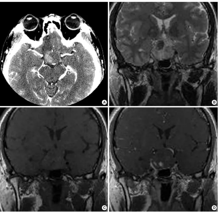
A 43-yr-old man presented with a sudden onset of severe headache, visual disturbance and left hemiplegia. He was lethargic and unable to walk on his own. The initial neuro- logical examination showed an impaired direct/indirect light reflex of the right eye and a right third nerve palsy, accompa- nied by anisocoria and ptosis. His motor power was grade II on the left side. Computed tomography (CT) scans of the brain showed an enlarged pituitary fossa containing a hem- orrhagic pituitary tumor (Fig. 1A). Magnetic resonance imag- ing (MRI) revealed a nodular mass, approximately 3×2×3 cm in size, located in the sella and suprasellar portion, accom- panied by compression of the optic chiasm (Fig. 1B-D). The mass compressed the bilateral cavernous sinuses, resulting in the obliteration of the cavernous portion of the right inter- nal carotid artery (Fig. 2A). A border zone infarct in the right
fronto-parietal region was also found (Fig. 2B).
The patient was initially treated with fluid replacement and steroids. Although the patient’s level of consciousness improved during the next 24 hr, the focal neurological signs persisted. Transsphenoidal tumor decompression was per- formed within four days of symptom onset. The patient’s vision improved immediately after the decompression, but the left hemiplegia persisted.
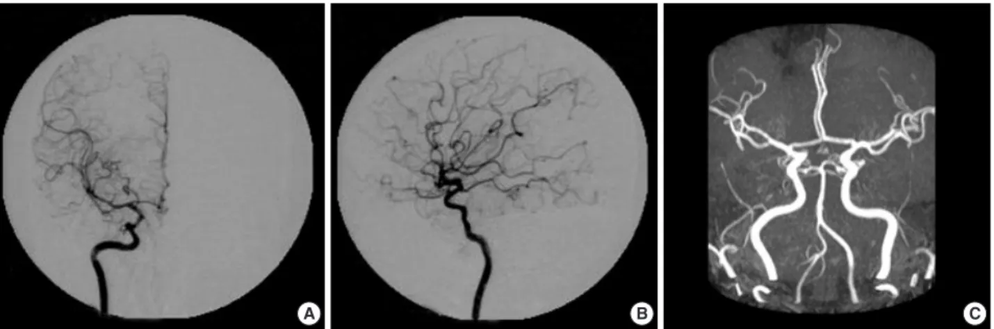
Pathological examination revealed an almost completely infarcted pituitary adenoma (Fig. 3). A conventional cere- bral angiography performed one week after the operation and MR angiography demonstrated the restoration of flow within the right internal carotid artery (Fig. 4). His left side motor power improved to grade IV+within six months after the operation.
DISCUSSION
The occurrence of cerebral ischemia, or infarct, in patients with pituitary apoplexy is rare, with only 13 cases reported thus far. The ischemic events were attributed to cerebral vasospasm in six cases (2-6) and to mechanical compression by tumor in seven cases (7-13). The pathophysiology of vasospasm following pituitary apoplexy remains unclear. How- ever, several hypotheses have been proposed to account for vasospasm following surgery for pituitary adenoma and other
1113
Department of Neurosurgery, St. Vincent’s Hospital, Suwon; Departments of Neurosurgery* and Hospital Pathology�, Kangnam St. Mary’s Hospital, The Catholic University of Korea, Seoul, Korea
Address for correspondence Yong-Kil Hong, M.D.
Department of Neurosurgery, Kangnam St. Mary’s Hospital, The Catholic University of Korea, 505 Banpo-dong, Seocho-gu, Seoul 137-701, Korea Tel : +82.2-590-2732, Fax : +82.2-594-4248 E-mail : [email protected]
Received : 23 July 2007 Accepted : 27 December 2007
parasellar tumors (14, 15), such as the presence of subarach- noid blood, the release of vasoactive chemical substances from the tumor, hypothalamic damage and dysfunction, and intra- operative manipulation. Bilateral involvement of the intrac- erebral arteries was common in cases of cerebral vasospasm.
In our case, the restoration of cerebral blood flow follow- ing the decompression of the apoplexy was confirmed angio- graphically. The characteristics of patients whose ischemic events were attributed to the mechanical compression of the cerebral arteries by a pituitary apoplexy are summarized in Table 1. Pituitary apoplexy was associated with both a cere-
bral angiography and a triple bolus test with the intravenous injection of luteinizing hormone-releasing hormone, thy- rotropin-releasing hormone and regular insulin (7, 11). The occlusion sites of the compromised vessels were primarily located in the supraclinoid or cavernous portions of the inter- nal carotid artery. Theoretically, the pressure of the pituitary apoplexy should be stronger than the intraarterial pressure for the mechanical compression. Intrasellar pressure mea- surements in patients with pituitary apoplexy were conduct- ed in a previous study (16). The measurements ranged from 25-58 mmHg, with a median pressure of 47 mmHg. More-
Fig. 1. Computed tomography scan (A) showing a heterogeneous mass of high density in the right side. Coronal magnetic resonance (MR) image showing heterogeneous high-signal intensity on the T2-weighted image (B), slightly elevated signal intensity on the T1-weighted image (C) and focal enhancement with gadolinium (D), suggesting a necrotic or hemorrhagic site.
A
C D
B
over, the tight dural attachment of the cavernous and clinoid segments in addition to the surrounding bony structures could create additional pressure and a barrier that occludes the compensatory space of the compromised vessels, resulting in further mechanical compression. Therefore, the primary goal of management is to reduce the intratumoral pressure.
Emergent decompression was performed in two cases (9, 11) of pituitary apoplexy associated with cerebral infarction.
The decompression of the internal carotid artery resulted in
a patent vessel, but the procedure was also likely to result in the hemorrhagic transformation of the infarct area. The com- bined effect of the infarct and the brain edema was usually the cause of mortality in patients who underwent emergent decompression. On the other hand, delayed decompression following conservative therapy with steroids was associated with better outcomes in patients with cerebral infarct. We performed delayed surgery following steroid administration in our patient, which resulted in the immediate improve-
Fig. 2. Non-visualization of the right internal carotid artery on MRA (A) and linear high signal intensity in the right fronto-parietal region on a diffusion-weighted image (B).
A B
Fig. 3. Microscopic examination showing the infarct region in which viable cells are seen only around the vascular channels (H&E, ×100 [A] and ×200 [B]).
A B
ment of the patient’s vision. The patient’s motor weakness steadily recovered with physiotherapy, and the patient is now capable of walking without assistance.
If the pituitary apoplexy is associated with cerebral ischemia rather than infarct, as in the case described by Bernstein et al. (7), early operative management to restore the flow in the carotid artery may result in the resolution of the neurological deficits. Motor weakness following pituitary apoplexy is rare, but clinicians should pay close attention to the initial images in order to differentiate between infarct and ischemia in cases of pituitary apoplexy.
REFERENCES
1. Dubuisson AS, Beckers A, Stevenaert A. Classical pituitary tumour
apoplexy: clinical features, management and outcomes in a series of 24 patients. Clin Neurol Neurosurg 2007; 109: 63-70.
2. Akutsu H, Noguchi S, Tsunoda T, Sasaki M, Matsumura A. Cere- bral infarction following pituitary apoplexy-case report. Neurol Med Chir (Tokyo) 2004; 44: 479-83.
3. Cardoso ER, Peterson EW. Pituitary apoplexy and vasospasm. Surg Neurol 1983; 20: 391-5.
4. D’Haens J, Baleriaux D, Mockel J, Flamment-Durand J, Brotchi J.
Ischemic pituitary apoplexy and cerebrovascular accident. Neu- rochirurgie 1983; 29: 401-5.
5. Itoyama Y, Goto S, Miura M, Kuratsu J, Ushio Y, Matsumoto T.
Intracranial arterial vasospasm associated with pituitary apoplexy after head trauma-case report. Neurol Med Chir (Tokyo) 1990; 30:
350-3.
6. Pozzati E, Frank G, Nasi MT, Giuliani G. Pituitary apoplexy, bilat- eral carotid vasospasm, and cerebral infarction in a 15-year-old Fig. 4. Anteroposterior (A) and lateral view (B) of a conventional angiography performed one week after the operation, and MRA (C) show- ing the restoration of cerebral blood flow.
A B C
*, Not described.
MCA, middle cerebral artery; ICA, internal carotid artery; ACA, anterior cerebral artery; TSA, transsphenoidal approach.
Authors Compromised
vessel
Radiological
finding Treatment Associated
factor Motor symptom
Age/sex Outcome
Schnitker et al. 65/M Left hemiplegia - Right MCA ND* Conservative Death
(1952) care
Sakalas et al. 9/M Lethargy - Left ICA ND craniotomy Good
(1973) (supraclinoid portion)
Rosenbaum et al. 77/M Left hemiparesis Angiography Right ICA Infarct Emergent Death
(1977) (supraclinoid portion) craniotomy
Majchrzak et al. 29/M Transient left - Right ACA Ischemia Delayed Good
(1983) hemiparesis craniotomy
Bernstein et al. 48/M Left hemiparesis Triple bolus test Bilateral ICA No evidence TSA Good
(1984) (cavernous portion) of infarct
Clark et al. 40/M Right hemiplegia - Left ICA Infarct Conservative Extensive
(1987) (supraclinoid portion) care infarct
Lath et al. 40/M Left hemiplegia - Right ACA Infarct Emergent Death
(2001) TSA
Present case 43/M Left hemiplegia - Right ICA Infarct TSA Good
(2007) (cavernous portion)
Table 1. Reported cases of mechanical compression of major vessels following pituitary apoplexy
11. Rosenbaum TJ, Houser OW, Laws ER. Pituitary apoplexy produc- ing internal carotid artery occlusion. Case report. J Neurosurg 1977;
47: 599-604.
lar pressure in patients with pituitary apoplexy: relation to pituitary function. J Clin Endocrinol Metab 2004; 89: 5649-54.
![Fig. 3. Microscopic examination showing the infarct region in which viable cells are seen only around the vascular channels (H&E, ×100 [A] and ×200 [B]).](https://thumb-ap.123doks.com/thumbv2/123dokinfo/5131700.89809/3.892.94.803.535.883/microscopic-examination-showing-infarct-region-viable-vascular-channels.webp)