INTRODUCTION
Invasive fungal infections play a key role in contributing to morbidity and mortality in hospitalized patients, especial- ly those who are profoundly immunocompromised such as patients on bone marrow transplantation (BMT). Although a variety of yeasts and molds can cause invasive fungal infec- tions, Candida and Aspergillus species account for the majori- ty of the infections. Recently, other less common fungal iso- lates, such as Zygomycetes, are recognized as opportunistic pathogens with a increasing frequency (1, 2).
Mucormycosis is an opportunistic infection caused by fungi of the order Mucorals, class Zygomycetes. Fungal spores are dispersed into the air from decaying material, and the portal of entry for human infections is via the inhalation route. Zygomy- cetes has a tendency for vascular invasion and tissue necrosis, with resultant hemorrhage and necrotic lesions. The major clinical diseases can be divided into five forms according to the site of involvement: rhinocerebral, pulmonary, gastroin- testinal, cutaneous, and disseminated mucormycosis. Dis- seminated mucormycosis is an extremely rare condition with high mortality rate and generally occurs in severely immu- nocompromised patients (3-5).
Several cases of mucormycosis were reported in Korea, in- cluding disseminated form after chemotherapy in a patient with acute lymphocytic leukemia (6). Now, we first report two cases of disseminated mucormycosis following BMT in
Korea. One was successfully treated with combination of sur- gery, conventional and liposomal amphotericin B with dis- continuing immunosuppressants and the other died despite surgery and amphotericin B. In the latter patient, immuno- suppressants could not be discontinued due to extensive graft- versus-host disease (GVHD).
CASE REPORTS CASE 1
A 35-yr-old man was admitted through emergency room due to sudden development of high fever and left flank pain.
Thirteen months before he had been diagnosed as acute my- elogenous leukemia (M5), and had received allogeneic BMT five months after the diagnosis. He was suffering from steroid- induced diabetes and was on combined immunosuppressive therapy with prednisolone (40 mg/day) and cyclosporine (300 mg/day) for extensive chronic GVHD.
On physical examination, there was tenderness on the left costovertebral angle with hepatosplenomegaly. At this time the leukocyte count (and neutrophil count) was within nor- mal range. Urinalysis revealed pyuria. A chest radiograph showed focal pneumonic infiltration in the right lower lung field. Empirical antibiotics with cefoperazone/sulbactam, amikacin was started. Blood, sputum, and urine cultures were repeatedly negative for fungi and bacteria. Chest and
Dong-Gun Lee, Jung-Hyun Choi, Su Mi Choi, Jin-Hong Yoo, Yoo-Jin Kim, Chang-Ki Min, Seok Lee, Dong-Wook Kim, Wan-Shik Shin, Chun-Choo Kim
The Catholic Hemopoietic Stem Cell Transplantation Center, College of Medicine, The Catholic University of Korea, Seoul, Korea
Address for correspondence Wan-Shik Shin, M.D.
Department of Internal Medicine, St. Mary's Hospital, 62 Yoido-dong, Yongdunpo-gu, Seoul 150-713, Korea
Tel : +82.2-3779-1151, Fax : +82.2-780-3132 E-mail : symonlee@catholic.ac.kr
403 J Korean Med Sci 2002; 17: 403-6
ISSN 1011-8934
Copyright � The Korean Academy of Medical Sciences
Two Cases of Disseminated Mucormycosis in Patients following Allogeneic Bone Marrow Transplantation
We describe two cases of disseminated mucormycosis following allogeneic bone marrow transplantation (BMT). Both patients were suffering from chronic graft-ver- sus-host disease (GVHD) and treated with prolonged administration of corticos- teroid. In both cases, the initial symptoms were high fever and left flank pain. In- volved organs were the spleen, right kidney and the right lung in one case, and the spleen and the brain in the other. The diagnosis was confirmed by pathology after splenectomy. One patient, in whom the immunosuppressesants could be discontinued, was treated with prolonged conventional and liposomal amphotericin B and 5-fluorocytosine. The other, in whom the immunosuppressants could not be discontinued due to extensive GVHD, was unresponsive to amphotericin B, and eventually died from the fungal infection. Although mucormycosis, especially the disseminated form thereof is infrequent, it should be considered in high-risk patients because early diagnosis and timely therapy combining antifungal drug or surgery and reduction of immunosuppression appear to improve the prognosis.
Key Words : Mucormycosis; Immunocompromised Host; Bone Marrow Transplantation; Amphotericin B
Received : 22 March 2001 Accepted : 13 July 2001
404 D.-G. Lee, J.-H. Choi, Y.-J. Kim, et al.
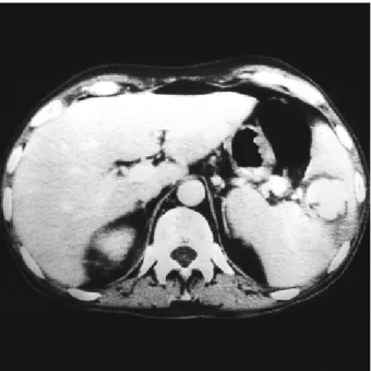
abdomen computed tomography (CT) scan showed pseudoa- neurysm in the spleen (Fig. 1), abscess in the right kidney, and pneumonic infiltration in right middle lobe of the lung.
On day 6, splenectomy was performed and showed charac- teristically broad, irregular, and non-septate hyphae. Cyclos- porine therapy was discontinued in an attempt to restore host defenses. Prednisolone was also tapered to physiologic dosage without aggravation of chronic GVHD. Amphotericin B with 5-fluorocytosine (75 mg/kg/day) was started to a dose of 1 mg/
kg/day and was later replaced by liposomal amphotericin B (4 mg/kg/day) because of deterioration of renal function. Total accumulated dosage of amphotericin B was 6.69 g (conven- tional amphotericin B, 0.79 g, liposomal amphotericin B, 5.9 g in 45 days).
He was successfully treated with combination of splenec- tomy, prolonged use of conventional and liposomal ampho- tericin B and 5-fluorocytosine, and discontinuing of cyclo- sporine and prednisolone. During the 1-yr period of follow- up after antifungal therapy, he was in good health without relapse of the infection.
CASE 2
A 39-yr-old man was admitted due to development of fever and left flank pain. Eight months before he had been diag- nosed as chronic phase of chronic myelogenous leukemia and had received allogeneic BMT three months after the diagno- sis. The post-BMT course was complicated by steroid-induced
diabetes, veno-occlusive disease, drug-induced hepatotoxicity, and cytomegalovirus infection. Two weeks before this event, the patient was readmitted due to extensive chronic GVHD involving the liver and gut, which was successfully treated with tacrolimus and prednisolone.
On physical examination, hepatosplenomegaly was detect- ed. Laboratory investigation showed liver failure with coagu- lation abnormality but leukocyte count was within normal range. Chest radiographs were normal. Cefoperazone/sulbac- tam, amikacin, and teicoplanin were started. Cultures of blood, sputum, and urine were all repeatedly negative. Abdo- men CT scan showed splenic abscess with peritoneal leakage.
On day 10, splenectomy was performed and histologic exami- nation revealed broad, irregular, and non-septate hyphae.
Splenic arterioles were thrombosed and contained the same hyphae (Fig. 2). Amphotericin B was started to a dose of 1 mg/kg/day and was later replaced by liposomal ampho- tericin B (4 mg/kg/day). Because chronic GVHD were ag- gravated during the course of tapering tacrolimus and pred- nisolone, immunosuppressants could not be tapered.
The patient's condition progressively deteriorated with lethargy, confusion, stiff neck, and subsequent coma (day 20).
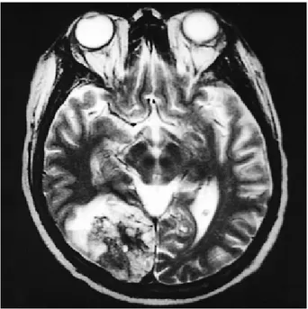
Cerebrospinal fluid did not show any leukocytes, and cul- tures for bacteria, fungus, and virus were all negative. The brain magnetic resonance scan revealed a lesion in the right occipito-parietal cerebral hemisphere (Fig. 3). The patient died on day 23 and the death was related with disseminated mucormycosis. He had received amphotericin B until his
Fig. 1.Abdominal CT reveals splenomegaly, and a well defined highly enhanced lesion with non-enhanced peripheral portion about 4×3 cm in diameter in the perihilum of the spleen, which is most likely the intrasplenic pseudoaneurysm with peripheral thrombi formation.
Fig. 2.Intramural thrombi and broad, non-septate, irregular hy- phae invading blood vessels in splenic tissue (hematoxylin-eosin staining, ×400, arrows).
Disseminated Mucormycosis Following BMT 405
death (total dose, conventional amphotericin B 0.15 g, lipo- somal amphotericin B 1.95 g in 13 days)
DISCUSSION
Mucormycosis is an uncommon filamentous mycosis which most frequently occurs in immunocompromised patients.
Although the epidemiological data on this fungal infection are at best scanty, it would appear that the incidence of this complication has increased in patients with hematological malignancies during the last decade probably due to the more severe and prolonged post-chemotherapeutic neutropenia caused by the use of more aggressive treatments (4). Morri- son et al. reported on the detection of mucormycosis infection in 0.9% during the post-BMT period (2). Several risk factors for mucormycosis such as prolonged neutropenia, poorly controlled diabetes, prolonged use of steroids, and impaired immune system were usually present in the BMT recipients, in whom the infections are likely to progress more rapidly (7, 8). Both of our patients were not neutropenic when mu- cormycosis developed. But they all were diabetics, using immunosuppressants including corticosteroids, and their immune system, such as cell-mediated immune responses, was impaired due to chronic extensive GVHD.
The spectrum of mucormycosis seen in the BMT popula- tion may be different from that seen in patients with diabetes and leukemia. The classic rhinocerebral type of infection is not common in the BMT population (2, 7). In either of our patients, sinonasal region was not involved, suggesting that
it may not be the main port of entry. In disseminated forms, mucor enters through the respiratory tract and invades the lung and arrives at other sites by embolic dissemination (1).
The most common site of spread is known to be the brain, but metastatic necrotic lesions have also been found in the spleen, heart, and other organs. Disseminated mucormyco- sis is usually diagnosed after the death of the patient from the infection. Occasionally metastatic cutaneous lesions per- mit an earlier diagnosis (3, 5). In our cases, the infection was first detected in the spleen and cutaneous lesions were not present. It would be reasonable to assume that other lesions also due to mucormycosis. But the concept should be remind- ed that more than one microorganism can simultaneously infect such immunocompromised patients like ours (9, 10).
Although we did not perform repeated biopsies and cultures in other sites, if there were clinical clues that suggest two or more different infectious processes (development of new le- sions while receiving therapy, progression of disease, or abnor- mal presentation of fungal infections, etc), it would be essen- tial to obtain specimens repeatedly from the affected sites.
The hallmarks of mucormycosis are vascular invasion and tissue necrosis. Typically, the fungi appear as broad (10 to 20 m in diameter), non-septate hyphae with branches occur- ring at right angles. We found the typical fungi in the splenic arterioles with intramural thrombi.
Oral antifungal prophylaxes are known to be unable to prevent mucormycosis (1, 2). Zygomycetes are resistant to triazole and nystatin, and whether the use of intranasal, aero- solized, or parenteral amphotericin B can play a role in pre- venting mucormycosis is still uncertain. Both patients in this report had received prophylactic fluconazole during the neutropenic period during the BMT and following 3 and 4 months, respectively.
Therapy of mucormycosis consists of rapid correction of the predisposing factors, surgical debridement of necrotic tissue whenever feasible, antifungal therapy, and adjunctive therapy such as hyperbaric oxygen, granulocyte colony stim- ulating factor, and granulocyte transfusion (3, 11, 12). Both of our patients underwent splenectomy and received conven- tional and liposomal amphotericin B.
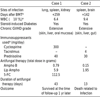
The differences between the two lie in the possibility of discontinuation of immunosuppressants, days after the BMT, the duration of antifungal therapy, total dosage of ampho- tericin B, and the difference of potency of immunosuppres- sant between cyclosporine and tacrolimus (Table 1).
In hematologic patients the most important good prog- nositc factor is the outcome of the hematologic disease (4).
The importance of reversing underlying factors is evident in the experience with mucormycosis. In the former of this two cases, discontinuation of immunosuppressants, lower poten- cy of immunosuppressants used, and longer days after BMT, all of which may mean the more possible immune restora- tion, were the reason for good prognosis. The prolonged use of amphotericin B is known to be correlated with a good
Fig. 3.Magnetic resonance scan of the brain. This T2-weighted scan shows an extensive right occipital lobe abscess with much surrounding edema.
406 D.-G. Lee, J.-H. Choi, Y.-J. Kim, et al.
prognosis (4, 7). However, one should not lose a sight of the fact that the correlation between the high total dosage, long duration of use of amphotericin B and the cure of patients could be simply due to the longer survival of these patients.
In these reports, favorable prognosis might be actually due to the longer survival of patient rather than having received the high total dosage of amphotericin B with longer dura- tion.
The use of lipid formulations of amphotericin B has recent- ly been implicated for the treatment of systemic fungal infec- tions to reduce the toxicity of conventional amphotericin B.
In a few cases, liposomal amphotericin B has been reported to be efficacious for the treatment of mucormycosis. How- ever, further studies are needed to assess whether these com- pounds are truly efficacious for the treatment of this systemic fungal infection (3, 11).
In conclusion, careful clinical and radiologic examinations are the key to early diagnosis of mucormycosis, especially in the disseminated form, in immunocompromised patients.
Early histologic confirmation certainly lead to early and pro- per treatment i.e., antifungal therapy and surgical approach.
Successful outcome is attributed to early diagnosis in con- junction with not only surgery or antifungal therapy, but cor-
rection of risk factors such as discontinuation or tapering of immunosuppressants.
REFERENCES
1. Bowden RA. Fungal infections after marrow transplantation. In:
Bowden RA, Ljungman P, Paya CV, eds. Transplant infections (1st ed). Philadelphia: Lippincott-Raven, 1998; 325-38.
2. Morrison VA, McGlave PB. Mucormycosis in the BMT population.
Bone Marrow Transplant 1993; 11: 383-8.
3. Gonzalez CE, Couriel DR, Walsh TJ. Disseminated zygomycosis in a neutropenic patient: successful treatment with amphotericin B lipid complex and granulocyte colony-stimulating factor. Clin Infect Dis 1997; 24: 192-6.
4. Pagano L, Ricci P, Tonso A, Nosari A, Cudillo L, Montillo M, Cenac- chni A, Pacilli A, Fabbiano F, Del Favero A. Mucormycosis in pa- tients with haematological malignancies: a retrospective clinical study of 37 cases. Br J Haematol 1997; 99: 331-6.
5. Cuverlier I, Vogelaers D, Peleman R, Benoit D, Van Marck V, Offner F, Vandewoude K, Colardyn F. Two cases of disseminated mucormy- cosis in patients with hematological malignancies and literature review. Eur J Clin Microbiol Infect Dis 1998; 17: 859-63.
6. Suh IW, Park CS, Lee MS, Lee JH, Chang MS, Woo JH, Lee IC, Rhu JS. Hepatic and small bowel mucormycosis after chemothera- py in a patient with acute lymphocytic leukemia. J Korean Med Sci 2000; 15: 351-4.
7. Gaziev D, Baronciani D, Galimberti M, Polchi P, Angelucci E, Gia- rdini C, Muretto P, Perugini S, Riggio S, Ghirlanda S, Erer B, Maiello A, Luearelli G. Mucormycosis after bone marrow transplantation:
report of four cases in thalassemia and review of the literature. Bone Marrow Transplant 1996; 17: 409-14.
8. Penalver FJ, Romero R, Fores R, Cabrera R, Briz M, Fernandez M.
Mucormycosis and hematopoietic transplants. Haematologica 1998;
83: 950-1.
9. Perfect JR, Schell WA. The new fungal opportunists are coming.
Clin Infect Dis 1996; 22(suppl 2): S112-8.
10. Morris A, Schell WA, McDonagh D, Chaffee S, Perfect JR. Pneu- monia due to Fonsecaea pedrosi and cerebral abscesses due to Emericella nidulans in a bone marrow transplant recipient. Clin Infect Dis 1995; 21: 1346-8.
11. Maury S, Leblanc T, Feuihade M, Molina JM, Schaison G. Success- ful treatment of disseminated mucormycosis with liposomal ampho- tericin B and surgery in a child with leukemia. Clin Infect Dis 1998;
26: 200-2.
12. St-Germain G, Robert A, Ishak M, Tremblay C, Claveau S. Infection due to Rhizomucor pusillus: report of four cases in patients with leukemia and review. Clin Infect Dis 1993; 16: 640-5.
Sites of infection lung, spleen, kidney spleen, brain
Days after BMT* +259 +142
WBC (×10 9/L)* 6.4 9.4
Steroid-induced Diabetes Yes Yes
Chronic GVHD grade Extensive Extensive
(skin, liver, oral mucosa) (skin, liver, gut) Immunosuppressants
used* (mg/day)
Cyclospirine 300 -
Tacrolimus - 4
Prednisoline 40 30
Antifungal therapy (total dose in grams)
Ampho B 0.79 0.15
Lip Ampho 5.9 1.95
5-FC 112.5
Duration of antifungal
therapy (days) 43 13
Outcome Survived at the time Death related to of follow-up 1 yr later Infection
Case 1 Case 2
Table 1.Characteristics of the patients with disseminated mucormycosis after bone marrow transplantation
BMT: bone marrow transplantation; GVHD: graft-versus-host disease;
Ampho B: amphotericin B; Lip Ampho: liposomal amphotericin B, 5- FC: 5-fluorocytosine.
*At the time of infection.
![[ 갑상선암 ]](data:image/gif;base64,R0lGODlhAQABAIAAAP///wAAACH5BAEAAAAALAAAAAABAAEAAAICRAEAOw==)