INTRODUCTION
Cerebrovascular disease (CVD) is a common cause of death or disability in Koreans. Ischemic stroke is a main cause of CVD and is well known that atherosclerotic stenosis of extra- cranial carotid artery is a major cause of ischemic strokes (1, 2). Prevalence of asymptomatic carotid atherosclerosis in Korea is not uncommon and is reported up to the 12.5% (3). In current practice, early detection of asymptomatic carotid disease and carotid endarterectomy is recommended before an occurrence of major stroke to reduce morbidity and mor- tality associated with cerebral infarction (4).
Framingham study (5) is a well-known, prospective, pop- ulation-based study for development of coronary artery dis- ease. Thereafter many epidemiologic studies reported various risk factors for development of cardiovascular disease (6, 7).
We conducted a retrospective study to determine the risk factors for development of asymptomatic atherosclerotic carotid stenosis (CS) in Korean population.
MATERIALS AND METHODS Data collection
The database of 21,400 subjects who underwent duplex ultrasonography of carotid artery on purpose of a regular check up at the Center for Health Promotion, Samsung Medical Center from March 1998 through November 2003 was col- lected and retrospectively analyzed. Among them, 2,805 subjects who had CS ≥30% or <10% as well as answered to questionnaire were included for present study. Subjects with non-atherosclerotic CS or past medical history of CVD did not exist in the our study population. The subjects were divided into 2 groups; Group I, CS <10% and Group II, CS
≥30%. Demographic data (age, sex, and body mass index [BMI], life style data [smoking, alcohol consumption, aspirin medication, VO2max, total daily calorie intake, and fat intake], coexisting medical conditions such as hypertension, diabetes mellitus [DM], hyperlipidemia, CVD, ischemic heart disease [IHD]) were retrieved from questionnaire and compared between group I and group II. Those who have hypertension,
Yun Jeong Lim, Young-Wook Kim*, Yeon Hyen Choe�, Chang-Seok Ki�, Sue Kyung Park�
Department of Internal Medicine, Dongguk University International Hospital, Dongguk University College of Medicine, Goyang; Division of Vascular Surgery, Department of Surgery*, Department of Radiology and Center for Imaging Science�, Department of Laboratory Medicine�, Samsung Medical Center, Sungkyunkwan University School of Medicine, Seoul; Department of Preventive Medicine�, Konkuk University College of Medicine, Chungju, Korea; �Division of Cancer Epidemiology and Genetics, National Cancer Institute of USA
Address for correspondence Young-Wook Kim, M.D.
Division of Vascular Surgery, Department of Surgery, Samsung Medical Center, Sungkyunkwan University School of Medicine, 50 Irwon-dong, Gangnam-gu, Seoul 135-710, Korea
Tel : +82.2-3410-3461, Fax : +82.2-3410-0040 E-mail : [email protected]
15
Risk Factor Analysis for Development of Asymptomatic Carotid Stenosis in Koreans
Many risk factors for atherosclerosis have been proposed to identify high risk indi- viduals. We conducted a retrospective study to determine the risk factors for devel- opment of carotid stenosis (CS) in Koreans. Database of 2,805 subjects who under- went a check up of carotid artery for health examination were analyzed. Stenosis (%) of common carotid artery or proximal internal carotid artery was examined with ultrasonography. Subjects were divided into 2 groups (Group I; CS <10%, Group II; CS ≥30%). We compared demographic, laboratory and clinical data between 2 groups to determine the risk factors of CS. One hundred ninety seven subjects (7.0%) were categorized as Group II. At age- and sex-adjusted multivariate analysis, dia- betes mellitus, hypertension, cerebrovascular disease, ischemic heart disease, hyperlipidemia, aspirin medication, current smoking, fasting glucose, total choles- terol, low density lipoprotein-cholesterol (LDL-C) and leukocyte count were signifi- cant risk factors of CS. At stepwise logistic regression analysis, age, hypertension, hyperlipidemia, LDL-C and leukocyte count were independent risk factors. At sub- group analysis by smoking, age and leukocyte count were independent risk factors in smoker and age and hypertension in nonsmoker.
Key Words : Carotid Stenosis; Atherosclerosis; Risk Factors
Received : 21 April 2005 Accepted : 21 July 2005
DM, hyperlipidemia, CVD or IHD had a reply to yes in the question (‘‘Do you have a experience of diagnosis about each disorder by doctor’’). Data including smoking, alcohol con- sumption (frequency and amount of alcohol), coexisting med- ical conditions and total amount of calorie and fat intake in diet surveys were collected from standardized questionnaire.
Smoking status divided into two groups (current smoking group and non-smoking group) by current smoking status.
Nonsmoking group is composed of past smoking or non- smoking. Alcohol consumption was divided into two groups by frequency and amounts of soju consumption in question- naire. Over or equal 3-4 frequencies a week and ≥80 g once alcohol consumption is defined as the group of heavy alcohol consumption. Below 3-4 frequencies a week or <80 g once alcohol consumption are classified as the other group.
Measurement of CS
CS was defined as percentage of maximal diameter reduc- tion at common carotid artery (CCA) or proximal internal carotid artery (ICA) on either side. The percentage of diam- eter reduction is calculated according to the European carotid surgery trial criteria (% stenosis=[diameter of carotid artery including the plaque - luminal diameter at the stenotic seg- ment/diameter of carotid artery including the plaque]×100).
CCA or ICA diameter reduction ≥30% of normal diameter at CCA or ICA was regarded as CS group (group II). Those who showed more than 30% of CS had also more than 1.2 mm intimomedial thickness.
To ascertain the precision in measuring % stenosis of carotid artery, we undertook a pilot study on 20 individuals. ICA and CCA diameter was measured 2 times on 20 individuals (right ICA and CCA, 10; left ICA and CCA, 10) by 2 examin- ers. We examined the inter-examiner bias between 2 exam- iners and intra-examiner bias between 2 examinations by same examiner. Our inter- and intra-examiner biases (mean
±SD) were 3.7±2.5% and 2.4±2.6% respectively. Intra- class correlation coefficient of Inter-examiner is 0.9639 (95%
confidence interval [0.9147, 0.9851]). Intraclass correlation coefficient of intra-examiner is 0.9787 (95% confidence inter- val [0.9491, 0.9912]). The difference between measured % stenosis of carotid artery obtained in repeated examination by a examiners was kept less than 10% in our practice.
All carotid examinations was performed with color flow ultrasonograpies (Antares, Siemens medical system, Germany;
LOGIQ 9, GE medical system, Milwankee, WI, U.S.A.) by registered vascular technists.
Laboratory assay and anthropometric measurement
Leukocyte counts, platelet counts, fasting glucose, total cholesterol, triglyceride, high density lipoprotein-cholesterol (HDL-C), low density lipoprotein-cholesterol (LDL-C), high sensitivity C-reactive protein (hs-CRP), Lipoprotein(a)(LP(a)),
homocysteine, fibrinogen and plasminogen activator inhi- bitor-I (PAI-I) were measured.
To measure peak oxygen uptake values (VO2max), the sub- jects performed symptom-limited graded exercise tests on the treadmill using the Bruce protocol. Peak oxygen uptake (mL/kg/min) was defined as the highest value recorded dur- ing the test. Height and weight were measured and the BMI (body mass index) was calculated as weight (kilograms) divided by height squared (square meters).
Statistical analysis
Logistic regression analysis was conducted by fixing the each group as dependent variable and the putative risk fac- tors as independent variables. Continuous variables such as age, BMI, VO2max, total daily calorie intake and fat intake were analyzed by t-test. Sex, smoking, alcohol consumption, aspirin medication, hypertension, DM, hyperlipidemia, CVD and IHD were analyzed by 2test. The age- and sex-adjust-
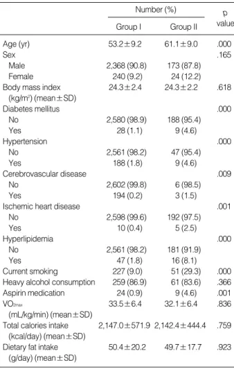
Number (%) Group I Group II
p value
Age (yr) 53.2±9.2 61.1±9.0 .000
Sex .165
Male 2,368 (90.8) 173 (87.8)
Female 240 (9.2) 24 (12.2)
Body mass index 24.3±2.4 24.3±2.2 .618 (kg/m2) (mean±SD)
Diabetes mellitus .000
No 2,580 (98.9) 188 (95.4)
Yes 28 (1.1) 9 (4.6)
Hypertension .000
No 2,561 (98.2) 47 (95.4)
Yes 188 (1.8) 9 (4.6)
Cerebrovascular disease .009
No 2,602 (99.8) 6 (98.5)
Yes 194 (0.2) 3 (1.5)
Ischemic heart disease .001
No 2,598 (99.6) 192 (97.5)
Yes 10 (0.4) 5 (2.5)
Hyperlipidemia .000
No 2,561 (98.2) 181 (91.9)
Yes 47 (1.8) 16 (8.1)
Current smoking 227 (9.0) 51 (29.3) .000 Heavy alcohol consumption 259 (86.9) 61 (83.6) .366 Aspirin medication 24 (0.9) 9 (4.6) .001
VO2max 33.5±6.4 32.1±6.4 .836
(mL/kg/min) (mean±SD)
Total calories intake 2,147.0±571.9 2,142.4±444.4 .759 (kcal/day) (mean±SD)
Dietary fat intake 50.4±20.2 49.7±17.7 .923 (g/day) (mean±SD)
Table 1.Risk factor analysis* with demographic, clinical and life style factors
*Age and sex-adjusted logistic regression analysis.
Group II is defined as carotid stenosis which had equal or over 30%
diameter reduction by ultrasonography and Group I is lesser than 10%.
ed relative risk to develop CS was calculated with odds ratio with 95% confidence interval (CI). Risk factors were exam- ined by a forward stepwise multiple logistic regression anal- ysis with entering the significant variables. Statistical signifi- cance was assumed at p<0.05. Statistical analyses were per- formed with SAS version 8.1 (SAS Institute Inc, Cary, NC, U.S.A.).
RESULTS
Demographic, clinical and life style factors
The prevalence of group II (CS ≥30%) was 7.0% (197/
2805) increasing its prevalence with age (p<.001) (Table 1).
But there was no significant association between 2 groups in sex or BMI. DM, hypertension, CVD, IHD and hyperlipi- demia were significantly more common in group II at age- and sex-adjusted logistic regression analysis. At age- and sex-adjusted logistic regression analysis, current smoker and
daily aspirin medication were more common in Group II.
Heavy alcohol consumption, VO2max, daily calorie intake or fat intake did not significantly associated with CS.
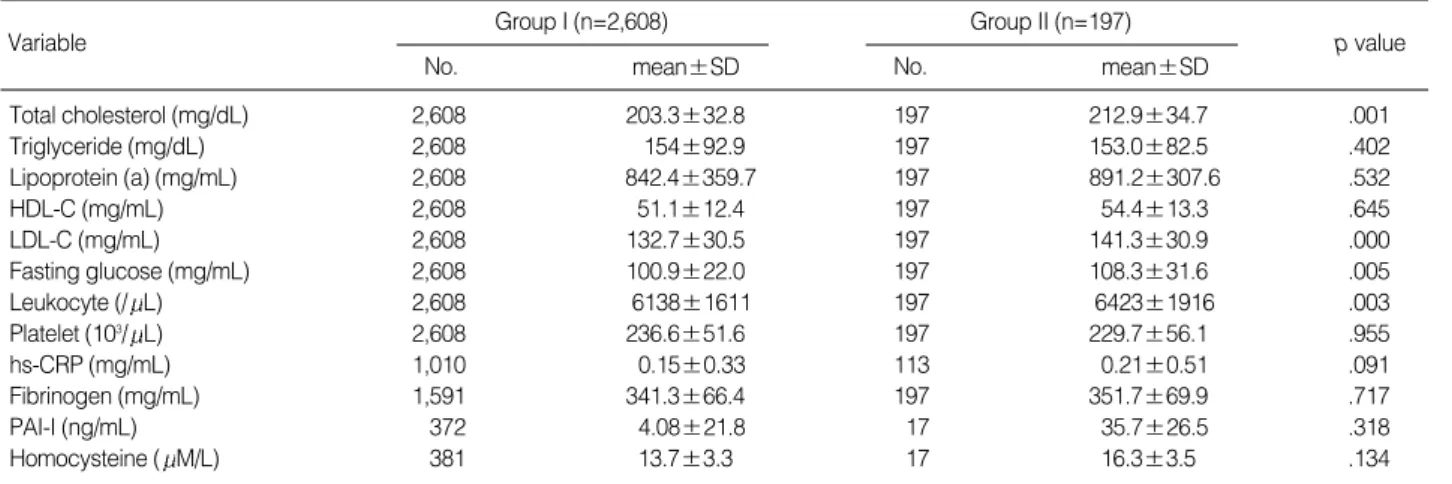
Laboratory variables
At age- and sex-adjusted logistic regression analysis, group II was associated with significantly higher levels of total choles- terol, LDL-C, fasting glucose and leukocyte count compared to Group I (Table 2). But there were no significant difference in serum level of triglyceride, HDL-C, homocysteine, PAI-1, hs-CRP, fibrinogen, LP(a)or platelet count between 2 groups.
Stepwise multiple logistic regression analysis
Finally, we conducted stepwise multiple logistic regres- sion analysis in which above significant variables were used as independent variables. Number of subjects entered into the stepwise logistic regression model were 113 in group II and 1007 in group I. Entered variables were age, sex, BMI, DM, hypertension, hyperlipidemia, CVD, IHD, daily aspirin
Variable Group I (n=2,608)
No. mean±SD
p value Group II (n=197)
No. mean±SD
Total cholesterol (mg/dL) 2,608 203.3±32.8 197 212.9±34.7 .001
Triglyceride (mg/dL) 2,608 154±92.9 197 153.0±82.5 .402
Lipoprotein (a) (mg/mL) 2,608 842.4±359.7 197 891.2±307.6 .532
HDL-C (mg/mL) 2,608 51.1±12.4 197 54.4±13.3 .645
LDL-C (mg/mL) 2,608 132.7±30.5 197 141.3±30.9 .000
Fasting glucose (mg/mL) 2,608 100.9±22.0 197 108.3±31.6 .005
Leukocyte (/ L) 2,608 6138±1611 197 6423±1916 .003
Platelet (103/ L) 2,608 236.6±51.6 197 229.7±56.1 .955
hs-CRP (mg/mL) 1,010 0.15±0.33 113 0.21±0.51 .091
Fibrinogen (mg/mL) 1,591 341.3±66.4 197 351.7±69.9 .717
PAI-I (ng/mL) 372 4.08±21.8 17 35.7±26.5 .318
Homocysteine ( M/L) 381 13.7±3.3 17 16.3±3.5 .134
Table 2.Risk factor analysis* with laboratory data
*Age and sex-adjusted logistic regression analysis.
Group II is defined as carotid stenosis which had equal or over 30% diameter reduction and Group I is lesser than 10%.
HDL-C, high density lipoprotein-cholesterol; LDL-C, low density lipoprotein-cholesterol; hs-CRP, high sensitivity C-reactive protein; PAI-1, plasminogen activator inhibitor-I.
CI, confidence interval; LDL-C, low density lipoprotein cholesterol- cho- lesterol.
*all variables in full model are as follows: age, sex, BMI, DM, hyperten- sion, hyperlipidemia, CVD, IHD, daily aspirin medication, interaction term between medical history of hypertension and hyperlipidemia, total choles- terol, LDL-C, fasting glucose and leukocyte count.
Variable* Odds ratio (95% CI) p value
Age 1.06 (1.04-1.09) .000
Hypertension 6.05 (2.98-12.29) .000
Hyperlipidemia 3.29 (1.50-7.21) .003
LDL-C 1.01 (1.00-1.02) .002
Leukocyte 1.19 (1.06-1.35) .004
Table 3.Stepwise logistic regression analysis
CI, confidence interval.
*all variables in full model are as follows: age, sex, BMI, DM, hyperten- sion, hyperlipidemia, CVD, IHD, daily aspirin medication, interaction term between medical history of hypertension and hyperlipidemia, total cholesterol, LDL-C, fasting glucose and leukocyte count.
Subgroup Variable* Odds ratio (95% CI) p value
Smoker Age 1.10 (1.05-1.16) .000
(n=153) Leukocyte count 1.44 (1.14-1.81) .002
Non-smoker Age 1.06 (1.04-1.09) .004
(n=67) Hypertension 3.65 (1.03-12.92) .045 Table 4.Stepwise logistic regression analysis after stratification by current smoking status
medication, interaction term between medical history of hypertension and hyperlipidemia, total cholesterol, LDL-C, fasting glucose and leukocyte count. The interactions among variables were not significant except interaction term between hypertension and hyperlipidemia. As a result, age, hyperten- sion, hyperlipidemia, LDL-C and leukocyte count were inde- pendent risk factors for development of CS (Table 3). Smok- ing was already recognized as significant independent risk factors by the age- and sex-adjusted logistic regression analy- sis. But we did not enter smoking into stepwise logistic reg- ression model due to great numbers (43.7%) of missing data regarding smoking history. Instead, we conducted an analy- sis on putative risk factors stratified by current smoking sta- tus. Subjects entered into stratification analysis were 38 in group II and 115 in group I for current smoker, and 21 in group II and 46 group I for non-smoker. When stepwise logistic regression analysis was performed after stratification by smoking, age and leukocyte count were independent pre- dictors of CS in current smoker while age and hypertension were independent predictors in non-smoker (Table 4).
DISCUSSION
A number of risk factors for atherosclerosis have been pro- posed to identify high-risk individuals to develop cardiovas- cular disease (7, 8). A few population-based studies revealed the impact of vascular risk factors on carotid atherosclerosis (9). The Framingham study reported that age, cigarette smok- ing, systolic blood pressure and high serum cholesterol were independent risk factors of carotid atherosclerosis (10). A num- ber of new candidate markers have been proposed as predic- tors of atherosclerosis (7). We examined various variables to find out risk factors for development of asymtomatic athero- sclerotic CS in Koreans.
Duplex ultrasonography is known as the most useful non- invasive method to detect CS and measure the degree of steno- sis (11). With improving non-invasive imaging technology with ultrasound, duplex ultrasonography has replaced carotid arteriography and became a screening tool in detection of extracranial carotid disease with high accuracy (11). We exam- ined the inter-examiner bias between 2 examiners and intra- examiner bias between 2 examinations by a same examiner.
Our inter- and intra-examiner biases (mean±SD) were kept less than 10% in our practice. On the basis of this pilot study, we defined the control group as CS <10% and the cut point of our study was equal or over 30% diameter stenosis of carotid artery at either side. Our study patients were mostly com- posed of mild (<50%) carotid stenosis and severe stenosis (>70%) is rare in our study population.
Old age and male gender have been well-known risk fac- tors to atherosclerosis (6). In our study, old age is a risk fac- tor, but male gender was not a risk factor. In present study, fasting glucose level was higher in stenosis group than in
control group. Fasting glucose level was under control in most of the known diabetes by use of oral hypoglycemic agent or insulin in our study. Higher level of total cholesterol and LDL- C as well as medical history of hyperlipidemia were found in stenosis group than in control group. Those who had a daily aspirin medication had a concurrent medical illness such as hypertension, CVD or IHD in substantial numbers of sub- ject. Our observation of more common daily medication of aspirin in stenosis group can be explainable by this reason that aspirin medication is not a risk factor for CS but merely a result of concurrent medical illness.
Inflammation is widely accepted that it works a central role in pathogenesis of atherosclerosis (12, 13). We also examined inflammatory markers such as leukocyte count, hs-CRP, serum fibrinogen, Lp(a), serum homocysteine and PAI-1 whether they work as risk factor to CS or not. Among various inflam- matory markers examined, higher leukocyte count was only independent risk factor of CS in present study. Recent report suggested that CRP is a strong predictor of future develop- ment of CVD (7). Unlike other inflammatory markers, CRP levels are stable over long periods, can be measured inexpen- sively with available high sensitivity assays, and have shown high specificity in predicting the risk of CVD (7, 13). Our data did not show any significant difference of hs-CRP level between 2 groups. Decisions regarding clinical use of CRP remain uncertain. Higher level of serum fibrinogen was re- ported as a significant independent predictor of recurrent cardiovascular events after adjustment for conventional risk factors (7, 13). But, they could not conclude whether fibrino- gen has a causal role in atherothrombosis or is merely a marker of the degree of vascular damage. Elevation of serum Lp(a), homocysteine and PAI-1 was reported to predispose to athero- sclerosis in the general population (7, 13). Present study demonstrated no significant difference of fibrinogen, Lp(a), PAI-1 or homocysteine between 2 groups. There were so many missing data in those inflammatory markers that we can not exclude selection bias.
Several reports indicate that inflammatory processes and modifiable risk factors interact with genetic factors to cause stroke (12-15). Smoking is well known as an independent modifiable risk factor of stroke (9, 14). In our study, smoking was recognized as a significant risk factor in age- and sex- adjusted logistic regression analysis. The limitation of our study is great numbers (43.7%) of missing data regarding smoking history. We did not enter smoking into stepwise logistic regression. Instead, we conducted an analysis on putative risk factors stratified by current smoking status. Although small or moderate amount of alcohol consumption was reported to be associated with reduced risk of stroke, excessive alcohol intake and alcohol binges were reported to increase risk of stroke and its recurrence (15). We could not find any associa- tion between heavy alcohol consumption and carotid stenosis.
The limitation of our study is inter-observer bias of mea- surement of carotid ultrasonography. The other limitation is
not general population based study but specific class com- posed of those who had more attention about health promo- tion than general population as well as high economic status based study. The aim of this analysis was to produce reliable estimates of relative risk of each variable.
In conclusion, age, hypertension, hyperlipidemia, higher leukocyte count and higher serum level of LDL-C were found to be independent risk factors of CS diagnosed by carotid duplex ultrasonography during the regular health examina- tion. Our result showed that screening carotid ultrasonogra- phy is needed in subjects who had many risk factors such as age, hypertension, hyperlipidemia, higher leukocyte count and higher serum level of LDL-C to prevent CVD.
REFERENCES
1. Kannel MB, Wolf PA, Verter J, McNamara PM. Epidemiologic assess- ment of the role of blood pressure and stroke: the Framingham study.
JAMA 1970; 214: 301-8.
2. Whisnant JP, Fitzgibbons JP, Kurland LT, Sayre GP. Natural history of stroke in Rochester, Minnesota, 1945 through 1954. Stroke 1971;
2: 11-22.
3. Park KC, Kim DI, Lee SJ, Lee BB, Lee CH, Kim DY, Heo JS, Kim YI. Prevalence of asyptomatic carotid atherosclerosis in Korea. J Korean Soc Vasc Surg 1999; 15: 228-33.
4. Executive Committee for the Asymptomatic Carotid Atherosclero- sis Study. Endarterectomy for asymptomatic carotid artery stenosis.
JAMA 1995; 273: 1421-8.
5. Kannel WB, Dawber TR, Kagan A, Revotskie N, Stokes J 3rd. Fac- tors of risk in the development of coronary heart disease--six year follow-up experience. The Framingham Study. Ann Intern Med 1961;
55: 33-50.
6. Kullo IJ, Ballantyne CM. Conditional risk factors for atherosclero-
sis. Mayo Clin Proc 2005; 80: 219-30.
7. Hackam DG, Anand SS. Emerging risk factors for atherosclerotic vascular disease: a critical review of the evidence. JAMA 2003; 290:
932-40.
8. Pearson TA, Mensah GA, Alexander RW, Anderson JL, Cannon RO 3rd, Criqui M, Fadl YY, Fortmann SP, Hong Y, Myers GL, Rifai N, Smith SC Jr, Taubert K, Tracy RP, Vinicor F. Centers for Disease Control and Prevention; American Heart Association. Markers of inflammation and cardiovascular disease: application to clinical and public health practice: A statement for healthcare professionals from the Centers for Disease Control and Prevention and the American Heart Association. Circulation 2003; 107: 499-511.
9. Fabris F, Zanocchi M, Bo M, Fonte G, Poli L, Bergoglio I, Ferrario E, Pernigotti L. Carotid plaque, aging, and risk factors. A study of 457 subjects. Stroke 1994; 25: 1133-40.
10. Fine-Edelstein JS, Wolf PA, O’Leary DH, Poehlman H, Belanger AJ, Kase CS, D’Agostino RB. Precursors of extracranial carotid atherosclerosis in the Framingham Study. Neurology 1994; 44: 1046- 50.
11. Sorof JM, Alexandrov AV, Garami Z, Turner JL, Grafe RE, Lai D, Portman RJ. Carotid ultrasonography for detection of vascular abnor- malities in hypertensive children. Pediatr Nephrol 2003; 18: 1020-4.
12. Ross R. Atherosclerosis--an inflammatory disease. N Engl J Med 1999; 340: 115-26.
13. Ridker PM, Stampfer MJ, Rifai N. Novel risk factors for systemic atherosclerosis: a comparison of C-reactive protein, fibrinogen, homo- cysteine, lipoprotein (a), and standard cholesterol screening as pre- dictors of peripheral arterial disease. JAMA 2001; 285: 2481-5.
14. Szirmai IG, Kamondi A, Magyar H, Juhasz C. Relation of laboratory and clinical variables to the grade of carotid atherosclerosis. Stroke 1993; 24: 1811-6.
15. Humphries SE, Morgan L. Genetic risk factors for stroke and carotid atherosclerosis: insights into pathophysiology from candidate gene approaches. Lancet Neurol 2004; 3: 227-35.