INTRODUCTION
Radiation has been used increasingly from discovery of x- ray over one hundred years ago in medicine, both in diagno- sis and treatment. Clinically, radiation is applied to whole body for bone marrow transplantation and to limited area of the body for the cure of local cancer. Radiation produces reac- tive oxygen species (ROS), which can lead to the damage of lipids, proteins, carbohydrates and nucleic acids, and destroy cancer cells in the treated area. However, normal cells in the surrounded area can also be injured by radiation itself or even in the other site by circulating ROS.
Intracellular ROS are tightly controlled by different anti- oxidant defense systems. It has been demonstrated during the last decade that one of the important components of the anti-oxidant defense is heme oxygenase (HO) (1, 2). HO-1 (HO, EC 1.14.99.3) is identified as the 32-kDa stress (heat shock) protein (HSP32) (3, 4), that is a microsomal enzyme to catalyze oxidative breakdown of heme molecule to carbon monoxide (CO), Fe2+, and biliverdin, which is subsequently reduced to bilirubin by biliverdin reductase (5, 6). Cytopro- tective role of HO-1 can be explained by the removal of free heme (a pro-oxidant) and the production of bilirubin, which
is considered as a potent anti-oxidant (1, 2, 7, 8). Carbon monoxide is thought to be an active participant in signal transduction and possesses potent anti-inflammatory prop- erties (9). Ferrous iron liberated in this reaction has pro-oxi- dant activity like free heme, however, intracellular chelatable iron in cytoplasm is sequestered by ferritin (10), preventing its participation in the oxidative processes.
The other key component involved in redox regulation of signal transduction and expression of anti-inflammatory anti- oxidant defense enzymes is reduced glutathione ( -glutamyl- cysteinylglycine, GSH) (11-13). Beside participation in sig- nal transduction, GSH plays extremely potent role in anti- oxidant defense because it possesses not only the direct radi- cal-scavenging ability but also is the essential component of glutathione peroxidase system, which eliminates different hydroperoxides without free radical production (12). In ani- mal tissues, GSH is the most abundant non-protein thiol (NPSH), comprising approximately 75-90% of NPSH (13- 15). Other widespread NPSH are cysteine and coenzyme A.
Radiation therapy (RT) is a common modality used for the treatment of thoracic malignancies, but impaired heal- ing of wounds in irradiated tissues or injury to surrounding normal lung can present clinical complications such as pneu-
Youngsoo Han*, Alexander Platonov*,�, Medea Akhalaia*,�, Yeon-Sook Yun* Jie-Young Song*
Laboratory of Radiation Immunology*, Korea Institute of Radiological & Medical Sciences, KAERI, Seoul, Korea; Laboratory of Radiation Biophysics�, Department of Biophysics, Biological Faculty, Moscow State University, Moscow, Russia
Address for correspondence Jie-Young Song, Ph.D.
Laboratory of Radiation Immunology, Korea Institute of Radiological & Medical Sciences, KAERI, 215-4 Gongneung-dong, Nowon-gu, Seoul 139-706, Korea Tel : +82.2-970-1309, Fax : +82.2-970-2402 E-mail : [email protected]
*This study was supported by Korea Institute of Science & Technology Evaluation and Planning (KISTEP) and Ministry of Science & Technology (MOST), Republic of Korea Government, through its National Nuclear Technology Program.
535
Differential Effect of -radiation-induced Heme Oxygenase-1 Activity in Female and Male C57BL/6 Mice
Ionizing radiation produces reactive oxygen species, which exert diverse biological effects on cells and animals. We investigated alterations of heme oxygenase (HO) and non-protein thiols (NPSH), which are known as two major anti-oxidant enzymes, in female and male C57BL/6 mice in the lung, liver, and brain after whole-body - irradiation with 10 Gy (1-7 days) as well as in the lung after whole-thorax -irradia- tion (WTI) with 12.5 Gy (1-26 weeks). Most significant alteration of HO activity was observed in the liver, which elevated 250% in males. NPSH level in female liver was increased on the 5th-7th days but decreased in males on the 3rd day. In the lung, the elevation of HO activity in both sexes and the pattern of NPSH change were similar to that of the liver. On the other hand, the increase of HO activity on the 16th week and the decrease of NPSH level on the 2nd week were observed only in male lung after WTI. This study shows that the liver is the most sensitive tissue to -irradiation-induced alterations of HO activity in both female and male mice. In addition, there exists significant differential effect of -irradiation on antiox- idant system in female and male mice.
Key Words : Heme Oxygenase; Non-Protein Thiols; Antioxidants; Liver; Lung; Brain; Mice; Gamma rays
Received : 8 March 2005 Accepted : 11 April 2005
monitis and accompanying fibrosis, occurring in up to 20%
of patients irradiated for lung cancer, breast cancer, lymphoma, or thymoma (16). The aim of this study was therefore to inves- tigate and compare the radiation-induced alterations of the important antioxidant enzymes such as HO and NPSH after whole-body -irradiation by lethal dose and to determine if any differences due to gender exist. In addition, especially long-term observation of the antioxidant levels in lung after local thorax -irradiation can give the information of radia- tion-induced side effects related with the alterations of these antioxidants.
MATERIALS AND METHODS Animals
Female and male C57BL/6 mice were purchased from Dae- han Biolink (Korea) and 7-8 weeks old at the beginning of experiments. The mice were housed in conventional animal house conditions with 12-hr light-dark cycle, temperature of 22±2℃, relative humidity of 50%, standard pelleted diet and tap water ad libitum. The animal care, handling, and experimental procedures were conducted in accordance with the guidelines approved by the Animal Care and Use Committee of the Korea Institute of Radiological & Medi- cal Sciences (KIRAMS).
Irradiation
Whole-body irradiation and local whole-thorax irradiation of the mice were performed using 60Co -rays from a Thera- tron 780 irradiation unit (Atomic Energy of Canada, Ltd., Canada). For whole-body irradiation, mice were placed into Plexiglas box and irradiated with a single dose of 10 Gy (0.5 Gy/min). For local whole-thorax irradiation, mice were anes- thetized by i.p. injection of a mixture of ketamine (70 mg/kg) and xylazine (6 mg/kg), and fixed in a supine position on a Plexiglas tray. The thoracic regions (1.5 cm in length in cephal- ic-tail direction) of mice were placed in the field defined by collimators and irradiated with a single dose of 12.5 Gy (0.76 Gy/min). Other parts of the bodies were additionally shield- ed with 5-cm lead blocks. Before tissue sampling, irradiated and control mice were sacrificed by cervical dislocation in 1, 3, 5, and 7 days after whole-body irradiation and in 1, 2, 4, 12, 16, and 26 weeks after whole-thorax irradiation.
HO activity assay
Immediately after sacrifice of mice by cervical dislocation, the cranial, peritoneal, and chest cavities of mice were opened, and the tissues (lung, liver, and brain) were quickly harvested and washed (except for brain) in cold 100 mM K-phosphate buffer (pH 7.4) containing 2 mM MgCl2(MgCl2-phosphate
buffer). The tissues were slightly dried by paper filter, minced with scissors, and homogenized in 4 volumes of MgCl2-phos- phate buffer on ice by a Tissue-Tearor with a 7-mm probe (BioSpec Products, Inc., WI, U.S.A.). The homogenates were centrifuged at 1,500×g for 10 min at 4℃, and the obtained supernatants were further centrifuged at 15,000×g for 15 min at 4℃. The 15,000×g supernatants were kept at 4℃ and used for determination of HO activity not later than 1- 1.5 hr after preparation. Protein concentration of the 15,000
×g supernatants was determined by the method of Schacterle and Pollack (17) with bovine serum albumin as a standard.
The reaction mixture for determination of HO activity includ- ed MgCl2-phosphate buffer, 412 mg of protein/mL of the 15,000×g supernatant, 1 mg of protein/mL of liver 105,000
×g supernatant as a source of biliverdin reductase, 2 mM glucose 6-phosphate, 1 U glucose-6-phosphate dehydroge- nase, 25 M hemin, and 1 mM NADPH in a final volume of 500 L in 1.7-mL Eppendorf microcentrifuge tubes. Reac- tion was initiated by the addition of NADPH, and the reac- tion mixtures were incubated in a water bath at 37℃in the dark for 10 min (liver), 20 min (brain), or 40 min (lung). To terminate the reaction and extract produced bilirubin, 500 L of chloroform were added to each sample, the samples were vortexed thoroughly for 30 sec, and centrifuged at 15,000
×g for 10 min. The lower chloroform layer was transferred to a spectrophotometer microcuvette to measure the difference in optical density between 464 and 530 nm ( OD464-530) using UV/Visible spectrophotometer Ultrospec 3100 pro (Biochrom, Ltd., England). The extinction coefficient ( 464-
530) of 40 mM/cm was used for calculation of bilirubin con- centrations (18). HO activity was expressed in picomoles of bilirubin produced per milligram of protein per hour.
As a source of biliverdin reductase, 105,000×g super- natant of liver homogenate was used (18). To prepare the supernatant, livers of overnight-fasted non-irradiated mice were perfused in situ with cold (4℃) 1.15% KCl, harvested, slightly dried by paper filter, minced with scissors, and ho- mogenized in 4 volumes of MgCl2-phosphate buffer on ice.
The homogenates were centrifuged at 1,500×g for 10 min at 4℃, and the pooled supernatant was further centrifuged at 15,000×g for 15 min at 4℃and then at 105,000×g for 1.5 hr at 4℃. The 105,000×g supernatant was diluted to 5 mg of protein/mL with MgCl2-phosphate buffer and stored at -20℃.
HO-1 mRNA expression determination
Semiquantitative reverse transcriptase polymerase chain reaction (RT-PCR) was used for the mRNA expression level determination. After thawing liver samples frozen in liquid nitrogen, RNA was purified from the liver using RNAzol (Tel-Test, Friendswood, U.S.A.), and RNA concentration was spectrophotometrically determined by absorbance at 260 nm.
One microgram of intact RNA was reverse-transcribed into
cDNA in the reaction mixture containing 50 mM Tris-HCl (pH 8.3), 3 mM MgCl2, 75 mM KCl, 2.5 g/mL pd(N)6 primer, 0.5 mM each of dNTP, and 10 U of AMV-RT (Amer- sham). Then produced cDNA was amplified by polymerase chain reaction (PCR) in the mixture containing 10 L cDNA template from RT reaction, 10 mM Tris-HCl (pH 8.3), 50 mM KCl, 1.5mM MgCl2, 0.5mM each of dNTP, 1.0 M of each primer, and 0.5 U Tag DNA polymerase (Takara, Tokyo, Japan). The oligonucleotide primers used in these experiments are as followings; HO-1 PCR primer (5′-primer 5′-AACAA- GCAGAACCCAGTC-3′, 3′-primer: 5′-TGTCATCTCC- AGAGT -GTTC-3′), -actin primer (5′-primer: 5′-TGG- AATCCTGTGGCATCCATGAAA-3′, 3′-primer: 5′-TAA- AACGCAGCTCAGTAACAGTCCG-3′). PCR was per- formed with a DNA thermal cycle (Hybaid, Ashford, U.K.) at 94℃for 30 sec, at 56℃for 30 sec, and at 72℃for 30 sec per cycle. The PCR products were visualized by electrophore- sis on 1% agarose gel in the presence of 0.5 g/mL ethidi- um bromide. Density of bands was measured using an image analyzer (Flour-S Multimager, Bio-Rad, CA, U.S.A.).
NPSH content assay
NPSH content in animal tissues was determined with DTNB by the method of Sedlak and Lindsay (19). Briefly, immediately after sacrifice of mice, the tissues (50-90 mg of liver, 100-150 mg of brain and lung) were quickly harvested, placed on ice, slightly dried by paper filter (except for brain), weighed, minced with scissors, and homogenized on ice in a mixture of 2 mL of 10% TCA and 2 mL of 0.02 M EDTANa2. The homogenates were centrifuged at 3,500×g for 20 min at 4℃. Then, 0.5 mL of the supernatant was mixed with 1 mL of 0.4 M Tris-HCl buffer (pH 8.9) containing 0.02 M EDTANa2, and 25 L of 0.01 M DTNB in methanol were added to each sample. OD was measured at 412 nm in 5 min.
Blank sample contained all the above-mentioned components, except animal tissue. NPSH contents were calculated using an extinction coefficient ( 412) of 13.1 mM/cm and expressed in micromoles per gram of wet tissue weight.
Statistical analysis
All experimental data were evaluated by GraphPad Prism software (San Diego, CA, U.S.A.). In the most of experiments, the significances of differences between irradiated and control groups were tested using Student’s t test or Mann-Whitney when the data failed to pass a normality test. The effect of irradiation on HO-1 mRNA level was analyzed by Kruskal- Wallis test. Differences among the groups were considered as statistically significant at p<0.05.
RESULTS
HO activity and NPSH level in different tissues of male and female control mice
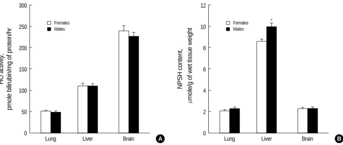
Comparison of HO activity and NPSH level in lung, liver and brain of male and female control mice demonstrated that HO activity was the highest in brain and the lowest in lung;
NPSH level was approximately same in lung and brain, and much higher in liver (Fig. 1). The significant difference between sexes was observed only in hepatic NPSH content: it was approximately 15% higher in males than in females.
HO activity in brain, liver and lung after whole-body irradi- ation
In brain, HO activity in males was slightly decreased on the 5th day but no alteration of that in females (Fig. 2). Sig-
HO activity, pmole bilirubin/mg of protein/hr 300
250
200
150
100
50
0
Fig. 1.HO activity (A) and NPSH level (B) in the lung, liver, and brain of control female and male C57BL/6 mice. Sixteen mice were used in each group. All data are shown as means±SEM. *p<0.05 compared to female mice.
Lung Liver Brain
Females Males
A
NPSH content, mole/g of wet tissue weight 12
10
8
6
4
2
0
Lung Liver Brain
Females *
Males
B
nificant elevation of HO activity (approximately 25-30%) in lung was observed on the 5th day in females and on the 7th day in males after irradiation. The most impressed radi- ation-induced alterations of HO activity occurred in livers
of both male and female mice. HO activity began to increase on the 3rd day after the irradiation in females and on the 5th day in males, and reached nearly 200% of the control level
Fig. 2.HO activity in the brain, lung and liver of female (A) and male (B) C57BL/6 mice after whole-body irradiation with 10 Gy. Each time point test group was compared with own control group. Four mice were used in each test and corresponding control group. All data are shown as means±SEM. *p<0.05, �p<0.01, �p<0.001 compared to corresponding control group.
Percent of control
250 225 200 175 150 125 100 75 50
0 1 2 3 4 5 6 7 8
Days after irradiation
Percent of control
275 250 225 200 175 150 125 100 75 50
0 1 2 3 4 5 6 7 8
Days after irradiation
Brain Lung Liver
Brain Lung Liver
* * *
*
*
*
�
�
A B
HO-1 mRNA level, relative units
20
15
10
5
0
Control 3 5 7
Days after irradiation
Fig. 3.HO-1 mRNA expression levels in the liver of female C57BL/6 mice after whole-body irradiation with 10 Gy. (A) The purified total RNA was converted to cDNA and amplified by reverse transcrip- tase polymerase chain reaction (RT-PCR). The amplified RT-PCR product was visualized on 1% agarose gel. (B) The band density was quantified in comparison to the -actin mRNA expression using image analyzer. Four mice were used in each test and control group.
All data are shown as means±SEM. *p<0.05, �p<0.01 compared to control group.
�
* HO-1
Control
-actin
3 5 7
A
B
Fig. 4.NPSH contents in the brain, lung and liver of female (A) and male (B) C57BL/6 mice after whole-body irradiation with 10 Gy.
Each time point test group was compared with own control group.
Four mice were used in each test and corresponding control group.
All data are shown as means±SEM. *p<0.05, �p<0.01 compared to corresponding control group.
Percent of control
175
150
125
100
75
50
0 1 2 3 4 5 6 7 8
Days after irradiation
Brain Lung Liver
*
*
�
A
Percent of control
150
125
100
75
50
0 1 2 3 4 5 6 7 8
Days after irradiation
Brain Lung Liver
* *
B
in females and 250% in males on the 7th day. To further con- firm whether the elevation of HO activity is accompanied with the increase of gene expression of HO-1, we performed the RT-PCR for HO-1 mRNA in liver. As shown in Fig. 3, mRNA expression of HO-1 was significantly elevated on the 5th day and persisted until 7 days after irradiation (8.7 and 8.0-fold compared to the control group respectively).
NPSH levels in different tissues and sexes after whole- body irradiation
NPSH level in the brain was not significantly changed in both male and female mice (Fig. 4). In case of the lung, NPSH level was significantly decreased on the 1st day after irradia- tion in males but increased in females. Like, irradiation induced different response of NPSH level in livers of males and females;
it was increased in females on the 5-7th days in sharp contrast
to decrease in males on the 3rd day. The alteration time of NPSH level in livers was shown later than that in lungs even the pattern of NPSH response was similar.
HO activity and NPSH level in the lung after whole-thorax irradiation
HO activity and NPSH level in the lung after whole-thorax irradiation at a dose of 12.5 Gy were evaluated during 26 weeks after exposure. As shown in Fig. 5, the whole-thorax irradia- tion resulted in different reaction of males and females. In females, there were no significant changes of both HO activity and NPSH level, although HO activity showed a tendency to increase on the 1st week and to decrease on the 12th week after irradiation. However, NPSH level was significantly de- creased on the 2nd week and HO activity was increased on the 16th week (by 45%) after irradiation in male mice.
HO activity, % of control
160
140
120
100
80
60 1 2 4 12 16 26
Weeks after irradiation A
Control Irradiation
HO activity, % of control
160
140
120
100
80
60 1 2 4 12 16 26
Weeks after irradiation B
Control Irradiation
*
NPSH content, % of control
160
140
120
100
80
60 1 2 4 12 16 26
Weeks after irradiation C
Control Irradiation
NPSH content, % of control
160
140
120
100
80
60 1 2 4 12 16 26
Weeks after irradiation D
Control Irradiation
*
Fig. 5.HO activity (A, B) and NPSH content (C, D) in the lung of female (A, C) and male (B, D) C57BL/6 mice after whole-thorax irradiation with 12.5 Gy. Each time point test group was compared with own control group. Four mice were used in each test and corresponding con- trol group. All data are shown as means±SEM. *p<0.05 compared to corresponding control group.
DISCUSSION
Cellular and tissue resistance against ionizing radiation depends on many endogenous parameters, including anti-oxi- dant systems, and their capacity for adaptive response. One of such defense systems is HO. There are numerous studies on high response of HO to various oxidant stimuli in differ- ent animal tissues (1, 2, 6, 9), however, only a few reports about HO response to ionizing radiation. Two-fold elevated HO activity was demonstrated in rat liver on the 5-7th days after 7 Gy whole-body X-irradiation (20). The transient dose- dependent 2 to 5.5 fold increase of HO-1 mRNA expression, HO-1 protein level and HO activity was shown in rat liver within a few hours after whole-body X-irradiation with doses varying from 4 to 21.7 Gy (21). Bilateral renal X-irradiation with 20 Gy dose increased HO-1 protein expression 6-10 fold in rat glomeruli on the 50th and 65th days (22). Gamma- irradiation with 0.1-10 Gy caused 2-4 fold increase of HO-1 mRNA expression in human skin fibroblasts in vitro (23).
In the present study, we investigated HO activity in the lung, liver and brain after whole-body irradiation. Brain exclu- sively possesses HO-2 which can not be induced by oxidative stimuli (9), whereas the lung and liver have a large fraction of stress-inducible HO-1 in their total HO pool (5, 24). It was interest to compare the radiation effects on these different organs from the light of HO activity, one of the main antioxi- dant systems. Furthermore, additional long-term investiga- tions of HO activity in the lung after whole-thorax irradia- tion with a single dose of 12.5 Gy were also performed. The reason of employing 12.5 Gy for whole-thorax irradiation was that it has often been used to induce the pulmonary injury in fibrosis-sensitive C57BL/6 mice (25, 26).
Our data showed that whole-body -irradiation with 10 Gy dose caused the significant increment of HO activity in the lung of both female and male mice. This increase of HO activity was preceded by the decrease of NPSH level on the 1st day after irradiation in males. It is known that the deple- tion of the main NPSH, GSH, can promote the expression of HO-1 either through the intensified accumulation of ROS or the direct influence of GSH or GSSG on signal transduc- tion pathways (11, 25). In our experiments, such interrela- tionship between NPSH level and HO activity was observed only in males, but not in females.
After whole-thorax irradiation with 12.5 Gy, HO activity in the lung of male mice was elevated almost 1.5-fold on the 16th week and NPSH level was significantly decreased as early time as 2nd week. However, no significant alterations of HO activity as well as NPSH level were shown in any time points in females. The appearance of increased HO activity in the irradiated lung of male mice was well accordance with other results (26, 27), supporting the view point of that HO-1 may have a universal protective role in inflammation. The presence of a low concentration of CO, a marker of HO activity, may protect against bleomycin-induced lung injury in mice
(28). Moreover, it is interesting to note that men are signifi- cantly less likely to develop severe radiation pneumonopathy than women who received chemoradiation (29).
The radiation-induced elevation of HO activity, HO-1 mRNA or protein expression in the liver after whole-body irradiation was previously reported in rats (20, 21). Here we found that the marked increase of HO activity in the liver occurred in several days after whole-body irradiation with lethal dose in both male and female mice, being slightly more expressed in males than in females. We suggest that this effect might be connected with stronger exposure of the liver to heme which was derived from damaged erythrocytes or desta- bilized membrane-bound tissue hemoproteins than other organs. Powerful pro-oxidant free heme causes a sharp burst of oxidative processes, particularly lipoperoxidation, and is known as a potent inducer of HO-1 (1). Earlier it was shown that the increase of the hepatic free heme level preceded the elevation of HO activity in the liver of X-irradiated rats (20).
The increase of HO activity in the liver was accompanied with significantly elevated the level of HO-1 mRNA, sug- gesting the presence of the second wave for the upregulation of HO-1 gene (on the 5-7th days) in addition to the first wave in early period (during several hours) after irradiation that was observed by other investigators (21). Unlike HO activity, the changes of NPSH level in the liver of male and female mice were markedly different; in males the increase of HO activity was preceded by the decrease of NPSH level, while there was no decrease of NPSH level in females and the increase of HO activity and NPSH level was occurred together.
In the brain, no increase of HO activity after whole-body irradiation was observed in both male and female mice, and there was even rather slight decrease in males on the 5th day.
It is known that the brain has higher basal level of HO activity than the liver or lung (9) (Fig. 1A), and also essentially dif- ferent composition of HO pool; in normal conditions HO-1 is almost absent and HO-2 is the dominant form of HO in the brain (9), whereas in the liver and lung HO-1 constitutes considerable part of total HO pool (5, 24). Unlike HO-1, HO-2 is not inducible by oxidative stress but may be induced by adrenal glucocorticoids (9). The high level of HO activity due to the large amount of HO-2 in the brain and the absence of HO-1 could interpret the resistance of the brain against radiation and negligible alterations of HO activity. The slight decrease of HO activity observed in the brain might have been caused by perturbation of hormonal status after irradiation.
In summary, to our knowledge, this is the first study to compare the radiation-induced effects on HO activity in dif- ferent tissues of female and male mice. Among the investi- gated tissues, the most pronounced changes of HO activity after lethal whole-body -irradiation were found in the liver, whereas the brain was the most resistant in this respect. After whole-thorax -irradiation with fibrosis-inducing dose, the significant increase of HO activity was observed only in males.
Probable mechanisms of the radiation-induced alterations of
HO system in the different tissues as well as sexual differences are investigated in further study.
ACKNOWLEDGEMENTS
We are grateful to Mr. Sin-Keun Kang, Mr. In-Sung Jung, and Ms. Ji-Young Shim for technical assistance.
REFERENCES
1. Ryter SW, Tyrrell RM. The heme synthesis and degradation path- ways: role in oxidant sensitivity. Heme oxygenase has both pro- and antioxidant properties. Free Radic Biol Med 2000; 28: 289-309.
2. Otterbein LE, Choi AM. Heme oxygenase: colors of defense against cellular stress. Am J Physiol Lung Cell Mol Physiol 2000;
279: L1029-37.
3. Taketani S, Kohno H, Yoshinaga T, Tokunaga R. The human 32- kDa stress protein induced by exposure to arsenite and cadmium ions is heme oxygenase. FEBS Lett 1989; 245: 173-6.
4. Keyse SM, Tyrrell RM. Heme oxygenase is the major 32-kDa stress protein induced in human skin fibroblasts by UVA radiation, hydrogen peroxide, and sodium arsenite. Proc Natl Acad Sci USA 1989; 86: 99-103.
5. Maines MD. Heme oxygenase: function, multiplicity, regulatory mechanisms, and clinical applications. FASEB J 1988; 2: 2557-68.
6. Abraham NG, Drummond GS, Lutton JD, Kappas A. The biologi- cal significance and physiological role of heme oxygenase. Cell Physiol Biochem 1996; 6: 129-68.
7. Stocker R, Yamamoto Y, McDonagh AF, Glazer AN, Ames BN.
Bilirubin is an antioxidant of possible physiological importance.
Science 1987; 235: 1043-6.
8. Clark JE, Foresti R, Green CJ, Motterlini R. Dynamics of haem oxy- genase-1 expression and bilirubin production in cellular protection against oxidative stress. Biochem J 2000; 348: 615-9.
9. Maines MD. The heme oxygenase system: a regulator of second messenger gases. Annu Rev Pharmacol Toxicol 1997; 37: 517-54.
10. Vile GF, Basu-Modak S, Waltner C, Tyrrell RM. Heme oxygenase 1 mediates an adaptive response to oxidative stress in human skin fibroblasts. Proc Natl Acad Sci USA 1994; 91: 2607-10.
11. Rahman I, MacNee W. Regulation of redox glutathione levels and gene transcription in lung inflammation: therapeutic approaches.
Free Radic Biol Med 2000; 28: 1405-20.
12. Dickinson DA, Forman HJ. Cellular glutathione and thiols metabolism.
Biochem Pharmacol 2002; 64: 1019-26.
13. Moran LK, Gutteridge JM, Quinlan GJ. Thiols in cellular redox sig- nalling and control. Curr Med Chem 2001; 8: 763-72.
14. Sedlak J, Hanus L. Changes of glutathione and protein bound SH- groups concentration in rat adrenals under acute and repeated stress. Endocrinol Exp 1982; 16: 103-9.
15. Sedlak J. Long-term effect of hypophysectomy on various fractions of sulfhydryl groups in thyroid, adrenal and some other organs in rats. Endocrinol Exp 1985; 19: 186-92.
16. Vujaskovic Z, Marks LB, Anscher MS. The physical parameters and molecular events associated with radiation-induced lung toxici- ty. Semin Radiat Oncol 2000; 10: 296-307.
17. Schacterle GR, Pollack RL. A simplified method for the quantitative assay of small amounts of protein in biologic material. Anal Biochem 1973; 51: 654-5.
18. Maines MD, Kappas A. Prematurely evoked synthesis and induc- tion of delta-aminolevulinate synthetase in neonatal liver. Evidence for metal ion repression of enzyme formation. J Biol Chem 1978;
253: 2321-6.
19. Sedlak J, Lindsay RH. Estimation of total, protein-bound, and non- protein sulfhydryl groups in tissue with Ellman’s reagent. Anal Biochem 1968; 25: 192-205.
20. Deev LI, Topchishvili GI, Akhalaia MI, Platonov AG. Effect of X- ray irradiation on the activity of key enzymes in heme biosynthesis and breakdown in the rat liver. Biull Eksp Biol Med 1985; 99: 681-3.
21. Suzuki K, Mori M, Kugawa F, Ishihara H. Whole-body X-irradia- tion induces acute and transient expression of heme oxygenase-1 in rat liver. J Radiat Res (Tokyo) 2002; 43: 205-10.
22. Datta PK, Moulder JE, Fish BL, Cohen EP, Lianos EA. Induction of heme oxygenase 1 in radiation nephropathy: role of angiotensin II. Radiat Res 2001; 155: 734-9.
23. Applegate LA, Luscher P, Tyrrell RM. Induction of heme oxyge- nase: a general response to oxidant stress in cultured mammalian cells. Cancer Res 1991; 51: 974-8.
24. Dennery PA, Rodgers PA, Lum MA, Jennings BC, Shokoohi V.
Hyperoxic regulation of lung heme oxygenase in neonatal rats.
Pediatr Res 1996; 40: 815-21.
25. Johnston CJ, Wright TW, Rubin P, Finkelstein JN. Alterations in the expression of chemokine mRNA levels in fibrosis-resistant and - sensitive mice after thoracic irradiation. Exp Lung Res 1998; 24:
321-37.
25. Lautier D, Luscher P, Tyrrell RM. Endogenous glutathione levels modulate both constitutive and UVA radiation/hydrogen peroxide inducible expression of the human heme oxygenase gene. Carcino- genesis 1992; 13: 227-32.
26. Rube CE, Uthe D, Schmid KW, Richter KD, Wessel J, Schuck A, Willich N, Rube C. Dose-dependent induction of transforming growth factor beta (TGF-beta) in the lung tissue of fibrosis-prone mice after thoracic irradiation. Int J Radiat Oncol Biol Phys 2000;
47: 1033-42.
26. Willis D, Moore AR, Willoughby DA. Heme oxygenase isoform expression in cellular and antibody-mediated models of acute inflammation in the rat. J Pathol 2000; 190: 627-34.
27. Rizzardini M, Terao M, Falciani F, Cantoni L. Cytokine induction of haem oxygenase mRNA in mouse liver. Interleukin 1 transcrip- tionally activates the haem oxygenase gene. Biochem J 1993; 290:
343-7.
28. Morse D. The role of heme oxygenase-1 in pulmonary fibrosis. Am J Respir Cell Mol Biol 2003; 29 (Suppl 3): S82-6.
29. Robnett TJ, Machtay M, Vines EF, McKenna MG, Algazy KM, McKenna WG. Factors predicting severe radiation pneumonitis in patients receiving definitive chemoradiation for lung cancer. Int J Radiat Oncol Biol Phys 2000; 48: 89-94.