INTRODUCTION
Diamond-Blackfan anemia (DBA) is a chronic congenital hematological disease characterized by a selective erythroid regenerative defect (1). The hematological features of DBA include anemia, reticulocytopenia and bone marrow erythrocy- topenia, and the main stay of treatment involves corticosteroid administration and blood transfusion. Although the majority of patients respond to steroids, some require long-term high- dose steroid therapy, and some of these patients develop steroid resistance and require chronic red cell transfusions.
Red cell enolase deficiency is an enzymopathy, which has been sparsely reported. To the best of our knowledge, the asso- ciation between DBA and red cell enolase deficiency has never been described in the literature. Rather, in DBA, red cell en- zyme activities including enolase were reported to be above the normal mean (2). In this report, we describe a case of DBA with accompanying red cell enolase deficiency in a child, who after successful bone marrow transplantation (BMT) showed resolution of anemia as well as normalization of red cell eno- lase activity.
CASE REPORT
A two-month-old baby girl was diagnosed as having DBA.
Her physical examination was normal except for signs of ane- mia, and there was no associated skeletal anomaly. At presen- tation, her hemoglobin was 2.7g/dL, white blood cell count 3,260/mL, platelet count 321,000/mL and reticulocytes 0.42%.
A peripheral blood smear showed macrocytic normochromic anemia with no evidence of hemolysis. A bone marrow eval- uation at 10 weeks of age showed a marked decrease in ery- thropoiesis and maturation arrest of red cell precursors (Fig.
1A). The estimated M:E ratio was 26.8:1. Serology for Par- vovirus B19 or Epstein-Barr virus revealed no evidence of active infection (3). After establishing the diagnosis of DBA, the patient was treated with corticosteroid and showed good initial response. However, at 3 yr of age, she became steroid resistant and soon became transfusion dependent. She had usually been transfused every 4 to 8 weeks and required chelat- ing treatment on a number of occasions. The anemia got pro- gressively worsened and reticulocyte counts were low most of the time. Yet, unexplained reticulocytosis was occasional- ly seen (Fig. 2).
At 4 yr old, a further evaluation of her chronic anemia inci- dentally revealed very low enolase activity (0.04 IU/g Hb;
Hb 4.0 g/dL) compared to normal control (normal control 5.19 IU/g Hb; Hb 13 g/dL) while erythrocyte adenosine deam- inase level (eADA) was within normal range (1.5 IU/g Hb;
normal control 1.6 IU/g Hb) (4). Other red cell enzyme activ- ities were normal or slightly increased. Her mother and father
626
Jeong A Park1,2, Yeon Jung Lim3, Hyeon Jin Park1, Sun Young Kong4, Byung Kiu Park1, and Thad T. Ghim1
Pediatric Oncology Center1, National Cancer Center, Goyang; Department of Pediatrics2, Inje University Haeundae Paik Hospital, Busan; Department of Pediatrics3, Chungnam University Hospital, Daejeon;
Department of Diagnostic Laboratory Medicine4, National Cancer Center, Goyang, Korea
Address for Correspondence Thad T. Ghim, M.D.
Pediatric Oncology Center, National Cancer Center, 111 Jungbalsan-ro, Ilsandong-gu, Goyang 410-769, Korea
Tel : +82.31-920-1653, Fax : +82.31-920-1520 E-mail : [email protected]
J Korean Med Sci 2010; 25: 626-9 ISSN 1011-8934
DOI: 10.3346/jkms.2010.25.4.626
Normalization of Red Cell Enolase Level Following Allogeneic Bone Marrow Transplantation in a Child with Diamond-Blackfan Anemia
We describe a girl with Diamond-Blackfan anemia with accompanying red cell eno- lase deficiency. At the age of 9 yr old, the patient received allogeneic bone marrow transplantation from her HLA-identical sister who had normal red cell enolase activi- ty. While the post transplant DNA analysis with short tandem repeat has continuous- ly demonstrated a stable mixed chimerism on follow-up, the patient remains trans- fusion independent and continues to show a steady increase in red cell enolase activity for over two and a half years following bone marrow transplantation.
Key Words : Anemia, Diamond-Blackfan; Erythrocyte Enzyme Deficiency; Red Cell Enolase Deficiency; Bone Marrow Transplantation
Received : 2 June 2008 Accepted : 14 November 2008
ⓒ 2010 The Korean Academy of Medical Sciences.
This is an Open Access article distributed under the terms of the Creative Commons Attribution Non-Commercial License (http://creativecommons.org/licenses/by-nc/3.0) which permits unrestricted non-commercial use, distribution, and reproduction in any medium, provided the original work is properly cited.
BMT for Diamond-Blackfan anemia (DBA) Accompanying Red Cell Enolase Deficiency 627
had normal hemoglobin levels and normal mean corpuscular volume (MCV), but their enolase activities were subnormal.
On the other hanad, the red cell enolase activity in her younger sister who subsequently donated marrow was normal (Table 1).
At the age of 7 yr, the patient underwent an allogeneic BMT from her human leukocyte antigen (HLA) identical sister. The conditioning regimen was comprised of intravenous busul- fan 3.2 mg/kg/day over 4 days followed by intravenous cyclo- phosphamide 60 mg/kg/day for 2 days and rabbit antithy- mocyte globulin 2.5 mg/kg/day for 3 days. A total of 2.92
×108/kg unmanipulated BM nucleated cells (CD34+ cells:
3.58×10⁶/kg) were infused. Methotrexate and cyclosporine A were given to prevent graft versus host disease (GVHD).
Granulocyte colony-stimulating factor at 10 g/kg/day was administrated from day 5 to day 16. Neutrophil engraftment was achieved on day 15, and platelet engraftment on day 24.
No transplant related morbidity occurred, e.g., infection, acute GVHD or veno-occlusive disease. Bone marrow aspi- rates on day 28 showed active trilineage hematopoiesis (Fig.
1B) and mixed chimeric state (recipient DNA: 11.26%) on
Fig. 1. (A) Bone marrow aspirates before BMT showed normocellular marrow with markedly reduced erythropoiesis. (B) Bone marrow aspi- rates obtained 28 days after BMT showed active trilineage hematopoiesis accompanied with presence of erythropoietic precursors (arrows).
A B
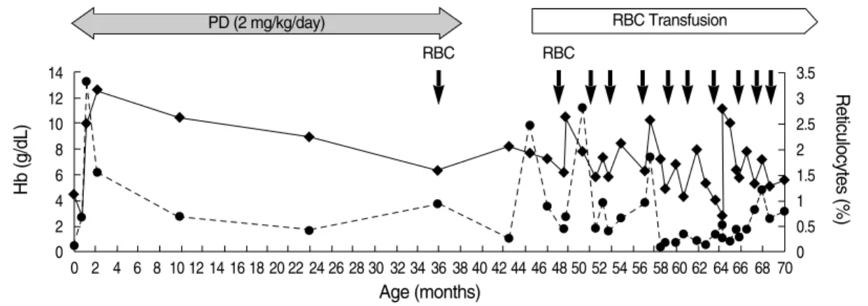
Fig. 2. This graph displays the changes of the hemoglobin level and reticulocytes counts. While on prednisolone from 2 months to 39 months of age, the hemoglobin levels increased with transient reticulocytosis, but later gradually decreased. Since 3 yr of age, she had been inter- mittently transfused, usually every 4 to 8 weeks. She eventually developed transfusion dependency.
Hb (g/dL)
14 12 10 8 6 4 2 0
Reticulocytes (%)
3.5 3 2.5 2 1.5 1 0.5 0 0 2 4 6 8 10 12 14 16 18 20 22 24 26 28 30 32 34 36 38 40 42 44 46 48 50 52 54 56 58 60 62 64 66 68 70
Age (months)
RBC RBC
RBC Transfusion
Reticulocytes (%) PD (2 mg/kg/day)
628 J.A. Park, Y.J. Lim, H.J. Park, et al.
multiplex short tandem repeat analysis (STR) performed with the AmpFlSTR Blue polymerase chain reaction (PCR) amplification kit (PE Applied Biosystems, Foster City, CA, USA) (5). As hoped for, the enolase activity (0.72 IU/g Hb;
normal control 3.4 IU/g Hb) on day 28 after BMT was higher than that of the low pre-transplant level (Fig. 3). At one year after BMT, her red cell enolase level had risen further to 6.54 IU/g Hb (normal control 7.76 IU/g Hb), and DNA analy- sis of the bone marrow aspirates showed a persistent stable mixed chimerism (recipient DNA 5.08%). She had no longer required any blood transfusion ever since the time of BMT.
DISCUSSION
Enolase deficiency is extremely rare, and its pathogenesis, inheritance and clinical manifestation have not been firmly established. Enolase deficiency is known to be associated with chromosome 1p locus mutations (1 pter-p36.13) and to cause chronic nonspecific hemolytic anemia (6). Lachant et al. (7) described four generations of a Caucasian family with heredi- tary red cell enolase deficiency. They reported that this dis- ease entity had a variety of clinical expression with autosomal dominant inheritance pattern. However, in our case, it doesn’t seem to correspond to the reported inheritance pattern.
DBA is also markedly heterogeneous with respect to patho- genesis, inheritance, and clinical manifestation. DBA is inher- ited in about 10-20% of cases, and genetic studies have iden- tified mutations in RPS19 (a ribosomal protein gene) in a quarter of cases (8). Recent studies have also suggested some other associated genes, i.e., chromosomes 8p, 10q (RPS24), 1q31 and 15 (RPS17) (9-11). However, in other cases, genetic linkage with DBA has yet to be identified. This fact suggests its genetic heterogeneity. Unfortunately, genetic screening for the patient and her family was not performed at the time of diagnosis although it could have shed some light in the understanding of the relationship between enolase deficien- cy and DBA.
Unlike the elevated erythrocyte enzyme activities report- ed both in hypoplasitic anemia and stress-induced environ- ment of erythropoiesis (2), our patient had a very low eno-
lase level, suggesting that DBA and enolase deficiency might also coexist. On the other hand, our patient may represent a hitherto undescribed disease entity expressing both defective erythropoiesis and enolase deficiency simultaneously.
Unlike our patient described here, the eADA level is known to be generally high in DBA patients. However, the role of eADA in the pathogenesis of DBA has not been established (12).
There’s a report of successful BMT in a child with a red cell pyruvate kinase (PK) deficiency (13). After transplant, the patient achieved a complete donor chimerism, normal hemo- globin level and normal red cell PK activity. On the other hand, our patient showed stable mixed chimerism. In non- malignant hematological disorders, it is believed that com- plete replacement of the recipient’s hematopoietic system is not necessarily required to improve the underlying disease state (14). Despite the danger of graft rejection in mixed chi- merism, it is known that even a small degree of mixed chime- rism may improve the disease status in some hematologic diseases (15, 16). To date, there are too few case reports of HSCT therapy results in RBC enzymopathies. However, as demonstrated in our case, mixed chimerism at an 89-95%
level may produce an acceptable outcome in the RBC eno- lase deficiency.
In summary, the case presented here uniquely illustrates that both red cell aplasia and red cell enzymopathy might coexist, and that successful marrow engrafment even with the mixed chimerism might correct both defective erythro- poiesis and the RBC enolase deficiency simultaneously.
ACKNOWLEDGEMENT
We thank the nursing staffs in the Hemtopoietic Stem Cell Transplantation Unit at the National Cancer Center, without whom this publication could have not been possible, and Dr.
Hee-Jin Kim for assaying the red cell enzyme levels as well as giving valuable comments regarding enolase enzymopathy.
Enolase (IU/g Hb)
Control (IU/g Hb)
Hb (g/dL)
MCV (fL)
MCH (pg)
Reticulo- cytes (%)
Patient 0.04 5.19 2.7 103.1 32.4 0.42
(pre BMT)
Father 0.09 3.22 17.8 91.5 32.3 0.95
Mother 0.32 3.22 12.9 32.5 34.4 0.65
Sister 6.4 4.2 12.6 80.8 28.1 1.56
Table 1. Red cell enolase levels of the patient, her family mem- bers, and of normal controls. This table also shows the CBC results of the family members
Fig. 3. This graph displays the observed erythrocyte enolase level increase that occurred after BMT.
Enolase (IU/g Hb)
9 8 7 6 5 4 3 2 1
0 pre-BMT post-BMT post-BMT post-BMT
D40 D166 D365
Patient Normal control
CBC, complete blood count; MCV, mean corpuscular volume; MCH, mean corpuscular hemoglobin; BMT, bone marrow transplantation.
BMT for Diamond-Blackfan anemia (DBA) Accompanying Red Cell Enolase Deficiency 629
REFERENCES
1. Da Costa L, Willig TN, Fixler J, Mohandas N, Tchernia G. Diamond- Blackfan anemia. Curr Opin Pediatr 2001; 13: 10-5.
2. Wang WC, Mentzer WC. Differentiation of transient erythroblastope- nia of childhood from congenital hypoplastic anemia. J Pediatr 1976;
88: 784-9.
3. Sung HJ, Kim SJ, Lee JH, Lee G, Lee KA, Choi CW, Kim BS, Kim JS. Persistent anemia in a patient with diffuse large B cell lymphoma:
pure red cell aplasia associated with latent Epstein-Barr virus infec- tion in bone marrow. J Korean Med Sci 2007; 22 (Suppl): S167-70.
4. Beutler E, Blume KG, Kaplan JC, Lo_hr GW, Ramot B, Valentine WN. International Committee for standardization in Haematology:
recommended methods for red-cell enzyme analysis. Br J Haematol 1977; 35: 331-40.
5. Wallin JM, Buoncristiani MR, Lazaruk KD, Fildes N, Holt CL, Walsh PS. TWGDAM validation of the AmpFISTR blue PCR amplification kit for forensic casework analysis. J Forensic Sci 1998; 43: 854-70.
6. Boulard-Heitzmann P, Boulard M, Tallineau C, Boivin P, Tanzer J, Bois M, Barriere M. Decreased red cell enolase activity in a 40-year- old woman with compensated haemolysis. Scand J Hematol 1984;
33: 401-4.
7. Lachant NA, Jennings MA, Tanaka KR. Partial erythrocyte enolase deficiency: a hereditary disorder with variable clinical expression.
Blood 1986; 65: 55A.
8. Perdahl EB, Naprstek BL, Wallace WC, Lipton JM. Erythroid fail- ure in Diamond-Blackfan anemia is characterized by apoptosis. Blood 1994; 83: 645-50.
9. Gazda H, Lipton JM, Willig TN, Ball S, Niemeyer CM, Tchernia G, Mohandas N, Daly MJ, Ploszynska A, Orfali KA, Vlachos A, Glad- er BE, Rokicka-Milewska R, Ohara A, Baker D, Pospisilova D, Web-
ber A, Viskochil DH, Nathan DG, Beggs AH, Sieff CA. Evidence for linkage of familial Diamond-Blackfan anemia to chromosome 8p23.3- p22 and for non-19q non-8p disease. Blood 2001; 97: 2145-50.
10. Cmejla R, Cmejlova J, Handrkova H, Petrak J, Pospisilova D. Ribo- somal protein S17 gene (RPS17) is mutated in Diamond-Blackfan anemia. Hum Mutat 2007; 23: 1178-82.
11. Quigley JG, Gazda H, Yang Z, Ball S, Sieff CA, Abkowitz JL. Inves- tigation of a putative role for FLVCR, a cytoplasmic heme exporter, in Diamond-Blackfan anemia. Blood Cells Mol Dis 2005; 35: 189-92.
12. Willig TN, Pe@rignon JL, Gustavsson P, Gane P, Draptchinskaya N, Testard H, Girot R, Debre@ M, Ste@phan JL, Chenel C, Cartron JP, Dahl N, Tchernia G. High adenosine deaminase level among healthy probands of Diamond Blackfan anemia (DBA) cosegregates with the DBA gene region on chromosome 19q13. The DBA Working Group of Societe d'Immunologie Pediatrique (SHIP). Blood 1998; 92:
4422-7.
13. Tanphaichitr VS, Suvatte V, Issaragrisil S, Mahasandana C, Veer- akul G, Chongkolwatana V, Waiyawuth W, Ideguchi H. Successful bone marrow transplantation in a child with red blood cell pyruvate kinase deficiency. Bone Marrow Transplant 2000; 26: 689-90.
14. Liesveld JL, Rothberg PG. Mixed chimerism in SCT: conflict or peace- ful coexistence? Bone Marrow Transplant 2008; 42: 297-310.
15. Wu CJ, Gladwin M, Tisdale J, Hsieh M, Law T, Biernacki M, Rogers S, Wang X, Walters M, Zahrieh D, Antin JH, Ritz J, Krishnamurti L. Mixed haematopoietic chimerism for sickle cell disease prevents intravascular haemolysis. Br J Haematol 2007; 139: 50-47.
16. Walters MC, Patience M, Leisenring W, Rogers ZR, Aquino VM, Buchanan GR, Roberts IA, Yeager AM, Hsu L, Adamkiewicz T, Kurtzberg J, Vichinsky E, Storer B, Storb R, Sullivan KM. Stable mixed hematopoietic chimerism after bone marrow transplantation for sickle cell anemia. Biol Blood Marrow Transplant 2001; 7: 665-73.