INTRODUCTION
Diseases that affect podocytes typically present with pro- teinuria, with or without nephrotic syndrome. However, it should be noted that not all cases with nephritic range pro- teinuria are caused by podocyte diseases, because the glomeru- lar filtration barrier also contains glomerular endothelial cells and glomerular basement membrane (GBM) (1). Moreover, fixed anionic charges on glomeruli are believed to form an important component of the glomerular filtration barrier. Thus several studies have been reported that the changes of anion- ic sites of the GBM might cause proteinuria (2-4). These stud- ies suggest that proteinuria can be induced by a combination of various factors, i.e., both structural and functional factors, and that it can be reduced by addressing the factors concerned.
Nonsteroidal anti-inflammatory drugs (NSAIDs) actively inhibit both cyclooxygenases (COX-1 and COX-2), and there- by block prostaglandin production. Anti-proteinuric effect of NSAIDs in nephrotic syndrome is presumed to be due to reductions in renal plasma flow and glomerular filtration rates.
Nevertheless, their use is limited by various side effects caused by COX-1 inhibition. Therefore the therapeutic applications of selective COX-2 inhibitor might result in more selective inhibition of inflammatory processes without influencing basal
COX-1-dependent prostaglandin synthesis (5, 6).
The 5-lipoxygenase (LOX) inhibitor, nordihydroguaiaret- ic acid (NDGA), is a typical lignin found in the creosote bush (Larrea tridentata or Larrea divarita) which has a worldwide distribution. These plants are used as traditional medicines for more than 50 illnesses, including rheumatism, diabetes, gallbladder and kidney stones, and sterility (7). LOX inhibitors have also been compared with other NSAIDs, COX inhibitors, and leukotriene inhibitors in terms of their anti-inflammato- ry, anti-thrombotic, and anti-platelet effects (8, 9). Recently it was reported that NDGA could reverse the oxidative stress, and reduce proteinuria in diabetic nephropathy rats (10).
Therefore we hypothesized that proteinuria might be relat- ed to both the COX and LOX pathways. The present study was designed to investigate the therapeutic efficacy of NDGA as compared with celecoxib in puromycin aminonucleoside (PAN) nephrosis rats.
MATERIALS AND METHODS Animals
Fifty five male Sprague-Dawley rats, which initially weighed
S183
Dong Won Lee*,�, Ihm Soo Kwak*,�, Soo Bong Lee*,�, Sang Heon Song*,�, Eun Young Seong*,�, Hyun Chul Chung� Byeong Yun Yang*, Min Young Lee�, and Mee Young Sol�
Department of Internal Medicine*, Pusan National University School of Medicine, Busan; Department of Internal Medicine�, Ulsan University Hospital, Ulsan;
Medical Research Institute�, Department of Pathology�, Pusan National University School of Medicine, Busan, Korea
Address for correspondence Ihm Soo Kwak, M.D.
Department of Internal Medicine, Pusan National University School of Medicine, 1-10 Ami-dong, Seo-gu, Busan 602-739, Korea
Tel : +82.51-240-7219, Fax : +82.51-254-3127 E-mail : [email protected]
DOI: 10.3346/jkms.2009.24.S1.S183
Effects of Celecoxib and Nordihydroguaiaretic Acid on Puromycin Aminonucleoside-Induced Nephrosis in the Rat
The selective cyclooxygenase-2 (COX-2) and 5-lipoxygenase (LOX) inhibitors might inhibit prostaglandin synthesis and reduce proteinuria. The present study was designed to investigate the anti-proteinuric effects of nordihydroguaiaretic acid (NDGA) as compared with celecoxib in puromycin aminonucleoside (PAN) nephrosis rats. Fifty five male Sprague-Dawley rats were divided into 4 groups; A, normal control; B, PAN group; C, PAN+COX-2 inhibitor (celecoxib) group; and D, PAN+5-LOX inhibitor (NDGA) group. After induction of PAN nephrosis through repeated injections of PAN (7.5 and 15 mg/100 g body weight), rats were treated with celecoxib, NDGA, or vehi- cle for 2 weeks. Twenty four hour urine protein excretions were significantly lower in PAN+celecoxib and PAN+NDGA groups than in PAN group. Serum creatinine (SCr) concentrations and 24 hr urine creatinine clearances (CCr) were not significantly different in the four groups. Electron microscopy showed that podocyte morphology was changed after the induction of PAN nephrosis and was recovered after celecox- ib or NDGA administration. Celecoxib significantly recovered the expressions of nephrin, CD2AP, COX-2, and TGF-β. NDGA also recovered TGF-βexpression, but did not alter the expressions of nephrin, CD2AP and COX-2. The present study suggest- ed that celecoxib and NDGA might effectively reduce proteinuria in nephrotic syn- drome without impairing renal function.
Key Words : Puromycin Aminonucleoside; Proteinuria; Celecoxib; Nordihydroguaiaretic Acid
Received : 23 July 2008 Accepted : 25 November 2008
250-300 g, were kept in individual metabolic cages with free access to standard chow and water. The rats were divided into 4 groups; A (n=10), normal control; B (n=15), PAN group;
C (n=15), PAN+celecoxib group; and D (n=15), PAN+
NDGA group. They were subjected to standard laboratory tests for 4 weeks before being sacrificed for kidney removal.
All experiments were performed in accordance with the NIH’s Guiding Principles in the Care and Use of Laboratory Animals.
Drugs and reagents
PAN (6-dimethylamino-9[3′amino-3′-deoxyribosyl]purine, Sigma Chemical Co., St. Louis, MO, U.S.A.) was prepared for i.p. injection (7.5 and 15 mg/100 g body weight). Cele- coxib (Celebrex�, Pfizer Inc., Seoul, Korea) was dissolved in drinking water for gavage (4 mg/100 g body weight/day).
NDGA (Sigma Chemical Co.) was prepared in dimethyl sul- foxide (DMSO, Sigma Chemical Co.) solution for s.c. injec- tion (1 mg/100 g body weight/day). All drugs administered i.p. or s.c. were prepared in a constant volume (0.1 mL/100 g body weight) at the beginning of each experiment.
Treatment schedule
After 3 days of acclimation, rats were injected i.p. with PAN 7.5 mg/100 g body weight (day 0), and then with 15 mg/100 g body weight 7 days later (day 7). Control animals received an identical volume of vehicle. On day 14, serum and 24 hr urine samples were obtained to confirm the onset of proteinuria in the nephrotic range.
After the induction of PAN nephrosis, rats were treated with celecoxib, NDGA, or vehicle from day 14 to 27. Fifteen nephrotic rats were gavaged daily with celecoxib (4 mg/100 g body weight/day), 15 were injected s.c. daily with NDGA (1 mg/100 g body weight/day), and 15 were injected s.c. with vehicle. Ten control rats were also injected s.c. with vehicle.
On days 21 and 28, serum and 24 hr urine samples were obtained to determine blood and urine chemistries. Twenty four hour urine creatinine clearances (CCr) was used as surro- gate of glomerular fitration rate (GFR) (Fig. 1).
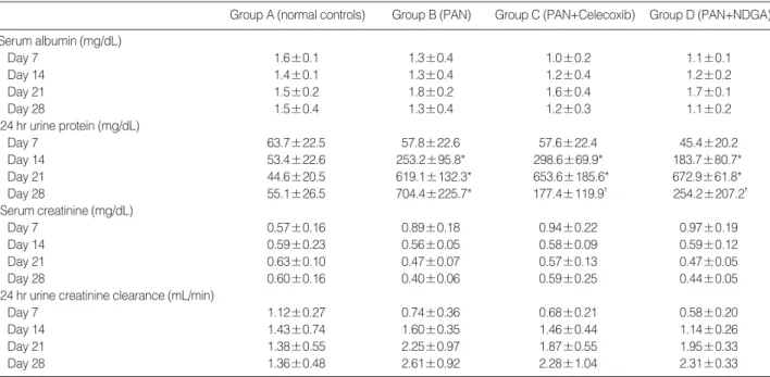
All values are expressed in mean±SD. *p<0.05 vs. group A, �p<0.05 vs. group B.
PAN, puromycin aminonucleoside; NDGA, nordihydroguaiaretic acid.
Group A (normal controls) Group B (PAN) Group C (PAN+Celecoxib) Group D (PAN+NDGA) Serum albumin (mg/dL)
Day 7 1.6±0.1 1.3±0.4 1.0±0.2 1.1±0.1
Day 14 1.4±0.1 1.3±0.4 1.2±0.4 1.2±0.2
Day 21 1.5±0.2 1.8±0.2 1.6±0.4 1.7±0.1
Day 28 1.5±0.4 1.3±0.4 1.2±0.3 1.1±0.2
24 hr urine protein (mg/dL)
Day 7 63.7±22.5 57.8±22.6 57.6±22.4 45.4±20.2
Day 14 53.4±22.6 253.2±95.8* 298.6±69.9* 183.7±80.7*
Day 21 44.6±20.5 619.1±132.3* 653.6±185.6* 672.9±61.8*
Day 28 55.1±26.5 704.4±225.7* 177.4±119.9� 254.2±207.2�
Serum creatinine (mg/dL)
Day 7 0.57±0.16 0.89±0.18 0.94±0.22 0.97±0.19
Day 14 0.59±0.23 0.56±0.05 0.58±0.09 0.59±0.12
Day 21 0.63±0.10 0.47±0.07 0.57±0.13 0.47±0.05
Day 28 0.60±0.16 0.40±0.06 0.59±0.25 0.44±0.05
24 hr urine creatinine clearance (mL/min)
Day 7 1.12±0.27 0.74±0.36 0.68±0.21 0.58±0.20
Day 14 1.43±0.74 1.60±0.35 1.46±0.44 1.14±0.26
Day 21 1.38±0.55 2.25±0.97 1.87±0.55 1.95±0.33
Day 28 1.36±0.48 2.61±0.92 2.28±1.04 2.31±0.33
Table 1. Changes in serum and 24 hr urine parameters in the four experimental groups
Fig. 1. Schematic of the treatment schedule. After three days of acclimation, test animals were injected intraperitoneally on day 0 with PAN 75 mg/kg, and then 7 days later with 150 mg/kg. Control animals were administered an identical volume of normal saline.
After inducing PAN nephrosis, all animals were treated with cele- coxib (40 mg/kg/day), NDGA (10 mg/kg/day), or vehicle up to Day 28. Serum and 24 hr urine samples were obtained on days 0, 7, 14, 21, and 28 ( ). All rats were sacrificed on day 28, and kidneys were removed immediately for histological examinations.
Normal controls
PAN
PAN+celecoxib
PAN+NDGA
Induction of
PAN nephrosis Celecoxib Vehicle
Day -3 0 7 14 21 28
PAN PAN Nephrectomy
7.5 mg/100 g 15 mg/100 g
Vehicle Saline
Accli- mation
NDGA
Kidney tissue preparation
On day 28, all rats were sacrificed under general anesthe- sia using i.p. injection of ketamine. Kidneys were removed immediately for histological examinations and for isolation of glomeruli. Kidney sections were fixed in 10% neutral-buffered formalin, blocked in paraffin wax, and stained with hema- toxylin-eosin (H&E) and periodic acid-Schiff (PAS) reagent.
To evaluate glomerular sclerotic changes, we observed at least 100 glomeruli in each section. For electron microscopy, kid- ney sections were fixed in 2.5% glutaraldehyde, postfixed in 1% osmium, and embedded in epoxy resin. Ultrathin sections were stained with uranyl acetate.
Isolation of glomeruli and reverse transcription-PCR Rats were perfused through left ventricles with saline. Kid-
neys were removed and cortices were finely minced, sieved through sequential sieves, and digested in collagenase. Total glomerular RNA was extracted using TRIzol�(Invitrogen, Carlsbad, CA, U.S.A.). The levels of the mRNAs of nephrin, CD2AP, COX-2, and TGF-βwere determined by reverse tran- scriptase-polymerase chain reaction (RT-PCR) using PCR Pre- Mix�(Bioneer, Daejeon, Korea) using the primers shown in Table 2.
Statistical analyses
All values are presented as means±SD. Repeated measures ANOVA and post-hoc analysis (Tukey test) were used for the analysis, and differences were considered significant when p values were <0.05.
RESULTS
Effects of celecoxib and NDGA on proteinuria
On day 7, seven days after the first PAN (7.5 mg/100 g body weight) injection, all rats in the treatment groups showed physiological proteinuria (63.7±22.5, 57.8±22.6, 57.6±
Gene Direction Sequence Size
GAPDH Forward GGCGTCTTCACCACCATGGAG 477 Reverse GCCTGCTTCACCACCTTCTTG
Nephrin Forward AGCCTCTTGACCATCGCTAA 293 Reverse GTCCTCGCCTTCAGCACCTG
CD2AP Forward CCAGCTCCAAAACCTGACC 496
Reverse GGGTTGGAGAATGTCCACC
COX-2 Forward CCAGTATCAGAACCGCATTGC 464 Reverse CAAGTTCTACCATGGTCTCCC
TGF-β1 Forward CAACAATTCCTGGCGATACC 468 Reverse GCTCCAAATGTAGGGGCAGGG Table 2. Sequences of PCR primers
Fig. 2. Effects of celecoxib and NDGA on 24 hr urine protein excre- tion in PAN-induced nephrosis rats. Twenty four hour urine protein excretion amounts were significantly lower in groups C and D on day 28 than in group B (*p<0.05 vs. group B). Group A, normal controls;
group B, a PAN group; group C, a PAN+celecoxib group; group D, a PAN+NDGA group.
24 hr urine protein (mg/day)
1,000
800
600
400
200
0
A B C D
Group
Day 7 Day 14 Day 21 Day 28
*
*
Fig. 3. Representative pathologic features of a PAN and a PAN+
celecoxib groups. (A) Light microscopic feature of a glomerulus in PAN group. Small segmental proliferations with matrix expansion and adhesions between the glomerular tuft and Bowman’s capsule were observed (PAS stain, ×300). (B) Electron microscopic fea- ture of mild foot processes changes (arrow) such as effacement, fusion and microvilli in PAN group. (C) Light microscopic feature of a glomerulus in PAN+celecoxib group. Slight expansion of mes- angial matrix was observed (PAS stain, ×300). (D) Electron micro- scopic feature of non-specific foot processes changes in PAN+
celecoxib group.
A B
C D
22.4, and 45.4±20.2 mg/24 hr, respectively) and increased weights (353.6±23.4, 338.6±28.8, 353.6±10.7, and 349.3
±20.9 g, respectively) versus their baseline weights (279.3
±10.6, 274.3±10.6, 291.4±8.0, and 290.7±12.4 g, res- pectively).
On day 14, seven days after the second PAN (15 mg/100 g body weight) injection, rats in groups B, C, and D exhibited pro- teinuria in the nephrotic range of 253.2±95.8, 298.6±69.9, and 183.7±80.7 mg/24 hr, whereas rats in group A showed physiological proteinuria of 63.7±22.5 mg/24 hr (p<0.05 vs.
group A). Mean serum albumin concentrations in groups B,
C, and D tended to be lower 1.3±0.4, 1.2±0.4, and 1.2± 0.2 g/dL, respectively, than in group A 1.4±0.1 g/dL.
On day 21, seven days after initiating celecoxib and NDGA treatment, group C and D rats still showed elevated levels of proteinuria (653.6±185.6 and 672.9±61.8 mg/24 hr, p<0.05 vs. group A).
On day 28, group C and D rats exhibited significant reduc- tions in proteinuria (177.4±119.9 and 254.2±207.2 mg/
24 hr), whereas group B rats showed a further increase in pro- teinuria (704.4±225.7 mg/24 hr, p<0.05 group C vs. group B and group D vs. group B, Table 1, Fig. 2).
Fig. 4. Effects of celecoxib and NDGA on nephrin, CD2AP, COX-2, and TGF-βmRNA expressions. (A) Nephrin mRNA expressions decreased after administering PAN in PAN group, and restored significantly in PAN+celecoxib group. (B) CD2AP mRNA expressions decreased in PAN group, and restored significantly in PAN+celecoxib group. (C) COX-2 mRNA expressions increased in PAN group, and restored significantly in PAN+celecoxib group. (D) TGF-βmRNA expressions increased in PAN group, and decreased significantly in PAN+cele- coxib and PAN+ NDGA groups. *p<0.05 vs. normal controls; �p<0.05 vs. PAN group.
A B
C D
Nephrin mRNA
1.4
1.2
1.0
0.8
0.6
0.4
0.2
0.0 A B C D
Group
*
�
COX-2 mRNA
1.4
1.2
1.0
0.8
0.6
0.4
0.2
0.0 A B C D
Group
*
�
CD2-AP mRNA
1.4
1.2
1.0
0.8
0.6
0.4
0.2
0.0 A B C D
Group
*
�
TGF-b mRNA
1.4
1.2
1.0
0.8
0.6
0.4
0.2
0.0 A B C D
Group
*
�
�
�
Effects of celecoxib and NDGA on renal functions
In groups C and D, SCr tended to decrease, and 24 hr urine CCr to increase. However there were no significant changes during the experiment (p>0.05, Table 1).
Effects of celecoxib and NDGA on renal morphology
Light microscopy revealed small segmental proliferations with matrix expansion and adhesions between the glomeru- lar tuft and Bowman’s capsule in group B, C, and D. How- ever, focal segmental or global sclerotic lesions were not ob- served. Electron microscopy revealed variable degrees of foot processes fusion, effacement, and microvillous transformation in group B, but not in groups C and D on day 28 (Fig. 3).
Effects of Celecoxib and NDGA on the expressions of nephrin, CD2AP, COX-2, and TGF-ββ
PAN significantly reduced the expressions of nephrin and CD2AP, and increased the expressions of COX-2 and TGF-β (p<0.05 vs. group A, respectively). However, celecoxib sig- nificantly restored the expressions of nephrin, CD2AP, COX- 2, and TGF-β(p<0.05 vs. group B, respectively). NDGA also restored TGF-βexpression (p<0.05 vs. group B), but did not alter the expressions of nephrin, CD2AP and COX-2 (Fig. 4).
DISCUSSION
The PAN nephrosis model used in the present study, is a well-described animal model of human idiopathic nephrotic syndrome, and has been used extensively to study the fun- damental processes of proteinuria (11-13). PAN is a nephro- toxin and may injure podocytes and alter the selectivity of the GBM. However, the precise mechanisms that underlie PAN-induced proteinuria are not well understood.
Sprague-Dawley rats that were injected with a single dose of PAN developed significant proteinuria several days later.
In PAN-induced nephrosis, the onset of proteinuria coincides with the development of focal defects in podocyte epithelium, which expose the outer surface of the GBM to the urinary space (14, 15). However these morphological and functional changes are recovered within 1-2 weeks after the induction of PAN nephrosis. Recent studies showed that the repeated injections of PAN induced specific focal glomerulosclerosis lesions, but with an incidence of only 10-30% (16-19). There- fore we attempted to repeat injections twice with an interval of seven days to maintain the proteinuria in the nephrotic range with the absence of self-reduction over the course of the exper- iments. In the present study, after the first PAN injection (7.5 mg/100 g body weight), no significant increase in protein- uria was observed, but the second injection (15 mg/100 g body weight) induced a significant and sustained increase in pro-
teinuria over a period of two weeks. However, no glomeru- losclerotic lesions were found in the present study.
In models of progressive glomerular injury, COX-2 inhibitors decrease proteinuria and retard progressive glomerulosclero- sis (20-22). LOX inhibitors have also been studied as poten- tial anti-inflammatory agents with anti-thrombotic and anti- platelet effects (8, 9). In the present study, anti-proteinuric effects of celecoxib and NDGA suggest that hemodynamic improvements are associated with eicosanoids in the arachi- donic acid pathway, and with anti-inflammatory and immune modulating effects. On the contrary to the previous reports, our study showed the sustained increase in 24 hr urine CCr in PAN-treated groups (B, C, and D). These findings are th- ought to be the results of hyperfiltration in the early stage of acute kidney injury caused by PAN injections. LOX inhibitors have been reported to have protective effects on ileal motor disturbances induced by endotoxins, to promote adaptation following massive bowel resection, and to promote correction of coronary spasms of immune origin (23-25). These findings suggest that LOX inhibitors can reduce proteinuria due to their anti-inflammatory and protective effects on vascular sm- ooth muscle.
PAN causes podocyte injury and reduces the expressions of nephrin and CD2AP. In the present study, celecoxib restored the expressions of nephrin and CD2AP, but NDGA did not alter their expressions. Furthermore, whereas celecoxib restored COX-2 expression, NDGA increased it through complemen- tary enhancing the COX pathway. It appears that overexpres- sion of COX-2 by NDGA predisposed podocyte injury, which negated its beneficial anti-inflammatory effect (26). PAN caused focal mesangial expansion and fibrosis, and increased the expression of TGF-β. TGF-βcan cause extracellular matrix (ECM) accumulation by enhancing the productions of colla- gen and fibronectin by glomerular mesangial cell, by suppress- ing the expression of ECM-degrading protease, and by increas- ing the synthesis of ECM protease inhibitors (27-29). In this study, we found that celecoxib and NDGA reduced TGF-β expression, which suggests the possible involvements of the COX and LOX pathways in TGF-β-induced ECM accumu- lation and proteinuria.
In conclusion, our results support the involvements of the COX and LOX pathways in PAN-induced proteinuria. Cele- coxib and NDGA could reduce proteinuria without impair- ing renal function, by promoting hemodynamic and anti-in- flammatory improvements. In particular, celecoxib might reduce proteinuria, restore the expressions of nephrin and CD2AP, and suppress the expressions of COX-2 and TGF-β. Further investigations should be conducted to elucidate the roles of these pathways in the pathogenesis of proteinuria, as this would help identify the anti-proteinuric effects of cele- coxib and NDGA in progressive glomerular disease.
REFERENCES
1. Shankland SJ. The podocyte’s response to injury: role in proteinuria and glomerulosclerosis. Kidney Int 2006; 69: 2131-47.
2. Mahan JD, Sisson RS, Vernier RL. Glomerular basement membrane anionic charge site change early in aminonucleoside nephrosis. Am J Pathol 1986; 125: 393-401.
3. Furness PN, Turner DR, Cotton RE. Basement membrane charge in human glomerular disease. J Pathol 1986; 150: 267-78.
4. Washizawa K, Kasai S, Mori T, Komiyama A, Shigematsu H. Ultra- structural alteration of glomerular anionic sites in nephrotic patients.
Pediatr Nephrol 1993; 7: 1-5.
5. Blume C, Heise G, Mhlfeld A, Bach D, Schrr K, Gerhardz CD, Gra- bensee B, Heering P. Effects of flosulide, a selective cyclooxygenase 2 inhibitor, on passive Heymann nephritis in the rat. Kidney Int 1999;
56: 1770-8.
6. Emery P, Vane J, Botting J, Botting R, eds. Pharmacology, safety data and therapeutics of COX-2 inhibitors, in improved non-steroidal anti-inflammatory drugs, COX-2 enzyme inhibitors, Hingham, MA:
Kluwer, 1996; 229-41.
7. Arteaga S, Andrade-Cetto A, Crdenas R. Larrea tridentata (Creosote bush), an abundant plant of Mexican and US-American deserts and its metabolite nordihydroguaiaretic acid. J Ethnopharmacol 2005;
98: 231-9.
8. Tries S, Laufer S, Radziwon P, Breddin HK. Antithrombotic and platelet function inhibiting effects of ML3000, a new anti-inflammatory drug with COX/5-LOX inhibitory activity. Inflamm Res 2002; 51: 129-34.
9. Salari H, Braquet P, Borgeat P. Comparative effects of indomethacin, acetylenic acids, 15-HETE, NDGA and BW755C on the metabolism of arachidonic acid in human leukocytes and platelets. Prostaglandins Leukot Med 1984; 13: 53-60.
10. Anjaneyulu M, Chopra K. Nordihydroguairetic acid, a lignin prevents oxidative stress and the development of diabetic nephropathy in rats.
Pharmacology 2004; 72: 42-50.
11. Frenk S, Antonowicz I, Craig JM, Metcoff J. Experimental nephrotic syndrome induced in rats by aminonucleoside. Renal lesion and body electrolyte composition. Proc Soc Exp Biol Med 1955; 89: 424-7.
12. Fishman JA, Karnovsky MJ. Effects of the aminonucleoside: puromycin on glomerular epithelial cells in vitro. Am J Pathol 1985; 118: 398- 407.
13. Bertram JF, Messina A, Ryan GB. In vitro effects of puromycin ami- nonucleoside on the ultrastructure of rat glomerular podocytes. Cell Tissue Res 1990; 260: 555-63.
14. Farquhar MG, Palade GE. Glomerular permeability: II. ferritin trans- fer across the glomerular capillary wall in nephrotic rats. J Exp Med 1961; 114: 699-716.
15. Furness PN, Harris K. An evaluation of experimental models of glomeru- lonephritis. Int J Exp Pathol 1994; 75: 9-22.
16. Grond J, Weening JJ, Elema JD. Glomerular sclerosis in nephrotic rats. Comparison of the long-term effects of adriamycin and aminonu-
cleoside. Lab Invest 1984; 51: 277-85.
17. Marinides GN, Groggel GC, Cohen AH, Border WA. Enalapril and low protein reverse chronic puromycin aminonucleoside nephropa- thy. Kidney Int 1990; 37: 749-57.
18. Liu S, Li Y, Zhao H, Chen D, Huang Q, Wang S, Zou W, Zhang Y, Li X, Huang H. Increase in extracellular cross-linking by tissue trans- glutaminase and reduction in expression of MMP-9 contribute dif- ferentially to focal segmental glomerulosclerosis in rats. Mol Cell Biochem 2006; 284: 9-17.
19. Hagiwara M, Yamagata K, Capaldi RA, Koyama A. Mitochondrial dysfunction in focal segmental glomerulosclerosis of puromycin ami- nonucleoside nephrosis. Kidney Int 2006; 69: 1146-52.
20. Wang JL, Cheng HF, Zhang MZ, McKanna JA, Harris RC. Selective increase of cyclooxygenase-2 expression in a model of renal ablation.
Am J Physiol 1998; 275: F613-22.
21. Wang JL, Cheng HF, Shappell S, Harris RC. A selective cyclooxyge- nase-2 inhibitor decreases proteinuria and retards progressive renal injury in rats. Kidney Int 2000; 57: 2334-42.
22. Cheng HF, Wang CJ, Moeckel GW, Zhang MZ, McKanna JA, Har- ris RC. Cyclooxygenase-2 inhibitor blocks expression of mediators of renal injury in a model of diabetes and hypertension. Kidney Int 2002; 62: 929-39.
23. Ezberci F, Tekin E, Onuk E. Protective effect of a lipoxygenase inhibitor, nordihydroguaiaretic acid, on the ileal motor disturbances induced by Serratia marcescens endotoxin in rats. Surg Today 2001; 31: 497- 501.
24. Kollman-Bauerly KA, Thomas DL, Adrian TE, Lien EL, Vanderhoof JA. The role of eicosanoids in the process of adaptation following mas- sive bowel resection in the rat. JPEN J Parenter Enteral Nutr 2001;
25: 275-81.
25. Marchenko HI, Kotsiuruba VM, Butovych IA, Sorochyns’kyi AE, Zrazhevs’ka VK, Tumanovs’ka LV. The correction of disorders in arachidonic acid metabolism in coronary spasm of an immune ori- gin. Frziol Zh 1994; 40: 81-7.
26. Cheng H, Wang S, Jo YI, Hao CM, Zhang M, Fan X, Kennedy C, Breyer MD, Moeckel GW, Harris RC. Overexpression of cyclooxy- genase -2 predispodes to podocyte injury. J Am Soc Nephrol 2007;
18: 551-9.
27. Poncelet AC, Schnaper HW. Regulation of human mesangial cell collagen expression by transforming growth factor-β1. Am J Physi- ol 1998; 275: F458-66.
28. Suzuki S, Ebihara I, Tomino Y, Koide H. Transcriptional activation of matrix genes by transforming growth factor-beta 1 in mesangial cells. Exp Nephrol 1993; 1: 229-37.
29. McKay NG, Khong TF, Haites NE, Power DA. The effects of trans- forming growth factor-beta 1 on mesangial cell fibronectin synthe- sis: increased incorporation into the extracellular matrix and reduced pI but no effect on alternative splicing. Exp Mol Pathol 1993; 59:
211-24.