Original Article
Feasibility Study of Patient Specific Quality Assurance Using Transit Dosimetry Based on Measurement with an Electronic Portal Imaging Device
Tae Seong Baek*, Eun Ji Chung*, Jaeman Son†, Myonggeun Yoon†
*Department of Radiation Oncology, National Health Insurance Co. Ilsan Hospital, Goyang, †Department of Bio-convergence Engineering, Korea University, Seoul, Korea
Copyright © 2017 Korean Society of Medical Physics
CCThis is an Open-Access article distributed under the terms of the Creative Commons Attribution Non-Commercial License (http://creativecommons.org/licenses/by- nc/4.0) which permits unrestricted non-commercial use, distribution, and reproduction in any medium, provided the original work is properly cited.
Introduction
Novel radiotherapy methods, including intensity modulated radiotherapy (IMRT), volumetric modulated arc therapy (VMAT) and heavy ion therapy, all of which are more advanced modalities than conventional 3D conformal radiation therapy, have recently been implemented clinically. These methods reduce exposure to radiation of a patient’s normal tissues and surrounding organs while increasing radiation dose to tumors.1-3) For
treatment purposes, the radiation dose calculated from a treatment planning system (TPS) should be accurate. In addition, care should be taken during radiation delivery because IMRT consists of a complex delivery system, with treatment beams having large monitor units (MUs), which may cause critical damage if errors occur during treatment.
Therefore, a quality assurance (QA) program is essential to verify the difference between dose distribution calculated from TPS and the actual dose distribution during the complex IMRT treatment procedure, which involves many
Progress in Medical Physics 28(2), June 2017 https://doi.org/10.14316/pmp.2017.28.2.54 pISSN 2508-4445, eISSN 2508-4453
This study was designed to measure transit dose with an electronic portal imaging device (EPID) in eight patients treated with intensity modulated radiotherapy (IMRT), and to verify the accuracy of dose delivery to patients. The calculated dose map of the treatment planning system (TPS) was compared with the EPID based dose measured on the same plane with a gamma index method.
The plan for each patient was verified prior to treatment with a diode array (MapCHECK) and portal dose image prediction (PDIP). To simulate possible patient positioning errors during treatment, outcomes were evaluated after an anthropomorphic phantom was displaced 5 and 10 mm in various directions. Based on 3%/3 mm criteria, the mean±SD passing rates of MapCHECK, PDIP (pre-treatment QA) for 47 IMRT were 99.8±0.1%, 99.0±0.7%, and, respectively. Besides, passing rates using transit dosimetry was 90.0±1.5% for the same condition. Setup errors of 5 and 10 mm reduced the mean passing rates by 1.3% and 3.0% (inferior to superior), 2.2% and 4.3% (superior to inferior), 5.9% and 10.9% (left to right), and 8.9% and 16.3% (right to left), respectively. These findings suggest that the transit dose-based IMRT verification method using EPID, in which the transit dose from patients is compared with the dose map calculated from the TPS, may be useful in verifying various errors including setup and/or patient positioning error, inhomogeneity and target motions.
Keywords: Linear accelerator, Transit dose, EPID, Gamma index Received 30 June 2017
Revised 10 July 2017 Accepted 10 July 2017
Corresponding author Myonggeun Yoon ([email protected]) Tel: 82-2-3290-5651 Fax: 82-2-921-6434
PMP
does not consider the heterogeneity in real patients, such as air cavities and bony structures,7,8) it cannot assure the accuracy of the actual dose distribution to patients.
Nevertheless, the estimated value from TPS and the measured value from QA are consistent. Portal dose image prediction (PDIP) using an electronic portal imaging device (EPID) was recently implemented clinically for pre-treatment QA.9,10) This QA method can also verify the difference between the dose distribution calculated from TPS and the dose distribution actually delivered, thus providing accurate IMB delivery. Furthermore, conventional pre-treatment IMRT QA methods do not provide information on actual beam delivery during treatment, since various errors, such as patient movement during treatment, cannot be determined. Therefore, current QA methods may be considered unacceptable in verifying the actual dose delivered to patients.11,12)
These drawbacks of current QA methods may be eliminated by transit dosimetry, measured using 2D detectors in the treatment room. To date, various studies have assessed transit dosimetry at the position of the EPID behind a patient.13-15) Comparisons of dose distributions by transit dosimetry in homogeneous solid water phantoms and heterogeneous anthropomorphic phantoms showed
delivered to patients by detecting random and systematic errors during treatment.
Materials and Methods
Eight patients undergoing IMRT were selected (Table 1).
During treatment, the transit dose of each treatment field was measured using an aS1000 EPID attached to a Varian Clinac iX linear accelerator (Varian Medical Systems, Palo Alto, CA) (Fig. 1). According to the vendor’s guidelines, EPID was calibrated for transit dose analysis with a dark field, a flood field and a diagonal profile correction at dmax
in water for a 40×40 cm2 open field. The EPID response was scaled such that 1 Calibrated Unit (CU) corresponded to 100 MU delivered by a 10×10 cm2 open field at 100 cm source-to-detector distance (SDD). The estimated non- uniform back-scatter pattern was corrected to remove any non-uniform backscattering pattern during the calibration process. The absolute dose of the transit beam was first measured with EPID in calibrated units (CU) and converted to the absorbed dose using the calibration curve.
The EPID system used in this study was an amorphous silicon flat panel imager type, with an active imaging area measuring 30×40 cm2, and a field of view at the isocenter
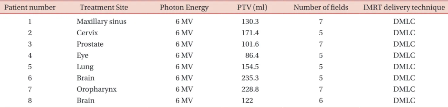
Table 1. Demographic and radiologic characteristics of the eight patients analyzed in this study.
Patient number Treatment Site Photon Energy PTV (ml) Number of fields IMRT delivery technique
1 Maxillary sinus 6 MV 130.3 7 DMLC
2 Cervix 6 MV 171.4 5 DMLC
3 Prostate 6 MV 101.6 7 DMLC
4 Eye 6 MV 86.4 5 DMLC
5 Lung 6 MV 154.5 5 DMLC
6 Brain 6 MV 235.3 5 DMLC
7 Oropharynx 6 MV 228.8 7 DMLC
8 Brain 6 MV 122 6 DMLC
DMLC: Dynamic Multileaf collimator.
from 25×33 cm2 to 16×22 cm2. This EPID has a 1,024×768 pixel matrix, 0.392 mm of pixel pitch, and an energy range of 4−25 MV. The transit doses were measured using 6 MV beams at a source-to-detector distance (SDD) of 150 cm.
Planned dose distributions were calculated with the Eclipse treatment planning system Ver 8.9. (Varian Medical Systems, Salt Lake City, UT, USA), using an anisotropic analytical algorithm (AAA). Conventional pre-treatment QAs were determined using MapCHECK (Sun Nuclear, Melbourne, FL) and Portal Dose Image Prediction (PDIP).
MapCHECK has 1527 diode detectors with 7.07 mm uniform spacing across an area of 32×26 cm2. Each detector
has active area of 0.8×0.8 cm2, a thickness of 2.0 g/cm2 and a thickness of backscatter of 2.75 g/cm2. To simulate possible setup error situation during treatment, transit doses were measured after displacing the heterogeneous anthropomorphic phantom from its original position by 5 and 10 mm in the superior to inferior (SI), inferior to superior (IS), right to left (RL), and left to right (LR) directions.
The gamma evaluation method was used to compare doses calculated from TPS with measured doses, using 3%
dose difference (DD) and 3 mm distance-to-agreement (DTA) criteria.18)
Table 2. Pre-treatment passing rates (%) of results measured with MapCHECK (M) and PDIP (P) based on the gamma index for the eight included patients.
Patient No.
Field number
Dosimetric tool 1 2 3 4 5 6 7 Mean
1 M 100.0 99.8 100.0 100.0 99.7 97.7 100.0 99.6
P 99.5 99.3 96.0 99.4 99.6 98.6 95.7 98.3
2 M 99.1 100.0 100.0 99.6 100.0 99.7
P 99.9 96.9 99.7 99.9 99.9 99.3
3 M 100.0 99.8 100.0 98.9 100.0 99.8 100.0 99.8
P 99.9 99.8 100.0 97.0 98.9 97.9 99.9 99.1
4 M 100.0 100.0 100.0 100.0 100.0 100.0
P 99.9 98.7 100.0 98.4 99.9 99.4
5 M 99.8 100.0 99.1 100.0 100.0 99.8
P 99.7 100.0 98.5 99.9 99.9 99.6
6 M 100.0 100.0 100.0 100.0 100.0 100.0
P 99.9 99.9 100.0 99.9 99.9 99.9
7 M 98.9 100.0 99.8 100.0 99.4 100.0 99.8 99.7
P 97.7 100.0 99.0 99.4 98.8 98.9 97.4 98.7
8 M 99.1 98.9 100.0 100.0 100.0 99.4 99.6
P 96.4 95.8 95.2 99.8 98.8 98.7 97.5
a b
Fig. 1. EPID measurements of transit doses during treatment at the posterior surfaces of Patients (a) 1 (maxillary sinus) and (b) 3 (prostate).
respectively, showing that the doses measured on the homogeneous phantom (i.e., MapCHECK) and on the fluence map with EPID were well matched with doses calculated from TPS.
Fig. 2A and B show the calculated and measured dose maps, respectively, of transit doses passing through field 3 of patient 1 in Table 2. For this field, the passing rate was 90.5%, indicating that the transit dose map measured with the EPID was relatively well matched with the dose distribution calculated by the TPS. Table 3 shows the
dosimetry than conventional pre-treatment IMRT as a QA tool. To determine whether transit dosimetry could detect random errors during treatment, radiation was deliberately delivered with a setup error and the changes in the transit dose distribution were evaluated.
Discussion
This study compared the transit doses of 47 IMRT fields measured with an EPID with the doses calculated from
a b
Fig. 2. Transit doses in field 3 from Patient 1 (a) calculated from TPS and (b) measured using EPID.
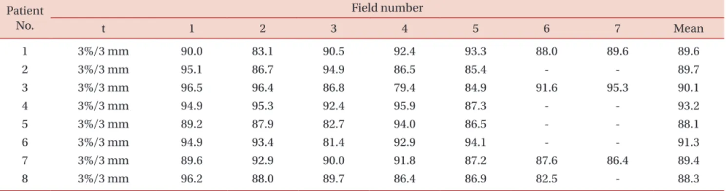
Table 3. Passing rates of results measured using transit dosimetry based on the gamma index values for the eight patients.
Patient No.
Field number
t 1 2 3 4 5 6 7 Mean
1 3%/3 mm 90.0 83.1 90.5 92.4 93.3 88.0 89.6 89.6
2 3%/3 mm 95.1 86.7 94.9 86.5 85.4 - - 89.7
3 3%/3 mm 96.5 96.4 86.8 79.4 84.9 91.6 95.3 90.1
4 3%/3 mm 94.9 95.3 92.4 95.9 87.3 - - 93.2
5 3%/3 mm 89.2 87.9 82.7 94.0 86.5 - - 88.1
6 3%/3 mm 94.9 93.4 81.4 92.9 94.1 - - 91.3
7 3%/3 mm 89.6 92.9 90.0 91.8 87.2 87.6 86.4 89.4
8 3%/3 mm 96.2 88.0 89.7 86.4 86.9 82.5 - 88.3
TPS. Although the measured and calculated transit doses were relatively well matched, the passing rates in some treatment fields were lower than in others. For example, the mean passing rate of five IMRT fields in patient 5 was 88.1±3.7%, much lower than the mean passing rate of 93.2±3.2% for five IMRT fields in patient 4. Reduced passing rates observed in actual patients were likely due to
the inaccuracy of TPS calculations of inhomogeneity. That is, although the calculation algorithm of the TPS worked well for homogeneous phantoms, it was less accurate for inhomogeneous materials such as the human body. Fig. 3 presents an example of a GI map, showing the high passing rates with MapCHECK and PDIP but a relatively lower passing rate with EPID data of an actual patient.
a b
1.58 1.4 1.2 1 0.8 0.6 0.4 0.2 0.0 2.5
2 1.5 1 0.5
0c d
Fig. 3. Dose maps calculated in field 3 of Patient 2 by (a) MapCHECK and (b) PDIP and measured by transit dosimetry at (c) 3%/3 mm and (d) 5%/3 mm.
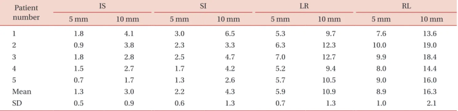
Table 4. Decreases in passing rates for phantom movement compared with the original passing rate without movement.
Patient number
IS SI LR RL
5 mm 10 mm 5 mm 10 mm 5 mm 10 mm 5 mm 10 mm
1 1.8 4.1 3.0 6.5 5.3 9.7 7.6 13.6
2 0.9 3.8 2.3 3.3 6.3 12.3 10.0 19.0
3 1.8 2.8 2.5 4.7 7.0 12.7 9.9 18.4
4 1.5 2.7 1.7 4.2 5.2 9.4 8.0 14.4
5 0.7 1.7 1.3 2.6 5.7 10.5 9.0 16.0
Mean 1.3 3.0 2.2 4.3 5.9 10.9 8.9 16.3
SD 0.5 0.9 0.6 1.3 0.7 1.3 1.0 2.1
verifying setup and/or patient positioning errors caused by patient movements and resulting in inter- or intra- fractional target motions.
Conclusion
This study used EPID to evaluate transit dosimetry based IMRT QA. While the IMRT QA results measured with MapCHECK and PDIP agreed well with the calculated dose, the failure rate was noticeably higher for actual patients. These findings suggest that conventional IMRT QA using homogeneity phantom may not be sufficiently accurate if inhomogeneities are present in the beam path. Our experimental results indicate that transit dosimetry may provide more accurate QA of IMRT plans than conventional IMRT QA and may also be used as a monitoring tool to verify dose delivery during treatment.
Acknowledgements
This work was supported by the Nuclear Safety Research Program through the Korea Foundation of Nuclear Safety (KOFONS), granted financial resource from the Nuclear Safety and Security Commission (NSSC), Republic of Korea (No. 1305033).
Conflicts of Interest
The authors have nothing to disclose.
Availability of Data and Materials
All relevant data are within the paper and its Supporting Information files.
1987;13:1255-60.
3. Morrill SM, Lane RG, Wong JA, Rosen II. Dose‐volume considerations with linear programming optimization.
Med Phys. 1991;18:1201-10.
4. Low DA, Moran JM, Dempsey JF, Dong L, Oldham M.
Dosimetry tools and techniques for imrt. Med Phys.
2011;38:1313-38.
5. Olch AJ. Dosimetric performance of an enhanced dose range radiographic film for intensity-modulated radiation therapy quality assurance. Med Phys. 2002;29:2159-68.
6. Ma C, Jiang S, Pawlicki T, Chen Y, Li J, Deng J et al. A quality assurance phantom for imrt dose verification.
Phys Med Biol. 2003;48:561.
7. Berry SL, Sheu R-D, Polvorosa CS, Wuu C-S. Implemen- tation of epid transit dosimetry based on a through-air dosimetry algorithm. Med Phys. 2012;39:87-98.
8. Saitoh H, Fujisaki T, Sakai R, Kunieda E. Dose distribution of narrow beam irradiation for small lung tumor. Int J Radiat Oncol Biol Phys. 2002;53:1380-87.
9. Van Esch A, Depuydt T, Huyskens DP. The use of an asi- based epid for routine absolute dosimetric pre-treatment verification of dynamic imrt fields. Radiother Oncol.
2004;71:223-34.
10. McDermott L, Wendling M, Van Asselen B, Stroom J, Sonke J-J, Van Herk M et al. Clinical experience with epid dosimetry for prostate imrt pre-treatment dose verifica- tion. Med Phys. 2006;33:3921-30.
11. Van Esch A, Vanstraelen B, Verstraete J, Kutcher G, Huyskens D. Pre-treatment dosimetric verification by means of a liquid-filled electronic portal imaging device during dynamic delivery of intensity modulated treatment fields. Radiother Oncol. 2001;60:181-90.
12. McDermott LN, Wendling M, Sonke J-J, van Herk M, Mijnheer BJ. Replacing pretreatment verification with in vivo epid dosimetry for prostate imrt. Int J Radiat Oncol
Biol Phys. 2007;67:1568-77.
13. Berry SL, Polvorosa C, Cheng S, Deutsch I, Chao KC, Wuu C-S. Initial clinical experience performing patient treatment verification with an electronic portal imaging device transit dosimeter. Int J Radiat Oncol Biol Phys.
2014;88:204-09.
14. Oldham M, Sakhalkar H, Guo P, Adamovics J. An investi- gation of the accuracy of an imrt dose distribution using two-and three-dimensional dosimetry techniques. Med Phys. 2008;35:2072-80.
15. Sabet M, Rowshanfarzad P, Vial P, Menk FW, Greer PB.
Transit dosimetry in imrt with an a-si epid in direct detection configuration. Phys Med Biol. 2012;57:N295.
16. Baek TS, Chung EJ, Koh EK, Seo J, Yoon M. Evaluation of the accuracy of dose delivery for imrt based on transit dosimetry. Health Phys. 2014;107:200-5.
17. Baek TS, Chung EJ, Son J, Yoon M. Feasibility study on the verification of actual beam delivery in a treatment room using epid transit dosimetry. Radiat Oncol. 2014;9:273.
18. Low DA, Harms WB, Mutic S, Purdy JA. A technique for the quantitative evaluation of dose distributions. Med Phys.
1998;25:656-61.